Exposures Exam II
 Call Kai
 Learn
 Practice Test
 Spaced Repetition
 Match
 Flashcards
 Knowt Play
1/83

There's no tags or description
Looks like no tags are added yet.
Name | Mastery | Learn | Test | Matching | Spaced | Call with Kai | Chat |
|---|
No analytics yet
Send a link to your students to track their progress
84 Terms
Ways to Reduce Patient Exposure
proper body or part immobilization
motion reduction techniques
collimation (appropriate beam limitation devices)
adequate filtration of the x-ray beam
gonadal or other specific area shielding
suitable technical exposure factors in with computer-generated digital images
appropriate digital image processing
eliminate repeats
Effective Communication
when verbal and nonverbal messages are understood as intended
Immobilization: Consequences, Minimization, Types + Examples
Consequences: repeated & additional exposure
Minimized by: proper body or part immobilization & use of motion reduction techniques
Types of motion: voluntary & involuntary
children, conditions, dementia
Protective Shielding (Parts & Steps)
Parts to Shield: eye lens, breasts, gonads & thyroid
Steps: collimate THEN shield
JRCERT on Protective Shielding
do not use shielding for abdominopelvic radiography exams when it could interfere with diagnostic value and may lead to repeats
Gonadal Shielding + Types
Female gonads receive ~3x more exposure during exams than males
Types
flat contact - like a blanket
shadow - piece of metal placed right below tube
shaped contact - cup-like shape (may be held in place by suitable carrier)
clear lead
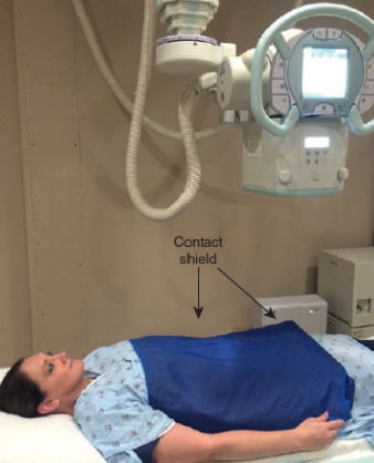
What kind of shielding is this?
Flat contact shield

What kind of shielding is this?
Shadow shield

What kind of shielding is this?
Clear lead shield (shadow shield)

What kind of shielding is this?
Shaped contact shield

What kind of shielding is this?
Clear lead shield
Specific Area Shielding
Shielding used for radiosensitive organs and tissues other than reproductive organs
use clear lead shadow shield for breasts & gonads
used in conjunction with PA than AP
minimizes the number of potentially deleterious x-ray induced mutations expressed in future generations
Repeat Images
“double dose” to skin & possibly gonads
Repeat Analysis Program + Reasons
By determining # & reasons of repeats, existing problems & conditions in an imaging department will be identified
Reasons
mispositioning
incorrect centering of CR
motion during exposure
incorrect collimation
presence of external foreign bodies
postprocessing artifacts
Unnecessary Radiologic Procedures
benefit vs. risk lays on the referring physician’s responsibility to order it
exams performed in the absence of definite medical indications (no benefit for the patient in terms of useful information gained)
Unnecessary Radiologic Procedures
chest X-ray upon automatic hospital admission
chest X-ray for preemployment physical
lumbar spine exam for preemployment physical
chest X-ray for routine health checkup
chest X-ray for TB screening
whole-body CT
Specifying Radiation Amount Received from Diagnostic Imaging Procedures
entrance skin exposure (ESE)
includes skin & glandular
most frequently used because the simplest to determine
use of TLDs to measure skin directly
bone marrow dose (estimated)
gonadal dose
GSD
Genetically Significant Dose
used to assess impact of gonadal dose
equivalent to EqD to gonads that, if received by every human, would expect to bring identical gross genetic injury to total population
GSD for U.S. population: 0.20 mSv
FGP
Fluoroscopically Guided Positioning
ASRT DOES NOT CONDONE
causes excessive unneccessary exposure
Protecting Potentially Pregnant Patients
ask patient the date of last mentrual period (LMP)
fine if within 10 days
ordering physician may order pregnancy test (if receiving substantial pelvic iradiation) & obtain results BEFORE pelvis is irradiated
ordering physician (or patient) determines to do or not to do X-ray examination
ACR’s Position on Abdominal Radiologic Exams on Female Patients
Abdominal exams requested after full consideration of patient’s clinical status including pregnancy cannot be postponed or be scheduled
Protecting KNOWN Pregnant Patients
if physician feels it’s best to undergo radiologic examination, perform without delay
take special effort to minimize dose of radiation in lower abdomen & pelvic region
restrictively select minimal exposure factors for useful image
precisely collimate
if patient’s lower abdomen & pelvic region don’t need to be included, SHIELD
also shield ovaries & uterus less than 5 cm from any edge of field
Position of NCRP Concerning Risk & Fetal Exposure with Regard to Pregnancy Termination
negligible risk if 5 or less cGy to the fetus BUT chance of malformations is significantly increased if more than 15 cGy
diagnostic imaging should NOT be blamed for a therapeutic abortion
Irradiating an UNKNOWN Pregnant Patient
usually done in emergency scans!
first, list the particulars of the X-ray exam in as much detail as possible
X-ray unit(s) used
projections taken
# of images associated with each exam
each projection’s technical factors (kVp, mAs & IR size)
SID for each projection
patient’s AP or lateral dimensions at site of each projection
if fluoroscopic, the ~ kVp, mA & especially duration
if spot films, the # taken, the kVp, mA & ~ exposure time
Absorbed EqD Determination to Embryo & Fetus Procedure & Responsibilities
collect necessary details on summary form & relate to RSO/medical physicist
determine embryo-fetus absorbed EqD
calculations include measurements of radiation output on X-ray unit(s)
dose estimate of ~EqD from exam is supplied to physician
refer to sample cases used to estimate ~EqD to the embryo-fetus
Pediatric Considerations During Radiographic Imaging
children are much more vulnerable to late somatic effects AND genetic effects than adults
require smaller radiation doses than adults
motion reduction methods
very short exposure times & select high mAs
effective immobilization
gaining cooperation during procedure
use specially designed rooms for pediatric exams with entertainment & distraction devices
communication is important!!
gonadal shielding & gonadal dose
collimation
Image Gently Campaign
initiative of the Alliance for Radiation Safety in Pediatric Imaging
goal: raise awareness about methods for lowering radiation dose during pediatric medical imaging examinations
Image Wisely Campaign
objective: lowering the amount of radiation used in medically necessary procedures (all ages)
Procedures That Increase Radiographer’s Exposure Risk from Scatter Radiation
general fluoroscopy
interventional procedures that emply high-level control (HLCF)
mobile exams
C-arm fluoroscopy
Why should no radiographer approach an EfD of 50 mSv?
By keeping with ALARA & diligent supervision of personnel cumulative radiation exposure records
EfD Review! (excludes…, Occupational CumEfD & annual limits)
Excludes
personal medical exposure
natural background exposure
Occupational CumEfD: should not exceed their age x 10 mSv
Frequent general population individuals: 1 mSv annually
Infrequent general population individuals: 5 mSv
Allowance for Larger EqD for Radiation Workers
radiation-related workforce is small compared to whole population
amount of radiation received by this workforce can be larger than the public without alteration in the genetically significant dose (GSD)
EqD still minimized to reduce potential for somatic damage & genetic damage
Dose-Reduction Methods & Techniques (7)
Patient is source of scattered radiation (greatest occupational hazard; Compton interactions)
@ 90° angle to primary X-ray beam at 1 m, scattered intensity is ~ 1/1000
filtration benefits patient & reduces scatter
removes non-useful low-energy photons that interact with patient’s body → increases scatter → increase radiographer EqD
protective apparel (from secondary/scatter & leakage)
minimum of 0.25mm, standard of 0.5mm (fluoro) & 1mm
wraparound is best!
technical exposure factors
eliminate repeats
digital image contrast & brightness can be manipulated AFTER capture
patient restraint (immobilization)
use mechanical restraining devices if necessary & possible
try to ask non-occupationally exposed persons (not pregnant women)
radiographers should never stand in primary/useful beam during exposure
when holding patient
use film holder for IR (never hold!)
Dose Reduction Technical Exposure Factors
use higher kVp - increases mean photon ENERGY of photons in beam
as average energy increases, % of forward-scatter increases AND side-scatter decreases
use lower mAs - decreases photon beam INTENSITY/# of photons
fewer photons to produce Compton scatter
Imaging Department Protocol for Pregnant Personnel (+ duties & EqD limit) (7)
Should be able to continue duties without interruption & follows policies to protect themselves from radiation
EqD limit designed to significantly restrict total lifetime risk of leukemia and other malignancies in person exposed in utero
voluntary “declaration” of the pregnancy
facility officially recognizes pregnancy
RSO provides counseling & furnishes waist monitor (under any aprons)
pregnant technologist must read & sign form acknowledging that she has received counseling & understands safety practices
dosimeter companies provide a separate monthly report that trackers worker & embro-fetus exposure
maternity protective aprons
0.5 mm Pb/eq over entire length & width
1 mm Pb/eq
0.5 mm Pb/eq wraparound
work schedule alteration
work schedules designed to distribute risk evenly to all employees
no reassign needed because of increased risk of others
not to exceed 0.5 mSv monthly & 5.0 mSv for entire gestation
Time Principle of Radiation Protection for Personnel
Time: decreasing length of time spent in room where X-radiation is being produced
directly proportional
most fluoroscopy equipment have 5-min timers
Distance: standing @ greatest possible distance from energized X-ray beam
most effective means of protection
inverse square law
Shielding: interposing radiation-absorbent shielding material between worker & source
used when not possible to use time and/or distance principles
lead & concrete art the most common structural protective barrier materials
protective shielding of appropriate thickness
accessory devices: aprons, gloves, eyeglasses & thyroid are made of lead-impregnated vinyl
Distance Principle of Radiation Protection for Personnel
Distance: standing @ greatest possible distance from energized X-ray beam
most effective means of protection
inverse square law
Shielding Principle of Radiation Protection for Personnel
Shielding: interposing radiation-absorbent shielding material between worker & source
used when not possible to use time and/or distance principles
lead & concrete are the most common structural protective barrier materials
protective shielding of appropriate thickness
accessory devices: aprons, gloves, eyeglasses & thyroid are made of lead-impregnated vinyl
effectiveness depends on: atomic number, density & thickness
qualified medical physicist determines exact protection requirements of protective structural shielding
Inverse Square Law
expresses relationship between distance & intensity (quantity) of radiation
“The intensity of radiation is inversely proportional to the square of the distance from the source”
I1/I2 = (D2)2/(D1)2
What does attenuate mean?
Stops X-rays
Primary Protective Barrier
Purpose: to prevent direct, or unscattered, radiation from reaching personnel or general public on other side of barrier
Location: perpendicular to the undeflected line of travel of the X-ray primary beam
Specifications for 120 kVp beam…
1.6 mm (1/16 in.) Pb
extends 2.1 m (7 ft) upward from floor if tube is 1.5-2.1 m (5-7 ft) from wall in question
Secondary Protective Barrier
Purpose: protects against secondary radiation (leakage & scatter)
Location: any wall or barrier that is never struck by primary beam (primary can be hit too)
Specifications
consist of 0.8 mm (1/32 in.) Pb
should overlap primary protective barrier by ~1.27 cm (1/2 in.)
Other Information
personnel must remain completely behind barrier
observation window w/ lead in barrier typically consists of 1.5 mm Pb/eq (~30%)
exposure of radiographer will not exceed 1 mSv/week or 0.02 mSv/week in well-designed facility
exposure cord should be short enough to only be operated when radiographer is completely behind the control-booth barrier
Control-Booth Barrier
Purpose: to protect radiographe from secondary radiation (leakage & scatter)
Location: in X-ray rooms housing permanent/nonportable radiographic equipment
Specifications
must extend 2.1 m (7 ft) upward from floor (permanently secured)
Other Information
should scatter 2+ times before reaching any area behind this barrier
Clear Lead-Acrylic Secondary Protective Barrier
Purpose: permits panoramic view to observe patient more completely
Location: mobile
Specifications
impregnated with ~30% Pb by weight
can extend 2.1 m (7 ft) upward from floor
available in Pb/eq from 0.3-2 mm
Other Information
shatter resistant
Clear Lead-Acrylic Overhead Protective Barrier
Purpose: can be used as overhead X-ray barrier to provide open view during special procedures & cardiac catheterization
Specifications
typically offers 0.5 mm Pb/eq
Accessory Protective Devices
Aprons
physical weight concerns
can be impregnated with tin
for 100 kVp, must be 0.25 mm Pb/eq
0.5 mm Pb/eq is most widely recommended
Gloves
0.25 mm Pb/eq
Thyroid
guards during general fluoroscopy & X-ray special procedures
minimum of 0.5 mm Pd/eq
Eyeglasses
reduce scatter radiation to eye lens
minimal 0.35 mm Pb/eq
X-Ray Tube Housing Cables
Radiographers should avoid handling or severely bending the high-tension cables connected to the (+) & (-) terminals of the tube, ESPECIALLY during any procedure
Fluoroscopy: Personnel Protection
stand as practically far away from patient and ONLY move closer when assistance is required
apron: no less than 0.5 mm Pb/eq (wraparound is best!)
gloves: no less than 0.25 mm Pb/eq
thyroid: no less than 0.5 mm Pb/eq
protective curtain: no less than 0.25 mm Pb/eq
bucky slot: no less than 0.25 mm Pb/eq
stand behind physician or behind booth unless assistance needed
Fluoroscopy: Dose-Reduction Techniques
adequate beam collimation
adequate filtration
control of technical exposure factors
appropriate source-to-skin distance (SSD)
use of cumulative timing device
diagnostic-type protective X-ray tube housing
Fluoroscopy: Remote Control Systems
provides personnel best radiation protection opportunity
permits personnel to remain outside of fluoroscopic room at console behind barrier
added ISL protection
Fluoroscopy: Bucky Slot Shielding Device
must automatically cover Bucky slot opening in table side during standard fluoroscopic exam when tray is positioned at the foot of table
protects personnel at gonadal level
Mobile Exams: Protection
use mobile X-ray unit remote control if available!
for non-remote-controlled, cord must be at least 2 m (~6 ft) from patient, tube & useful beam
should stand 90° to beam-scattering object (patient) when distance & shielding have been accounted for
C-Arm Fluoroscopy: Protection
patterns of exposure direction are less predictable
physicians who operate equipment may have less training & experience of an experienced technologist
exposure from scatter radiation (probably because it’s from the side of patient)
exposure rate near entrance surface (X-ray tube side) of patient EXCEEDS exposure rate caused by scatter near exit surface (image intensifier side)
C-Arm Fluoroscopy: Need for Protective Apparel for All Personnel & Monitoring
procedures needing C-arm to be energized for long periods of time increase radiation exposure
personnel that routinely operate C-arm must wear Pb apron & neck/thyroid 0.5 Pb/eq
appropriate monitoring of imaging personnel is MANDATORY
C-Arm Fluoroscopy: Positioning
best to place tube under table & image intensifier over table for increased radiation safety
helps contain scatter radiation on floor
C-Arm Fluoroscopy: Exposure Reduction for Personnel
minimize beam-on time
be aware of cumulative timer
use image storage device (e.g. last image hold)
position image intensifier end close to the patient
collimate
use distance whenever possible
use magnification feature sparingly (should be used ONLY on performing physician request)
HLCF: Importance of Safety
extended length of these procedures
large number of digital images that are taken
frequent use of high-level-control (boost) mode of operation
can range up to 20-40 cGy/min
exposure rate may significantly exceed rate used in routine fluoroscopy
HLCF: Radiographer Should Verify Dose-Reducing Features Are Available & In Good Working Order
high-quality low-dose fluoroscopy mode
pulsed beam operation
collimation
filtration
removable grids
“roadmapping”
time-interval differences
last-image-hold mode
HLCF: How the Interventional Physician Can Reduce Radiation Exposure
decreasing procedure duration (& beam-on time)
taking fewer digital images
reducing continuous fluoroscopic mode compared to pulsed mode
keeping protective curtain on image intensifier or scatter shield during procedure
regularly using last-image-hold feature
HLCF: Extremity Monitoring
performing physician hands & forearms must be monitored
subject to large radiation exposure if protocol not followed
physicians need to be aware of NCRP dose limits for extremities
annual EqD limit: 500 mSv for skin
wear gloves!!
Diagnostic X-Ray Suite Protection Design
every room in X-ray department must be equipped with radiation-absorbed barriers to reduce EqD to radiographers, other personnel & the public to levels safe by federal & international bodies
Reasons for Over-Shielding
to meet necessary radiation protection standards by taking all factors into account (designer)
designer plans conservatively to satisfy potentially more strict rules + ALARA = over-shielding
more expensive to add more shielding after suite done than during its initial construction
Radiation Source Categories in an X-Ray Room
primary radiation
scatter radiation (secondary)
leakage radiation (secondary)
Diagnostic X-Ray Suite Protection Design Calculation Considerations
Workload (W) - radiation output-weighted time that the unit is actually delivering (weekly) (e.g. min/week)
Inverse Square Law - intensity of the beam is lessened as the distance from its source increases
Use Factor (U)/beam direction factor - quantity introduced to select fractional contact time that most structures in a diagnostic X-ray suite are struck by radiation to some degree
Occupancy Factor (T) - modify shielding requirement from fraction of work week with the space beyond the barrier is occupied
leakage radiation penetrates through X-ray tube walls or through collimator sides when beam is on
maximum permissible leakage exposure rate at 1 m from the target of a diagnostic X-ray tube in all directions cannot exceed 100 mR/hour when tube is being operated continuously at its maximal permitted kVp and mA combination
Use Factors Recommended by the ICRP
U=1 (full use) - in routine X-ray room
U=1/4 (partial use) - nonroutine doors & walls
U=1/16 (nonroutine ceilings)
Suggested Occupancy Factors
T=1 - fully occupied
T=1/2 - exam & treatment rooms
T=1/5 - hallways, patient rooms & staff rest rooms
T=1/8 - hallway doors
T=1/20 - public toilets, unattended rooms, storage, seating outdoors, patient holding areas, etc.
T=1/40 - outdoors, stairways, attics, unattended elevators, janitor closets, etc.
Controlled & Uncontrolled Areas
Weekly maximum permitted equivalent dose (MPED)
controlled: 1000 μSv or 1 mSv
uncontrolled: 20 μSv or 0.02 mSv
Radiation Warning Signage
must be posted in any room or area where radioactive materials or sources are used or stored
beam-on indicator sign
should be self-illuminating whenever X-ray equipment is energized
Characteristics
lightweight
durable
detects even small exposures consistently
outside influences shouldn’t affect performance
should be reasonably inexpensive to purchase & maintain
permit facilities to use large numbers of monitors in a cost-effective manner
Requirement for Personnel Monitoring
personnel dosimetry - monitoring of eqD to occupational person (regularly exposed)
ensure occupational exposure levels are kept well below annual EfD limits
create awareness of various radiation exposure monitoring devices & their functions
required when radiation workers are likely to risk receiving 10% or more of annual occupational EfD limit (50 mSv/5rem) monthly
with ALARA, most facilities issue dosimetry devices for anyone who could receive 1% of annual occupation EfD (~0.05 mSv/50 mrem) in any month
Purpose of Personnel Dosimeter
provides indication of working habits & conditions of diagnostic imaging personnel
determines occupational exposure by detecting & measuring exposure over time
does NOT protect wearer from exposure
Placement of Personnel Dosimeters
wear on collar level anteriorly
with apron, wear outside of apron at collar level, anteriorly
if pregnant, put secondary monitor under Pb apron at abdominal level
What is Monitored by the Radiation Safety Program?
Deep Dose Equivalent (DDE) mrem
exernal whole-body exposure at 1+ cm tissue depth
dose to internal organs
Shallow Dose Equivalent (SDE) mrem
external exposure of skin at or below 0.007 cm tissue depth
Eye (lens) Dose Equivalent (LDE) mrem
dose to eye lens at 0.3 cm tissue depth
Radiation Safety Program Monitoring Periods
monthly
quarterly
year to date
lifetime
OSL
Optically Stimulated Luminescence
uses Al2O3 detector
detector lumeneces when struck by a laser (proportional to amount of radiation exposure)
sensitivity range 5 Kev-40 Mev
Pros
accuracy down to 1 mrem or 10 μSv
ideal for diagnostic imaging personnel (especially for the pregnant)
can be worn up to a 1 year
temperature and heat resistance
provides permanent record
allows for reanalysis
Cons
exposure cannot be immediately determined (1 month)
only records in body area where it’s worn
not effective if not worn
TLD
Thermoluinescent Dosimeter
uses LiF crystals as sensing material
LiF attracts & stores e- when exposed
emits light when heated (light intensity proportional to radiation exposure)
Extremity Dosimeter
TLD ring badge worn as second monitor when hands required near primary beam
laser-etched cover contains ID
reusable TLD element of the dosimeter
Pros
not affected by heat or humidity
worn up to 3 months
TLD crystals can be reused
crystals interact with radiation just like tissue - makes doses more accurate
Cons
no permanent legal record
high initial cost
not effective if not worn
PIC
Pocket Ionization Chamber (dosimeter)
pen-sized ionization chamber attached to small capacitator
charge created when chamber exposed, charge is read by external device or on PIC itself
Pros
small
immediate readout
Cons
no permanent legal record
expensive
lost readings
fragile-damage can lead to false high readings
not effective if not worn
Digital Ionization Dosimeter
small device that provides immediate measurement & long term tracking
plug into computer & use dosimetry software
Pros
light weight
instant access to reports
no wait time for mailing dosimeters
can be dropped/scratched without harm
Cons
not effective if not worn
Radiation Report
monthly report of deep, shallow & eye dose for each badge
report follows the imaging professional throughout their career
Annual Dose Limits for Radiation Workers Review
Whole Body Dose: 50 mSv/year
Cumulative: 10 mSv x age in years
Equivalent Limits
Eye Lens: 150 mSv
Localized Area of Skin, Hands & Feet: 500 mSv
Embryo & Fetus Exposures
Monthly: 0.5 mSv
Entire Gestation: 5.0 mSv
Radiation Survey Instruments for Area Monitoring (+ Categories)
most common have Geiger-Müller tube as detector
simlest form indicates presence of radiation about background level
do not provide a cumulative exposure reading
Categories
without readout scale
with readout scale
ionization-chamber based
Types of Area Monitoring Instruments
instruments repond to charged particles produced by radiation (radiographic & fluroscopic) interacting with and ionizing the gas in the detector
measure either quantity of electrical charge resulting from the gas ionization rate at which electrical charge is produced
Gas-Filled Detectors Include…
ionization chamber-type survey meter (cutie pie)
also used for radiation protection surveys
medical physicists connect to electrometers to perform standard measurements required by state, federal, and health accredited organizations
proportional counter
Geigger-Müller (GM) Survey meter
Requirements for Area Monitoring Instruments
portable
durable
reliable
interacts with radiation just like tissue (for more accuracy)
detects all common types of ionizing radiation
radiation energy shouldn’t affect detector response
cost effective
calibrated annually to ensure accurate operation (periodically to meet state & federal requirements)
Ionization Chamber-Type Survey Monitor
Cutie Pie
measures both exposure rate and cumulative exposure
measures X-radiation, gamma radiation & beta radiation
very good at measuring fluoroscopic applications as well as radioactive isotope
Geiger-Müller (GM) Detector
used primarily in nuclear medicine facilities
sensitivity allows for detection of individual particles
good for detecting areas of contamination