Biodiversity Lab Final
 Call Kai
 Learn
 Practice Test
 Spaced Repetition
 Match
 Flashcards
 Knowt Play
1/73

Earn XP
Description and Tags
Final exam flashcards! Happy studying!
74 Terms
Opisthokonta
Monophyletic clade that falls under supergroup Unikonta which includes animals, fungi, nucleariids and some protists (choanoflagellates).
What organism is the closest living eukaryote relative of animals?
Choanoflagellates — have collar cells (choanocytes) which are similar to those found in sponges, cnidarians, and echinoderms.
Choanocytes
Collar cells that contain a central flagellum which is surrounded by a collar of microvilli. These cells help with moving water for filter feeding.
Fungi
Absorptive heterotrophs that obtain energy from organic carbon through absorption.

Spores
Reproduction method fungi use; come from the fruiting body (mushroom visible). Dispered to produce new mycelia.
Hyphae
Fungal network of threads, main body of the fungus.
Mycelium
The complete feeding network of hyphae.
How are fungi classified?
Through the sexual reproductive structures they form.
Mucoromycota (or zygomycota)
The reproductive structure for this group of fungi is the zygosporangia, which becomes a zygote, e.g. Rhizopus stolonifera (bread mold).
Ascomycota — “Sac fungi”
The reproductive structure for this group of fungi is the asci, which becomes ascospores, e.g. Saccharomyces cervisiae (yeast), Peziza sp., Sordaria fimicola. They can reproduce sexually or asexually.
Basidiomyocota
The reproductive structure for this group of fungi is the Basidia, which becomes basidispores, e.g. Mushrooms (edible), Coprinus sp., Bracket/shelf fungus.
Rhizopus sp. Asexual Reproduction (Mucoromycota)

Rhizopus sp. Sexual Reproduction (Mucoromycota)

Peziza Fungus Reproductive System (Ascomycota)
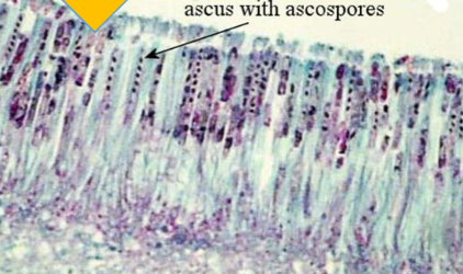
Coprinus sp. Basidiocarps and Basidiospores (Basidomycota)

Coprinus sp. Basidiocarps and Basidiospores (Basidomycota) — Zoomed In

Mycorrhizae
Symbiotic relationship between a fungus and a plant; the plant gives the fungus carbohydrates and the fungus, in return, increases the mineral and water absorption for the plant. About 90% of land plants have mycorrhizal fungi.
Lichens
Symbiotic relationship between multiple fungi, cyanobacteria, or green algae. The cortex and medulla layers are made of fungus, and then a layer of cyanobacteria or algae is encased by the fungal layers. Lichens aren’t a moss; they are found on every continent and can survive extreme conditions.
Crutose Lichens
Flat lichens that adhere tightly to the surface of rocks or tree bark.

Foliose Lichens
Lichens that are more leaf-like, and their edges general curl away from the substrate. They are typically found growing on the trunks or branches of trees.

Fruticose Lichens
Lichens with a more 3-dimensional, shrubby growth.

Metazoa
Kingdom Anamalia
Eumetazoa
Organisms with true tissues.
Parazoa
Organisms with no true tissues.
Phylum Porifera — Sponges
Organisms that are asymmetrical and lack true tissues; they have some specialized cells.
How does sponge feeding work?
Choanocytes in the body wall draw water through the Ostia
Food particles are trapped in microvilli collars and picked up by Amoebocytes
Amoebocytes digest food and deliver nutrients to sponge
Water exits through the Osculum

Ostia
Small pores that let water into the sponge.
Amoebocyte Cell
Cells specialized for digesting food and delivering nutrients to the sponge; pick up what choanocytes trap.
Choanocyte Cells
Cells that draw water in and trap food particles using their microvilli collars. Have flagella.
Osculum
Main hole in sponge where water exits.
Epithelial Cell
Cells that form the outer covering of the sponge.
Spicule
Small structures made of calcium carbonate or silica that form the sponge’s skeleton, providing support and defense from predators.
Cnidarians
Organisms with radial symmetry, nematocysts, and either a polyp or medusa life stage (some go through both forms at different life stages).
What germ layers do cnidarians and ctenophora have?
They are diploblastic, so they have ectoderm and endoderm (between the two layers is mesoglea). They also have a gastrovascular cavity capable of extracellular digestion and a nerve net composed of interconnected neurons (but no brain).
Cnidarian Clades
Clade Hydrozoa
Clade Anthozoa
Clade Scyphozoa
Protosomes
Organisms that’s blastopore becomes the mouth during development.
Blastula
Hollow ball of cells that developed from the embryo.
Archenteron
Part of the cells pinching inwards to form a tube; next stage after blastula formation.
Blastopore
The opening of the archenteron; in protostomes, the blastopore becomes the mouth, and in deuterostomes, it becomes the anus.
Lophotrochozoans — Clade Lophotrochozoa
Organisms that have either a lophophore, which is a feeding apparatus made of ciliated tentacles that surround a mouth, or a trochophore, which is a larval stage.

Flatworms — Phylum Platyhelminthes
Bilateral symmetrical organisms that posses a head region with sense organs (cephalization). They have an incomplete gastrovascular cavity (only one opening), a basic nervous system, and are hermaphroditic creatures.
Cephalization
The formation of a head in an organisms.
Tapeworm (Taenia pisoformis)
Phylum Platyhelminthes
Proglottids
Segments of a tapeworm, immature ones are closest to the scolex (head region).
Mature Proglottids
Largest segments that contain male and female sex organs; are in the “middle” of the worm
Gravid Proglottids
Segments toward the end of the worm. They ae filled with eggs and eventually break off the worm so the embryos can be transferred to the host.
Phylum Rotifera (Rotifers)
Small Organisms that have a complete digestive system, a corona, and a mastax.
Corona
Ciliated structure that looks like a rotating wheel; used for movement and feeding.

Mastax
Muscular pharynx and jaws for chewing food particles.

Phylum Brachiopoda
Organisms that possess a true body cavity, posses lophophores for feeding and respiration, and resemble clams (which do not have a lophophore).
Phylum Annelida
Organisms with a body form made of many segments.
Errantia
Phylum Annelida, free living predators
Sedentaria
Phylum Annelida, sedentary or slow-moving organisms, e.g. tube worms, earthworms, leeches.
External Worm Anatomy
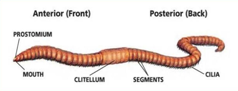
Internal Worm Anatomy

Pharynx
Acts as a muscular pump to suck in food; secretes mucus to moisten food and make it easier to swallow.
Crop
Located behind the esophagus, it temporarily stores food before digestion. This allows the worm to ingest food quickly and digest it later.
Gizzard
Located directly behind the crop, it grinds food into smaller, digestible pieces. It breaks down tough organic matter, making it easier for enzymes to act.
Seminal Vesicles
Store sperm in worm.
Seminal Receptacles
Receive and store the sperm of another worm, later used to fertilize the worm’s own eggs.
Earthworm Dissection Process
Dirt/food —> Mouth —> Pharynx —> Esophagus —> Crop —> Gizzard —> Intestines —> Anus —> Waste
Phylum Mollusca
Organisms with a muscular foot (used for movement), visceral mass (contains most of the internal organs), and a mantle (drapes the visceral mass and makes the shell if one is present), e.g. snails, slugs, oysters, clams, octopi, squids
Phylum Mollusca - Class Polyplacophora
Organisms known as chitons, which have a reduced head, dispersed sensory organs, and a strong foot.
Phylum Mollusca - Class Cephalopoda
Organisms are that active predators; some members have demonstrated. high levels of intelligence. They have a head surrounded by tentacles that are a modification of the foot, e.g. squid, octopus, cuttle fish, nautilus.
External Anatomy of a Squid
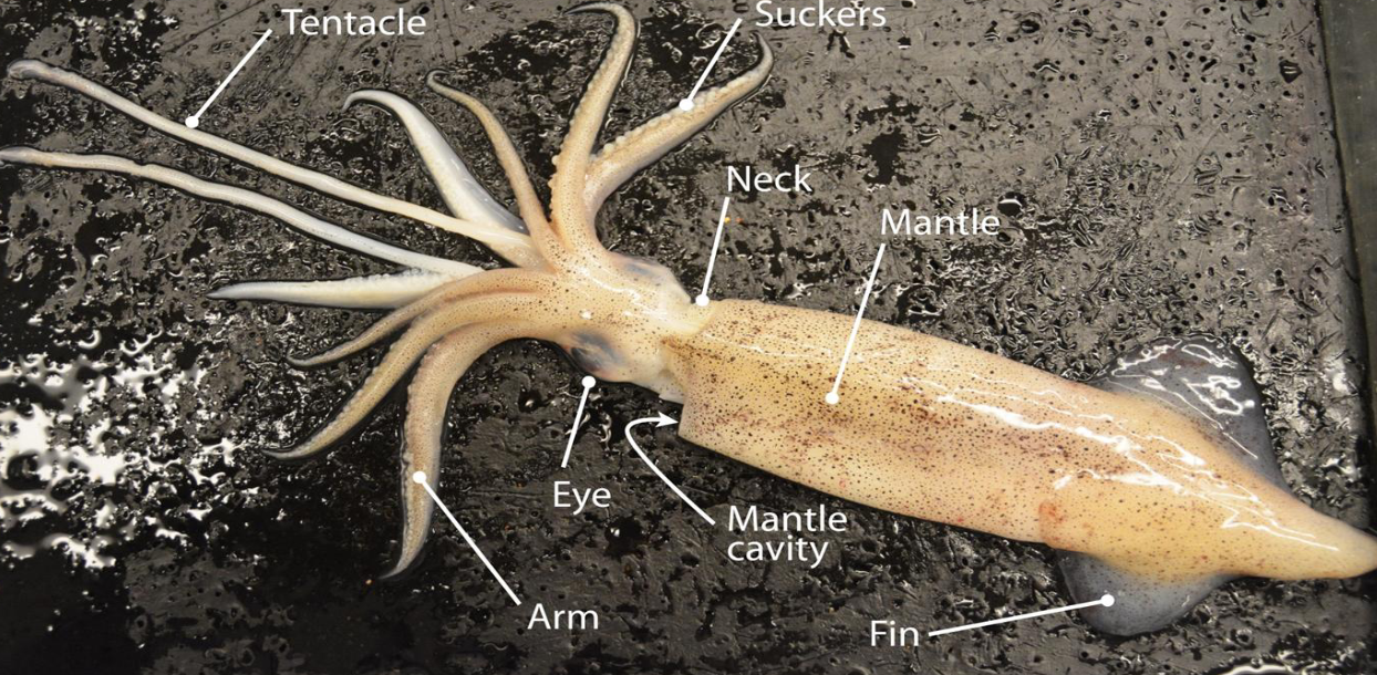

What kind of squid is this?
Male squid

What kind of squid is this?
Female squid
Siphon
Structure use to help push water over the gills and propel the squid through the water. It also expels waste products and pushes ink out of the body.
Chromatophores
Specialized cells that allow the squid to change colors.
Ink Sac
Used to expel ink through the siphon to deter predators.
Pen
Vestigial internal shell made of chitin. Helps hold the squid’s organs in place.
Class Bivalvia
Ecologically and commercially important organisms; they filter water by capturing particles from water column and ingesting sediment, e.g. clams, oysters, scallops.
Clam Basic Structure

Clam Internal Structure
