Nervous System: Neural Tissue 1 - PHYL 301
 Call Kai
 Learn
 Practice Test
 Spaced Repetition
 Match
 Flashcards
 Knowt Play
1/68

There's no tags or description
Looks like no tags are added yet.
69 Terms
Nervous System
Controls and regulates the activity of the body
Provides swift but brief responses
Endocrine System
Controls and regulates the activity of the body
Slower, but longer-lasting and multi-organ responses
Overview of Nervous System
Includes all nervous (neural) tissue
Central Nervous System (CNS)
Brain
Spinal cord
Peripheral Nervous System (PNS): all nervous tissue outside the CNS
Cranial nerves
Spinal nerves
Central Nervous System (CNS)
Brain
Spinal Cord
Peripheral Nervous System (PNS)
All nervous tissue outside the CNS
Cranial Nerves
Spinal Nerves
What are the 3 Nervous System Functions
Sensation (input)
Integration (processing)
Response (output)
Pathway: Stimulus → Receptor → interneurons → effector → response
What is sensation & sensation?
Detects stimuli (changes inside/outside body)
Receptors detect stimuli
Stimuli
Changes/events in the internal or external environment
What is integration?
Interneurons recieve sensory signals
Processes
Interprets and compares stimuli with current and past situations
May send signals (if needed) to cause a response/effect
What is Response (Motor Activity)?
Sensory or integrative signals can activate effectors (e.g., muscles, glands)
Produce a response that has an effect on the internal or external environment
What is the Central Nervous System (CNS)’s Functions?
Integrates, processes, and coordinates input & output
Sensory input: stimuli → CNS
Motor output: CNS → organs & tissues
Origin of intelligence, memory, learning, and emotion
Main sources of interneurons
What is the Peripheral Nervous System(PNS)’s Functions?
Provides sensory information to the CNS and carries motor commands away from the CNS
Divided into:
Afferent division
Brings sensory information to the CNS
Receptors detect stimuli
Efferent division
Carries motor commands to muscles and glands
Effectors respond to CNS commands
Afferent division
Brings sensory information to the CNS
Towards brain/spinal cord
Receptors detect stimuli
A = Arrives; Afferent → Axial Skeleton
Efferent division
Carries motor commands to muscles and glands
Away from brain/spinal cord
Effectors respond to CNS commands
Efferent causes Effects
E = Exits; Exit CNS → effectors
What are the two branches of the PNS’s efferent division, and what does each control?
Somatic nervous system (SoNS*)
skeletal muscle
Autonomic nervous system (ANS)
Sympathetic division/nervous system (SyNS*)
Parasympathetic division/nervous system (PSNS*)
smooth muscle, cardiac muscle, glands, and adipose tissue
What are the three branches of the PNS’s afferent division, and what does each control?
Somatic sensory receptors
skin, muscles, joints
Visceral sensory receptors
internal organs
Special sensory receptors
vision, hearing, etc.
Describe the nervous system as a control system with the following
components: sensory receptors, afferent pathways, control (integrating)
center, efferent pathways, and effector (target) organs
1. Sensory receptor: A receptor in your skin detects potentially damaging heat.
2. Afferent pathway: A sensory neuron carries the signal to the spinal cord.
3. Integrating center: Interneurons in the spinal cord process the signal and activate motor neurons.
4. Efferent pathway: Motor neurons carry commands to your arm muscles.
5. Effector (target organ): Your arm muscles contract, pulling your hand away.
Neurons
Transfer and process information via action potentials
Consist of a soma (cell body), axon, dendrites, perikaryon, dendritic spines, and axon terminals
Neuroglia
Support nervous system function
Protect the neuron
Cells with specialized support roles
Do NOT send action potenials
~50% brain mass
Order the signal travels through a neuron
DCBA
Dendrites → Cell Body → Axon → Axon Terminals

Axon
(typically) long neuron extension; carries impulses from cell body
Dendrites
Recieves input, branches
Cell Body
Contains the nucleus and organelles
Axon terminals
Output; Affects another neuron, or muscle
Ends of telodendria
Axon hillock
Where the axon initial segment connects to cell body; where cell body and axon attaches
Axolemma
plasma membrane of axon
Axoplasm
Cytoplasm of axon
Axoplasmic transport: movement of material between the cell body and the axons
Collaterals
side branches of main axon
“-lat” = middle
Telodendria
End branches of axon & collaterals
“-endria” = Later

Ependymal Cells
Form ependyma: a cellular lining
Line Central Nervous System (CNS) cavities
Lines brain & spinal cord
Produce cerebrospinal fluid (CSF)
Cilia circulate CSF
Microglia
Specialized phagocytes (eats stuff)
Protect against pathogens
Dispose of pathogens, debris, & waste
Immune defense and waste removal
Astrocytes
Largest & most abundant glial cells
Form the blood-brain barrier (BBB) around blood vessels
Oligodendrocytes
Form myelin sheaths in CNS
Can myeliante segments on mulitple axons
White matter
Areas rich in myelinated axons
White = “wrapped”, fatty creamy/yellow phospholipids
Gray matter
Areas lacking myelinated axons
Mostly neuronal cell bodies and dendrites
Ganglion
Cluster of neuronal cell bodies in Peripherial Nervous System (PNS)
What forms Peripheral nerves
PNS neuronal axons bundled together
Satellite Cells: PNS Neuroglia (glial cells)
Surround & protect cell bodies
Schwann Cells: PNS Neuroglia (glial cells)
Surround & myelinate axons
Covers one segment of axon
Schwann cells are also called neurolemmocytes
Consists of: Axolemma/neurolemma
What is a myelin sheath made of?
Made of multiple phospholipid membrane layers
Contains proteins embedded in myelin
PLPs - proteolipid proteins
MBPs - myelin basic proteins
What is myelin, what does it do?
Lipid-rich, insulating sheath made of tightly wrapped layers of glial cell membrane around an axon
Speeds up action potential conduction; The action potential is regenerated at gaps called nodes of Ranvier, so it appears to jump from node to node
Saltatory conduction
How does myelination differ in the CNS and PNS?
Both: Wraps around repeated and packed
CNS: An oligodendrocyte sends out several extensions; Each extension wraps an axon segment, so one cell can myelinate segments on multiple axons
PNS: A Schwann cell encloses an axon segment in a groove, then wraps its membrane around that segment; nucleus and most of its cytoplasm stay outside the compact myelin; One myelinating Schwann cell covers one segment of one axon.
COPS Myelin Mnemonic
CO = CNS & Oligodendrocytes
PS = PNS & Schwann Cells
Officer Maya Lin and pet Swan
Strucural classification: Neuron classification
Based on the number of processes extending from the cell body
Functional classification: Neuron classification
Sensory
Interneuron (connects sensory & motor; processes sensory information)
Motor
Anaxonic: Strucural classification of Neuron
Many processes, but difficult to differentiate axons and dendrites
Rare in humans; found only in the CNS
“ana” = without; without axon

Bipolar: Strucural classification of Neuron
Cell body between dendrite and axon
Unmyelinated axons
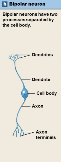
Pseudounipolar: Strucural classification of Neuron
Cell body off to one side of the axon
“Pseudo” – starts as bipolar, becomes “unipolar”
Most common sensory neuron type
Fast detection (Signal doesn’t have to go through cell body)

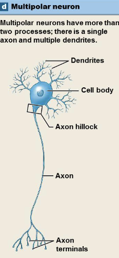
Multipolar: Strucural classification of Neuron
Single axon and multiple dendrites (typically)
Most common type in the CNS
Most common motor neuron type
What is a stimulus, and how does it reach the CNS as a signal?
Change in the internal or external environment by a receptor
Receptor detects the stimulus
Sensory neuron carries the signal from the PNS toward the CNS
An afferent fiber is the axon of that sensory neuron
Somatic sensory neurons: Sensory (Afferent) Neurons
Detect external stimuli in skin, muscles, & other organs
Interoceptors: monitor internal organ activity
Visceral sensory neurons: Sensory (Afferent) Neurons
Detect internal stimuli from organs
Exteroceptors: provide information about the external environment •
Proprioceptors: monitor position and movement
Interoceptors
monitor internal organ activity
Exteroceptors
provide information about the external environment
Proprioceptors
monitor position and movement
Motor neurons
Send information from CNS → PNS
Efferent fibers: Axons of motor neurons
What are the two motor divisions?
Somatic nervous system (SoNS): somatic motor neurons innervating skeletal muscle
Autonomic nervous system (ANS): visceral motor neurons innervating organs and glands
Somatic nervous system (SoNS)
Somatic motor neurons innervating (controlling) skeletal muscle
Autonomic nervous system (ANS)
visceral motor neurons innervating (controlling) organs and glands
Preganglionic & (post)ganglionic fibers: axons carry impulses (autonomic motor commands) from CNS → ANS tissues & organs
Interneurons
Located entirely in the CNS
Connect sensory & motor neurons
Analyze sensory input
Coordinate motor outputs
Can be excitatory or inhibitory
Synapse
Junction between a neuron and another cell
Two major types of synapses
Chemical synapse (vesicular): release of neurotransmitters
Electrical synapse (nonvesicular): ion movement via gap junctions
Electrical synapse
Membranes of both cells connected with gap junctions
Allows ions to flow from one cell to the next
Diffusion of ions changes membrane potential; may push postsynaptic cells to threshold
Less common than chemical synapses
Faster impulses; can travel in either direction
Chemical Synapses
Utilize neurotransmitters
Slower than electrical synapses
More regulation & control than electrical synapses
Make up vast majority of synapses (vs. electrical)
Which is more common in the nervous system?
Chemical Synapses
What is a synapse, and how do chemical and electrical synapses differ?
A synapse is a junction where a neuron communicates with another neuron or a target cell, such as a muscle or gland.
At a chemical synapse, the sending cell releases a neurotransmitter that crosses a gap and binds to receptors on the receiving cell. Transmission is typically one-way.
At an electrical synapse, ions flow directly between cells through gap junctions. Transmission is very fast and often goes both ways
What structures make up a chemical synapse?
Structure | Location and role |
|---|---|
Axon terminal (synaptic knob) | The end of the presynaptic neuron where neurotransmitter is released. |
Voltage-gated calcium channels | In the terminal membrane. They open when an action potential arrives, letting Ca²⁺ enter. |
Synaptic vesicles | Small sacs in the presynaptic terminal that store neurotransmitter. |
Synaptic cleft | The narrow gap between the cells that the neurotransmitter crosses. |
Neurotransmitter receptors | On the postsynaptic cell. They bind the neurotransmitter and trigger a response. |
What's the order of chemical synaptic transmission?
Calcium channels open in the presynaptic axon terminal
Exocytosis of neurotransmitters into synapic cleft
Neurotransmitters bind to receprots on the postsnyaptic membrane
Ion channels open in the postsynaptic membrane