Basics of Nucleic Acids and Proteins
 Call Kai
 Learn
 Practice Test
 Spaced Repetition
 Match
 Flashcards
 Knowt Play
1/9

There's no tags or description
Looks like no tags are added yet.
10 Terms
Chargaff’s Rule
in double-stranded DNA, the amount of adenine equals thymine (A = T) and the amount of guanine equals cytosine (G = C), meaning the total number of purines equals the total number of pyrimidines (A + G = T + C)
Watson and Crick (1953)
They found that DNA consists of two long, anti-parallel strands twisted around each other like a spiraling staircase or twisted ladder.
The outer sides of the ladder are made of alternating sugar and phosphate molecules.
The rungs of the ladder consist of four nucleotide bases that pair up in a specific way: adenine (A) pairs with thymine (T), and cytosine (C) pairs with guanine (G).
They realized that because the strands are complementary, each strand can act as a template to build a new strand during cell division. This explained how genetic information is copied and passed down.
Conformation
The spatial arrangement of atoms in a protein
Peptide bond is rigid and planar:
Each peptide bond has some double-bond character due to resonance and cannot rotate
The degradation of the peptide bond takes place through hydrolysis, thus requires the presence of water molecules.
The degradation reaction is very slow as the amide bond between the amino acids is stabilized by the partial double bond.
Because of the partial double bond between carbon and nitrogen molecule:
Carbon atom generates a slight positive charge
In the presence of water, the OH– ions of water attack the carbon atom, which results in degradation of the peptide bond
As a result of this, the peptide molecule is cleaved into two units; one unit with the carboxyl group and another with the amino group
The degradation of the peptide is an exergonic reaction that releases about 8-16 Kjol/mole of energy.
Because the protein degradation reactions are very slow, they are usually catalyzed by proteolytic enzymes like proteases and peptidases
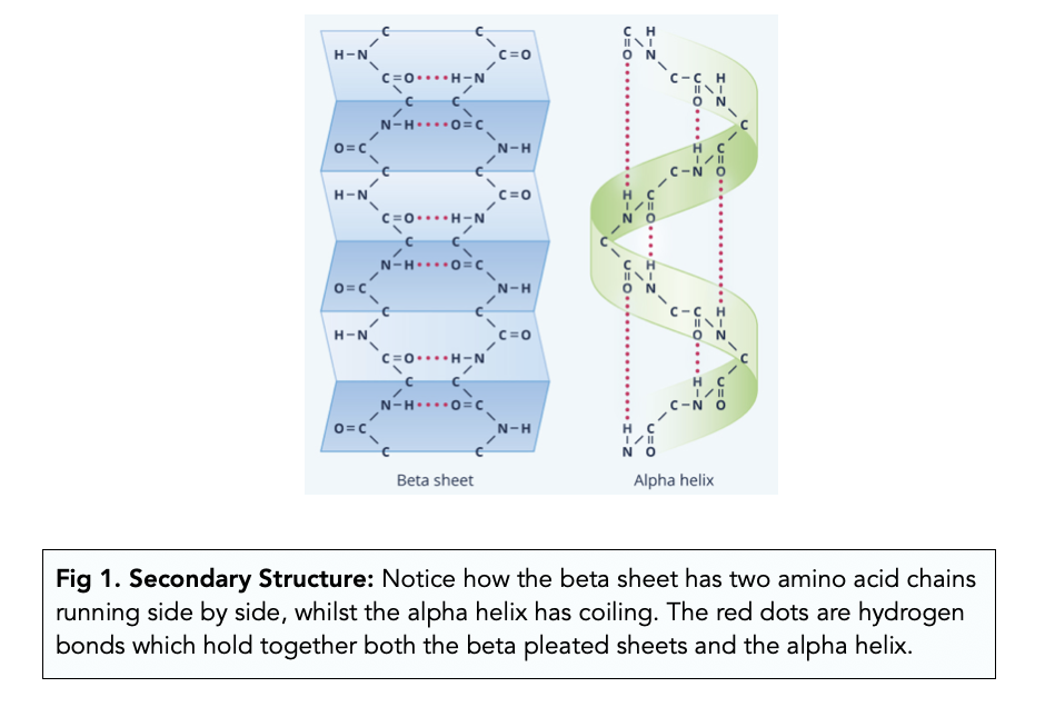
Secondary Structure
Alpha-helices and beta-sheets/strands
Held together by hydrogen bonds
Structural Domain
an element of the protein's overall structure that is stable and often folds independently of the rest of the protein chain
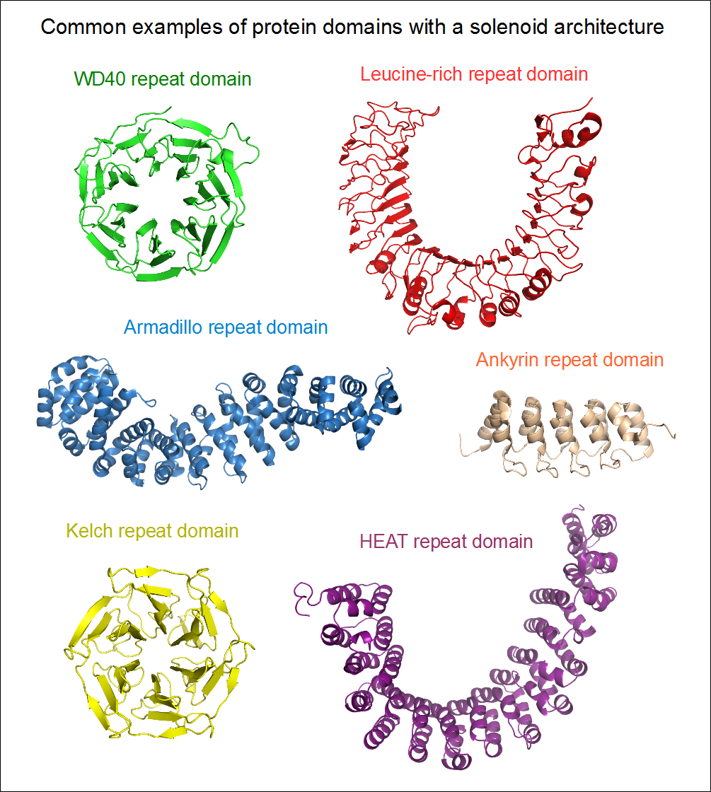
Protein Motifs
small regions of protein three-dimensional structure or amino acid sequence shared among different proteins

Supersecondary Structure
refers to a combination of secondary structure elements, such as beta-alpha-beta units or the helix-turn-helix motif
Protein Fold
refers to a general aspect of protein architecture, like helix bundle, beta- barrel, Rossman fold or other "folds" provided in the Structural Classification of Proteins database