Cell Physiology Lecture 1 - Intro, Membranes, Cell junctions, Organelles
 Call Kai
 Learn
Learn Practice Test
 Spaced Repetition
 Match
Match Flashcards
 Knowt Play
1/50

There's no tags or description
Looks like no tags are added yet.
Name | Mastery | Learn | Test | Matching | Spaced | Call with Kai | Chat |
|---|
No analytics yet
Send a link to your students to track their progress
51 Terms
What is a cell
Structural and functional unit of a living organism
All cells contain these 3 components:
A plasma membrane
cell organelles - compartmentalize the cell and perform specific functions
cytoplasm - suspend the organelles
4 Functions of the plasma membrane
Physical barrier between ICF and ECF fluids - maintains difference in ion composition and homeostasis
Cell-to-cell communication - contains receptors that bind signaling molecules
Structural support - contain connections composed of proteins which anchor cells to each other or the ECM
Transport - selectively permeable
Biological membrane
Cell/plasma membrane that surrounds organelles
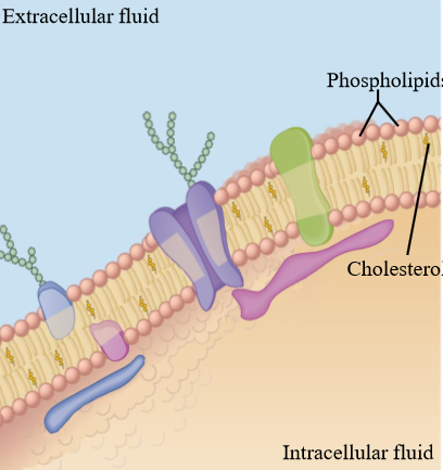
Composition of a biological membrane
Phospholipid bilayer - 2 layers of phospholipids with embedded prteins
Structure of a phospholipid
Ampipathic molecule
Polar head group: phosphate attached to glycerol and a glycerol backbone
Non-polar tail: 2 fatty acid chains composed of carbon and hydrogen
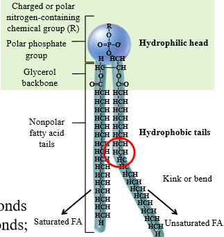
Unsaturated fatty acid chains
Contain double bonds, making a kink in the chain
Which part of the phospholipid is hydrophilic
The head group
Which part of the phospholipid is hydrophobic
The fatty acid tails
Structure of phospholipids in water
Forms a bilayer: Polar heads phase environment, non-polar tails form hydrophobic core. This forms the plasma membrane
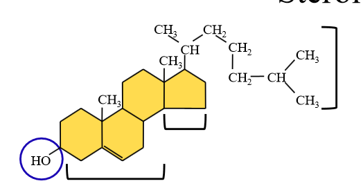
Cholesterol
Amphipathic steroid that maintains proper membrane fluidity
Almost 1 molecule of cholesterol per molecule of phospholipid
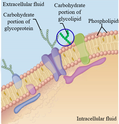
Glycolipids
Amphipathic lipids attached to carbohydrates, found on the outer leaflet of the plasma membrane
Form the glycocalyx - Layer of carbs linked to lipids/membrane proteins, allows cells to identify and interact with each other
Integral (intrinsic) membrane proteins
Amphipathic Proteins inserted into the phospholipid bilayer and span across the membrane
Often used as transported or channels
Outer proteins are hydrophilic, internal proteins (in membrane) are hydrophobic
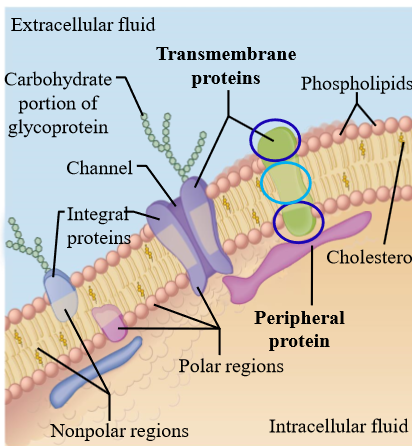
Peripheral (extrinsic) membrane proteins
non-amphipathic proteins that attach to the outside of the membrane, and do not associate with the non-polar tails inside the bilayer
Mostly extracellular and associate with cytoskeletal elements related to shape and mobility
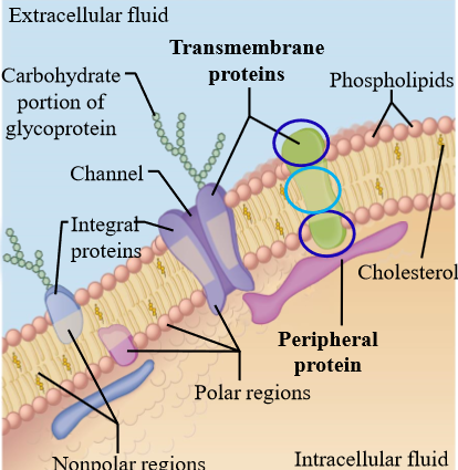
Glycoproteins
Carbohydrates attached to the peripheral membrane proteins that help form the glycocalyx
Cell Junctions
Specialized connections that stabilize interactions and promote communication betwen cells
3 types of cell junctions
Desmosomes, tight junctions, gap junctions
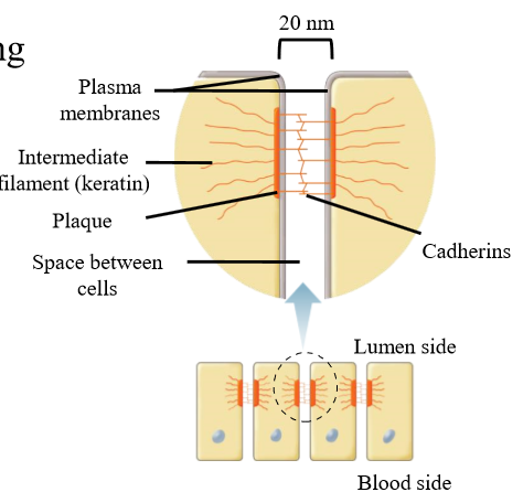
Desmosome function
Adhering junctions that anchor cells together that are subject to considerable stretching or mechanical stress
Maintain structural integrity of tissues, preventing cells from tearing apart from one another
Seen in heart muscle, uterus, and skin
What plaques (proteins) are desmosomes made out of
Plagues: On cytoplasmic surface - anchor cadherins
Cadherins: Link cells together by binding to plagues. Seen in the cell cleft
Intermediate filaments: Structural support Anchor cytoplasmic surface of desmosome to components inside the cell
TIght junction function
Form nearly impermeable junctions
Link cells together
Limit movement of molecules between cells - molecules must go through cells or through the plasma membrane
Limit integral protein and lipid movement to preserve function
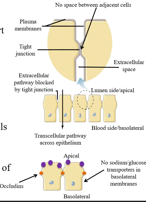
What are tight junctions made out of
Occludins
Gap Junction functions
Transmembrane, water-filled channels that electrically and metabolically couple cells. Also known as communicating junctions as molecules + ions can move from one cell to the next
Fast communication, low resistance
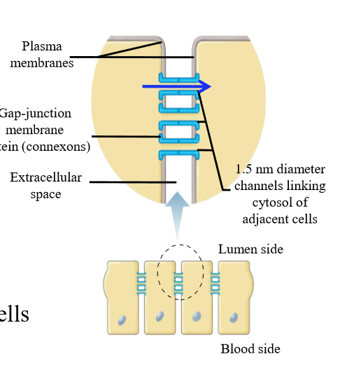
Electrical coupling
Allow ions to move between cells - size limited by diameter of connexons
Metabolic coupling
Allow small molecules such as nutrients to move between cells - size limited by diameter of connexons
What are gap junctions made out of
Connexons
Nucleus Function
Storage and transmission of genetic information required for protein synthesis
Parts of the nucleus
Chromatin - DNA and associarted proteins condense during cell division to form chromosomes
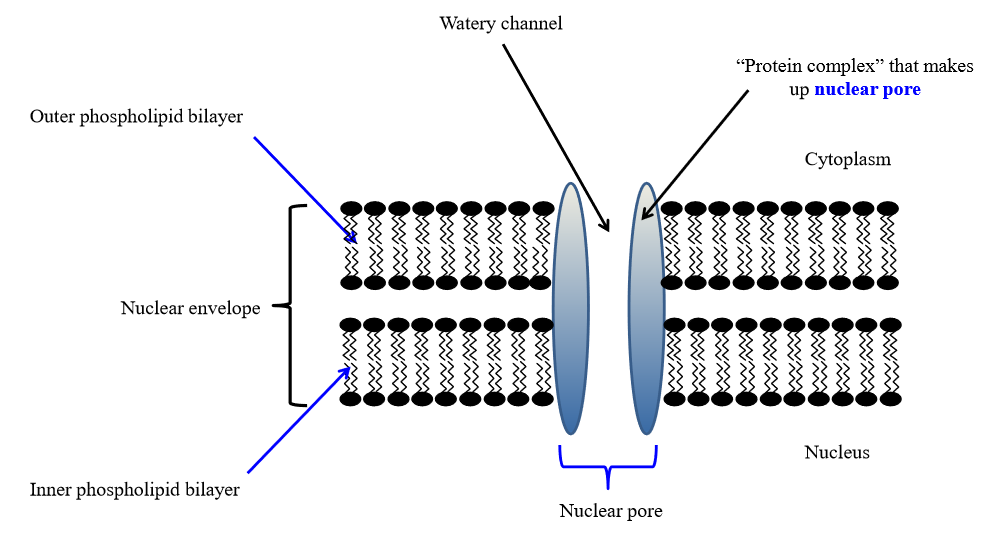
Nuclear envelope - doubled layered porous membrane to seperate nucleus from cytoplasm
Nuclear pores - pores in envelope that allow molecules to move in and out of the nucleus
Nucleolus - site of ribosomal RNA synthesis
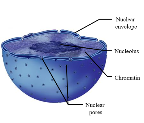
Structure of the nucleus
Nuclear envelope made out of 2 phospholipid bilayers with nuclear pores
DNA and RNA building blocks enter the nucleus
RNA and ribosomal subunits exist the nucleus
Most cells have one nucleus, except for:
RBCs - no nucleus
Muscle cells - multiple nuclei to satisfy high protein needs (ribosome synthesis)
Ribosome function
Protein synthesis - read and mRNA and assemble AAs to form proteins
Ribosome composition
Made of small and large subunits. Subunits must come together to form functional ribosome
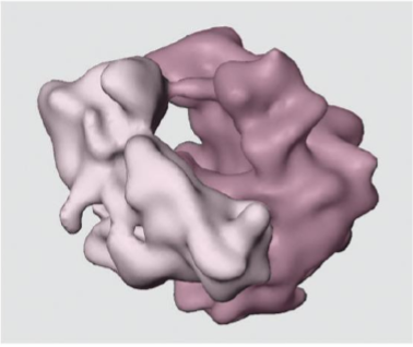
Location of ribosomes
Synthesized in nucleus
Found free in cytoplasm
Bound in the rough ER
Endoplasmic reticulum
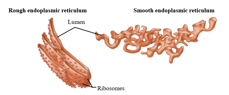
Fluid filled membranous system, with rough and smooth ER
Rough ER - flattened sacs with attached ribosomes
Smooth ER - branched tubular structure with no ribosomes
Function of the smooth ER
Synthesizes lipids, stores calcium (sarcoplasmic reticulum in muscles), detoxifies drugs by converting into water soluble form
Function of the rough ER
synthesizes proteins and performs post-translational modifications needed to produce functional proteins
Modifications include glycosylation and protein cleavage
High levels in cells needed to make secretory proteins (pancreatic cells)
Golgi apparatus structure
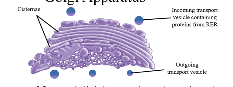
Composed of cisternae (flattened sacs)
Golgi apparatus function
Receives vesicles containing proteins synthesized in RER.
Post-translational modification
Sorts and packages proteins into vesicles
Fate of proteins put into vesicles by the golgi
Secreted from the cell
Become integral membrane proteins
Becomes proteins of the lysosome, ER, or golgi
Lysosome function
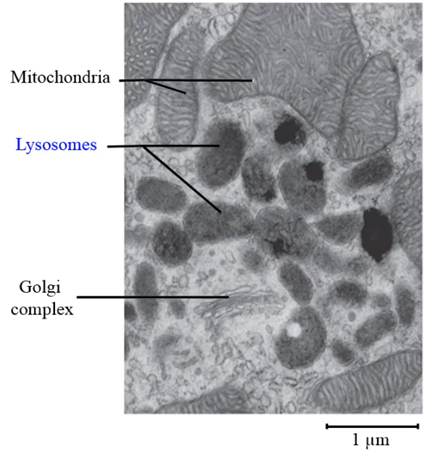
Contains hydrolytic enzymes that break large molecules into small subunits. Can recylce organelles or destroy viruses/bacteria brought into the cell by fusing to it.
Acidic environment activates enzymes (pH 5)
Breaks down proteins, nucleic acids, lipids, etc.
How does the lysosome have a lower pH than the rest of the cell
Protons pumps in membrane move protons inside the lysosome to lower pH
Peroxisosome function
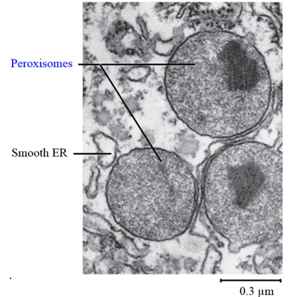
Contains oxidative enzymes, which use oxygen to remove hydrogen from molecules for breakdown. Creates hydrogen peroxide as a byproduct
Breaks down fatty acids, alcohol, and drugs
Hydrogen peroxide is toxic. How do peroxisomes remove hydrogen peroxide?
Peroxisomes have a catalase enzyme that converts hydrogen peroxide to water and oxygen
Function of mitochondria
Synthesizes energy by converting nutrients into ATP via the Kreb’s cycle, oxidative phosphorylation, and other metabolic pathways
Structure of the mitochondria
Contains double phospholipid membrane
Outer membrane - separates mt from cytosol
Inner membrane - folded into tubules called cristate
Matrix - contains enzymes responsible for cellular respiration
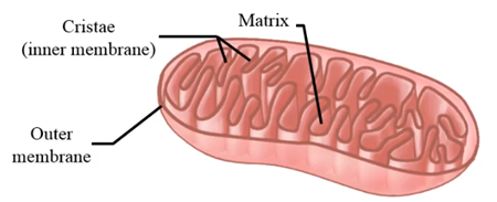
Which cells have more mt than normal
Cells that require lots of energy - skeletal muscle, heart muscle, liver, sperm
Important feature of Mitochondria
Has it’s own DNA. MtDNA comes from circular genomes of bacteria that were engulfed by eukaryotic cells
Cytoskeleton function
Maintain cell shape, maintain the position of organelles in the cell interior, and mediate cell + organelle motility
Is the cytoskeleton membrane bound?
No. No PL bilayer (same with ribosomes)
Are ribosomes membrane bound?
No. No PL bilayer (same with cytoskeleton)
Cytoskeleton composition
Composed of protein filaments or cytoskeletal filaments
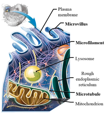
Cytoskeletal filament types
Microfilaments - made of actin
Determine cell shape and movement
Intermediate filaments - part of desmosomes
Support cell, and provide mechanical strength
Microtubules - made of tubulin
Suport for cell, movement of organelles