06heart anatomy and function
 Call Kai
 Learn
 Practice Test
 Spaced Repetition
 Match
 Flashcards
 Knowt Play
1/33

There's no tags or description
Looks like no tags are added yet.
34 Terms
[A] Where is the hearts position in human body and what is his function?
A little bit left in thorax between the two lobes of the lung
It is enclosed by heart sac (Pericardium)
Changes permanent it’s shape alternating between contraction (systole) and relaxation (diastole)

A
How is the heart structurally divided into chambers and circulatory systems?
Structure: The heart consists of 4 chambers (2 Atria: Right/Left Atrium; 2 Ventricles: Right/Left Ventricle) driving 2 circulatory systems (Pulmonary and Systemic Circulation).
A- What is the exact step-by-step pathway of blood flow through the heart chambers, valves and lungs?
Deoxygenated Flow (Right Heart): Vena cava into Right atrium (Atrium dextrum cordis) than pasing Right AV valve (Tricuspid valve) to Right ventricle
→ Systolic contraction ejects blood from right ventricle through Pulmonary semilunar valve into Pulmonary trunk/arteries → Lungs
After flowing through lung blood reaches →
Oxygenated Flow (Left Heart): via Pulmonary veins → Left atrium (Atrium sinistrum cordis), than passing Left AV valve (Bicuspid/Mitral valve), into Left ventricle
→ Passing the aortic semilunar valve blood is ejected in systole from left ventricle, into Aorta → Systemic organs
[B] What is needed in order to reach a coordinated heart contraction?
A certain fiber orientation because the heart muscle has no bone fixation (no origin-no insertion)
The different fiber orientations results in different pulling directions of myocardium

[A] How does the myocardial oxygen demand scale from baseline rest to maximal exercise load per 100g of left ventricle per minute?
Scaling Values (per 100g left ventricle/min):
Base ( Basis ): 0.8 ml O₂
Rest ( Ruhe ): 8.0 ml O₂
Maximal Load ( Maximale Belastung ): 80.0 ml O₂
Key Concept: Myocardial oxygen consumption increases 100-fold from base to maximal load, requiring an immediate proportional increase in coronary blood flow because the heart relies almost exclusively on aerobic energy.

[A] How does the cardiac muscle action potential differ from skeletal muscle, and what causes the difference?
Skeletal muscle: short, spike-like action potential, followed by a relatively short contraction.
Heart muscle: a much longer action potential (~300 ms) with a plateau, and its contraction also lasts longer
The prolonged plateau (~200–300 ms) is driven by sustained Ca²⁺ influx.

A What are the phases of the cardiac action potential, what is its refractory period, and what is its physiological role?
Phases: fast depolarization → plateau phase → repolarization.
Refractory period: It spans the whole action potential (~300 ms), with an absolute part first and a relative part at the end.
The absolute refractory period largely spans the duration of the mechanical contraction.
Physiological role: It prevents tetanic contractions (fused tetanus). Cardiac muscle cannot be tetanized by high-frequency electrical impulses, so the heart is guaranteed to relax between beats, which allows ventricular filling.

B - Which structures are labelde in the heart muscle cell?
Sarcomere (A-band, I-band, H, Z), mitochondria, sarcoplasm, sarcoplasmic reticulum, nucleus, myofibril, thin and thick myofilament, desmosome, and the Glanzstreifen (intercalated disk).
(Exam risk lower than for the conduction system)

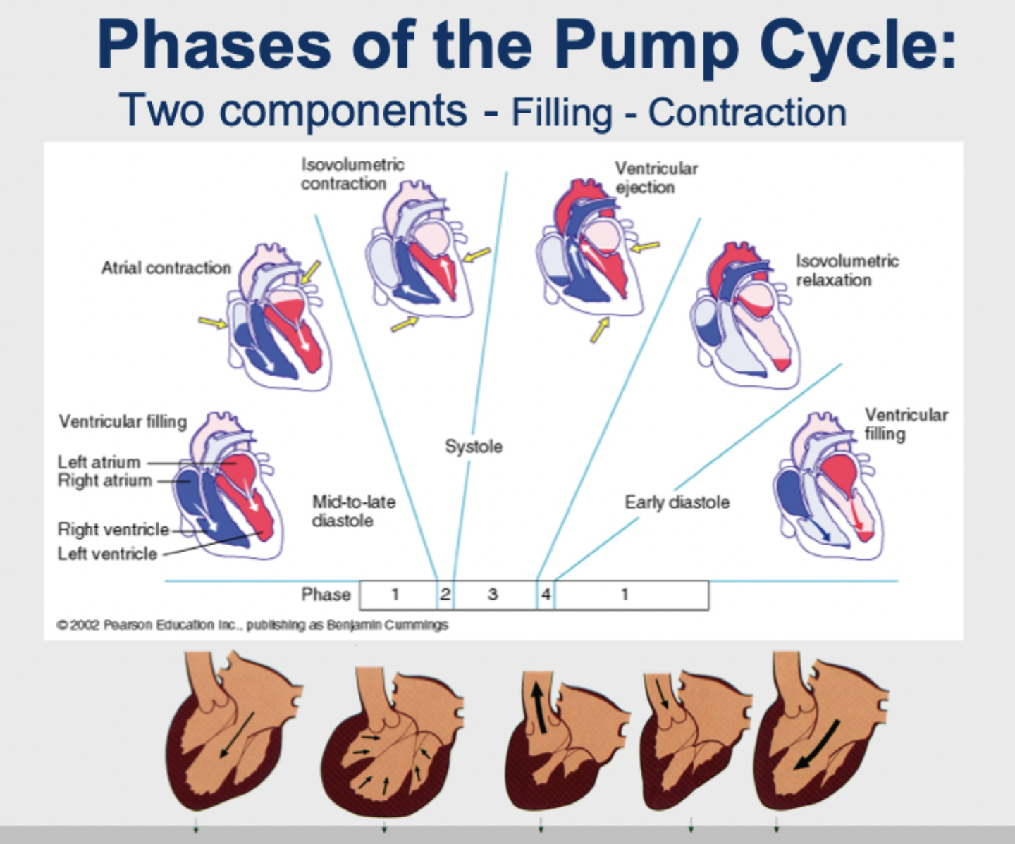
[A] What are the two components and the phases of the pump cycle?
The two components are filling and contraction. The phases are:
Ventricular filling (mid-to-late diastole, including atrial contraction; also early diastole)
Isovolumetric contraction
Ventricular ejection
Isovolumetric relaxation
Phase 2 and 3 make up systole; Phase 4 and 1 are diastole
[Supplement (not on the slide, standard physiology, please verify): In the two isovolumetric phases all valves are closed, so the volume stays constant. In ventricular filling the AV valves are open. In ejection the semilunar valves are open]
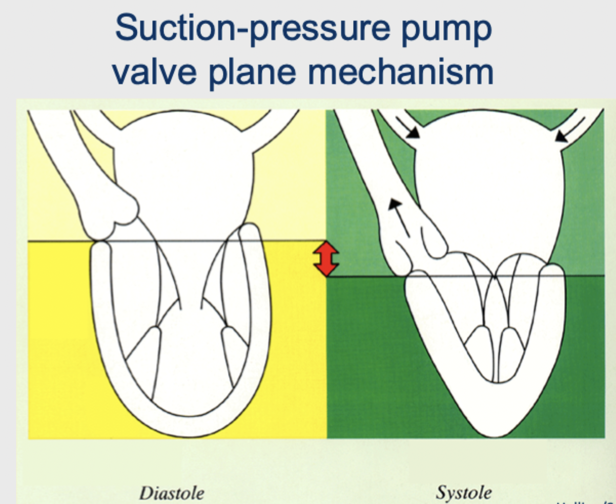
A - What is the valvulare plane and how does it make the heart a suction-pressure pump?
The plane in which all valves are arranged
Valves = essential for pump function
The valve plane is the interface between atrium and ventricle. It is strengthened by the heart skeleton (connective tissue that divides the atrial and ventricular muscles completely and serves as their origin). With its help the heart becomes a suction-pressure pump.
Supplement (not on the slides, please verify with a textbook or a fellow student's notes): In systole the valve plane moves toward the apex. The ventricle ejects blood (pressure), and at the same time the atria are enlarged, which draws blood in from the veins (suction). In diastole the valve plane moves back toward the base, which supports ventricular filling

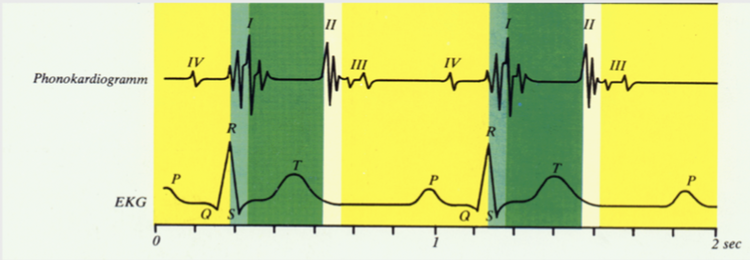
B/C - How do the heart sounds relate to the EKG and the cardiac cycle?
The phonocardiogram shows four heart sounds (I–IV). Sound I follows the QRS complex (beginning of systole) and sound II follows the T wave (end of systole). Sounds III and IV occur in diastole. The electrical events (EKG) precede the mechanical events (heart sounds).
Supplement (not on the slide): Sounds I and II are generally associated with valve closure
[A] What is the function of the valves, and what happens in systole?
The valves are responsible for the direction of blood flow in heart actions. They open and close in a defined rhythm to inhibit reflux of blood. In systole the atrioventricular valves are closed to inhibit backflow in atria (Vorhof) and consequently into the veins
A- Which valves types exist?
2 atrioventricular valves (also called sail valves): the right one is the tricuspid valve (3 sails), the left one the bicuspid or mitral valve (2 sails).
At each output of both ventricles there are semilunar valves
A- What is the valve plane and what os the heart skeleton?
The valve plane is regarded as the interface between atrium and ventricle. It is strengthened by the heart skeleton, which is connective tissue. The heart skeleton divides the muscles of atrium and ventricle completely and serves as the origin for the muscles.
A- What does the valve plane do for the heart?
With the help of the valve plane the heart becomes a suction-pressure pump
B- What supplies the heart with blood?
The right and left coronary artery with accompanying (begleitend) veins

B- What typical pattern of blood supply of heart via left and right coronary artery exist?
Three types
Intermediate (Intermediärtyp)
Left supply type (Linksversorgungstyp)
Right supply type (Rechtsversorgungstyp)
B - What regulates coronary blood flow?
Perfusion pressure
Autonomous control (sympathetic, α- and β-receptors, vagus)
Endothelial control
Metabolic control (adenosine, K⁺, H⁺, P_O2, P_CO2)
These act on the epicardial vessel and the resistance vessels.

B/C - How do the small coronary vessels run?
Transmurally, through the layers from endocardium via myocardium to epicardium

B - How do systole and diastole change with heart rate?
Both get shorter, but diastole shortens much more. At 50/min: diastole 0.76 s, systole 0.44 s. At 150/min: diastole 0.16 s, systole 0.24 s.

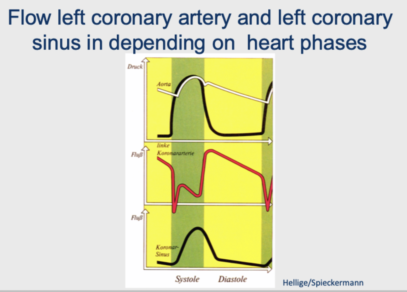
B - When does the left coronary artery Receiver its flow?
Flow is strongly reduced during systole and high in diastole. Coronary sinus flow shows the opposite pattern
C - What does the cross-section show?
Heart muscle cells, each surrounded by capillaries, with an occasional small arteriole
A - Why does depolarization spread so rapidly, and what are intercalated disks?
Depolarization spreads very rapidly throughout the heart, which facilitates a coordinated contraction pattern. Intercalated disks form junctions between adjacent cardiac muscle fibers. They contain a high concentration of gap junctions for rapid transmission of the action potential
(Picture shows also desmosomes: mechanically anchor adjacent cells to each other, so the fibers hold together during contraction, while the gap junctions carry the electrical current)

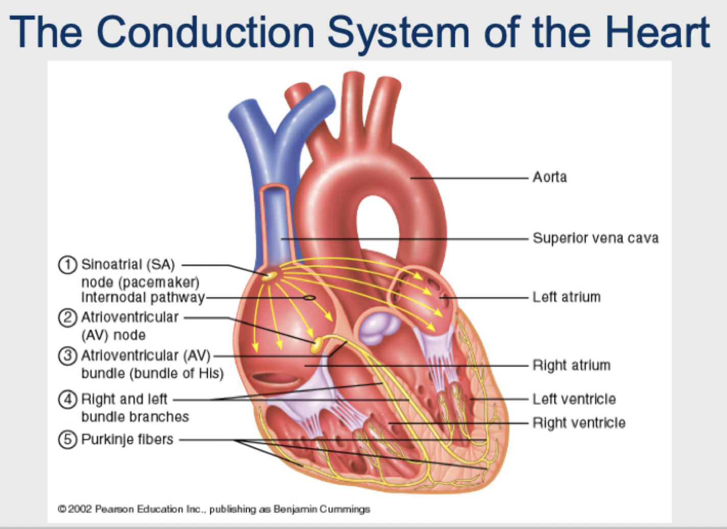
A - In which order does the impulse travel by the conduction system of the heart?
Sinoatrial (SA) node (pacemaker), where the action potential is initiated
AP travels toward the AV node
the impulse is conducteur to cells of AV node, with the AV delay
Impulses travels from AV Node through Atrioventricular (AV) bundle (bundle of His)
Impulses travels to right and left bundle branches
And from bundle branches to Purkinje fibers
B - What happens in skeletal muscle with rising stimulus frequency?
Single muscle twitches (5 Hz) → temporal summation (10 Hz) → unfused tetanus (25 Hz) → fused tetanus (50 Hz)
A - What influences heart rate, and what does the ANS regulate?
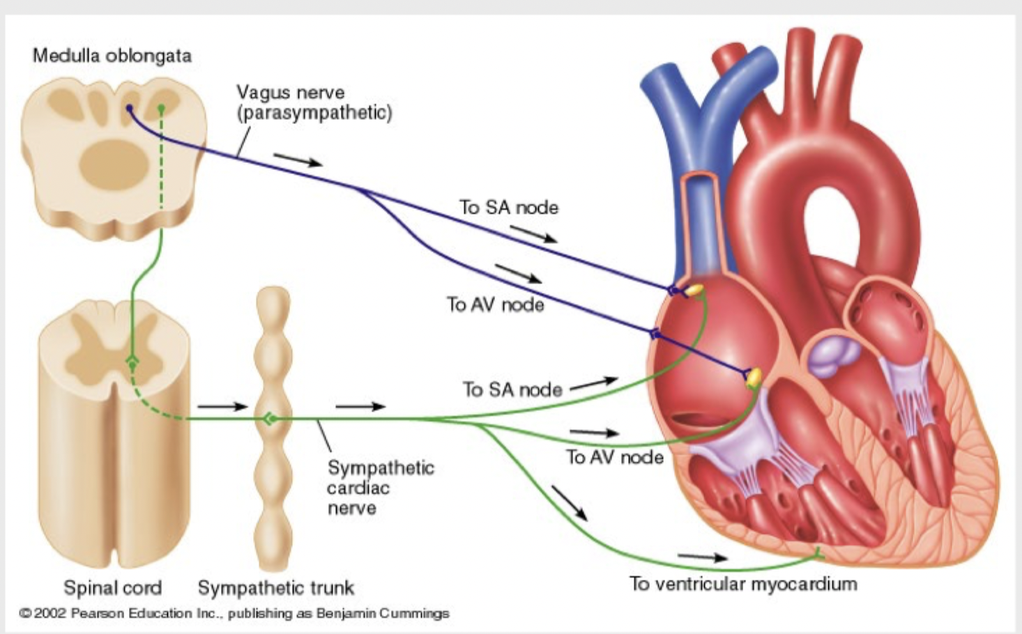
Three types of factors: sympathetic, parasympathetic and hormonal control. ANS fibers project to Almost every part of the heart: the SA node, AV node and ventricular myocardium. The ANS regulates both HR and SV (contractility)
A - Which nerves reach which structures?
The vagus nerve (parasympathetic, from the medulla oblongata) goes to the SA node and AV node. The sympathetic cardiac nerve (via spinal cord and sympathetic trunk) goes to the SA node, AV node and ventricular myocardium
A - What is an ECG (EKG)?
Electrocardiogram
A recording of the electrical changes that occur in the myocardium during the cardiac cycle. It is a graphic representation of the electrical activity of the heart, obtained by electrodes on the surface of the skin. Body fluids conduct the electrical activity that the electrodes detect
A - What are the waveforms of a normal EKG, and what can it show?
The waveforms are the P wave, P-R interval, QRS complex and T wave (also labels the PQ segment and the ST segment). The EKG detects only electrical events, such as arrhythmias or conduction blocks. It gives no information about the mechanical events of the heart

B - Which leads are shown?
Limb leads (I, II, III, aVR, aVL, aVF) and chest leads V1–V6. ⚠ Only a figure, so a lower exam risk.

B - How do central and peripheral adaptations differ?
Central (heart): 10–25% overall improvement, moderate decline with inactivity. Peripheral (muscle): 50–300% overall improvement, rapid decline with inactivity
B - Which parameters does aerobic training affect on cardiovascular function?
Heart rate, stroke volume, a-v O₂ difference, cardiac output, VO₂, systolic and diastolic blood pressure, coronary blood flow, brain blood flow, blood volume, plasma volume, red blood cell mass, heart volume.
A how do the three forms of myocardial hypertrophy differ?
Aerobic training: thicker walls and greater volume.
Strength training: thicker walls only.
Pathological: thicker but weaker walls.
The "athlete's heart" is a substantially enlarged heart (heart hypertrophy) compared to a normal, healthy heart. Heart mass figures (newborn 20 g, adult 300 g, athlete 500 g) are illustrative

B - What changes in the cardiovascular system with aging?
Blood: lower HCT; thrombi and emboli form more easily; blood pools in the legs.
Heart: lower efficiency and elasticity; atherosclerosis of coronary vessels; scar tissue forms.
Blood vessels: loss of elasticity; calcium deposits damage vessel walls.
Changes in heart function are minor at rest, more significant during exercise.
Hypertrophy of the left ventricle; maximum heart rate decreases.
Higher tendency for abnormal valve function and arrhythmias.
Higher O₂ consumption required to pump the same amount of blood.