Exam 2 Study Materials
 Call Kai
 Learn
 Practice Test
 Spaced Repetition
 Match
 Flashcards
 Knowt Play
1/127

Earn XP
Description and Tags
Learning objectives from chapters 5,6, and 24-28
128 Terms
B-DNA
More common form of DNA. a. Serves as the primary structure for genetic storage, replication and standard protein – DNA interactions.
A-DNA
Formed under dehydrating conditions, or in DNA-RNA and RNA-RNA duplexes. Shorter and wider than B-DNA.
Z-DNA
A high-energy, left handed helix favored by alternating purine-pyrimidine sequences and stabilized by high salt or negative supercoiling.
Electrostatic Interactions
Positively charged amino acids on the protein are attached to the negatively charged sugar-phosphate backbone of the DNA
Hydrogen Bonding
Specific amino acid side chains form hydrogen bonds with the chemical groups of the DNA bases and backbone
Histones
Highly alkaline, positively charged proteins found in the nuclei of eukaryotic cells and archaea that package, organize, and structure DNA
Nucleosomes
The basic repeating structural unit of chromatin that packages and organizes eukaryotic DNA inside the cell nucleus
Chromosomes
A thread-like structure made of protein and a single molecule of DNA that lives inside the nucleus of a plant and animal cells.
Meselson-Stahl Experiment
Proved that DNA replication is semi-conservative. They first labeled E.coli with heavy nitrogen and after many generations the E.coli DNA because very dense and heavy. Then they moved the bacteria into a medium with lighter nitrogen and allowed the cells to divide. Lastly, after each division they extracted the DNA and spun it in a cesium chloride density gradient to separate the molecules by weight. After the first generation they saw a hybrid band, which showed that both the heavy and the light DNA were being used in replication.
Equilibrium Density Gradient Ultracentriofugation
Separates particles strictly based on their buoyant density rather than their size or shape. This was important for meselson-stahl experiment because the DNA was treated with nitrogen that would make it more or less dense.
DNA Polymerase I:
DNA repair and removal of RNA primers
i. Has 5’-3’ exonuclease activity
ii. Has 3’-5’ exonuclease activity
DNA Polymerase III
Main enzyme for synthesis of new DNA strands.
i. Has 3’-5’ exonuclease activity
Replication Fork
Y shaped structure formed inside the nucleus or cell when a DNA double helix unwinds and separates into two single strands so it can be copied
Leading Strand
Strand of DNA that is synthesized continuously in the 5’ to 3’ direction, top strand.
Lagging Strand
Stand that is made discontinuously in short piecing moving away from the replication fork, causes fragments to be made
Okazaki Fragments
DNA Poly removes RNA primers and fills the gaps with DNA on lagging strand
DNA Ligase
Enzyme that joins DNA strands together by catalyzing the formation of phosphodiester bonds
RNA Synthesis
i. Initiation: RNA Poly binds to a specific DNA sequence called a promoter
ii. Elongation: RNA poly reads the template DNA strand in the 3’ to 5’ direction and builds a single-stranded RNA molecule in the 5’ to 3’ direction withing ribonucleoside triphosphates
iii. Termination: The enzyme reaches a specific termination sequence on the DNA, causing the RNA transcript and the polymerase to release from the DNA
Transcription Bubble
The unwound, single stranded region of DNA that forms when RNA poly melts the double helix during RNA transcription
Template Strand
Single strand of DNA that RNA poly reads to synthesize a complementary mRNA
Coding Strand (sense strand)
The DNA strand whose base sequence is identical to the newly synthesized messenger RNA
Transcription is what?
Highly Controlled
Lac Operon
A cluster of genes that controls the transport and breakdown of lactose. Used to turn on or off transcriptions depending on the amount of lactose in the cells environment.
Cis Peptide Bond
A rare peptide bond conformation that brings adjacent groups close together, resulting in steric clashing; it is occasionally observed when proline is present.
Trans Peptide Bond
The more common peptide bond conformation in which the amino acid R groups are positioned opposite and far away from each other, minimizing steric clashing.

Phi (Φ) Angle
The torsion angle describing rotation around the single bond between the nitrogen and the alpha carbon in a polypeptide backbone.

Psi (Ψ) Angle
The torsion angle describing rotation around the single bond between the alpha carbon and the carboxyl carbon in a polypeptide backbone.
Ramachandran Diagram
A 2D graph indicating the energetically allowed phi and psi backbone torsion angles for amino acid residues in a polypeptide.
N-terminus
The free amino (nitrogen) terminus of a polypeptide chain.
C-terminus
The free carboxyl (carbon) end of a polypeptide chain, serving as the growth end where incoming amino acids can be added.
Secondary Structure
The local conformation of a polypeptide backbone, governed by which backbone single bonds are rotatable around planar peptide units.
Alpha Helix
A helical secondary structure stabilized by hydrogen bonds between the N-H of the ith residue and the C=O of the (i−4)th residue with a distance of 2.8A˚ between the H and O, with side chains pointing outward and slightly downward.

Beta Strand
An extended polypeptide strand with repeating phi and psi angles that forms beta sheets through inter-strand hydrogen bonding between N-H and C=O groups.
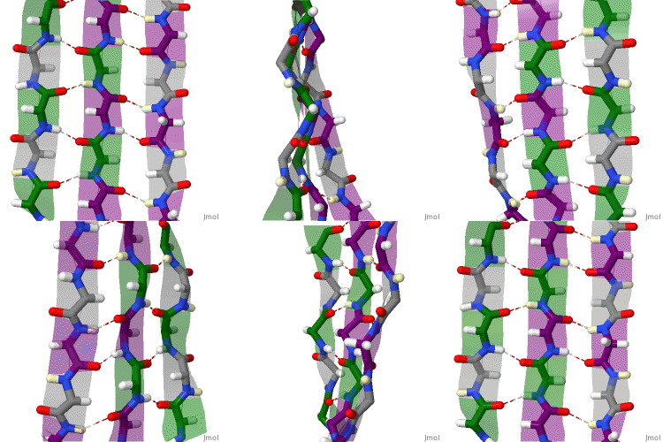
Parallel Beta Sheet
A beta sheet arrangement where adjacent hydrogen-bonded strands run in the same direction, having their N-termini on the same side and their C-termini on the same side.
Antiparallel Beta Sheet
A beta sheet arrangement where adjacent hydrogen-bonded strands run in opposite directions, with the N-terminus of one strand facing the C-terminus of the next.
Type I Turn
The more common turn geometry that easily accommodates standard L-amino acids to reverse the direction of a polypeptide chain.
Type II Turn
A less common turn geometry requiring tight sterics and specific geometric constraints to reverse the direction of a polypeptide chain.
Loops and Coils
Unstructured or irregularly shaped nonrepetitive segments that connect regular secondary structures or reside at a protein's N- or C-terminus.
Globular Proteins
Compact, water-soluble, roughly spherical proteins that fold tightly into round or glob-like shapes containing alpha helices, beta strands, and loops.
Supersecondary Structure
A compact 3D arrangement comprising multiple secondary structural elements that sits intermediate between the secondary and tertiary levels of protein structure.
Protein Domain
A self-stabilizing, compact 3D region of a polypeptide chain capable of folding and functioning independently from the remainder of the protein.
X-Ray Crystallography
A method used to determine the atomic structure of crystalline proteins via X-ray diffraction, typically utilizing vapor diffusion and sparse matrix screens to identify crystallization conditions.
Protein NMR
A spectroscopic technique used to assign chemical shifts and calculate 3D protein structures in solution based on molecular interactions without requiring crystals.
Hydrophobic Effect
The thermodynamic driving force that causes nonpolar groups to minimize contact with water and cluster in the protein interior, serving as the major driving force in protein folding.
Hydropathy Scale
A scale measuring the relative hydrophobicity or hydrophilicity of amino acid residues, indicating which regions of a globular protein are buried in the core or exposed to solvent.
Melting Temperature (Tm)
The characteristic temperature at which a folded protein denatures and unfolds, typically occurring below 100∘C for most proteins.
Chaotropic Agents
Substances like urea that denature proteins at high concentrations (e.g., 5–10M) by disrupting non-covalent interactions.
Fibrous Proteins
Elongated, water-insoluble structural proteins that provide mechanical strength, support, and protection to biological tissues.
Amyloid Fibrils
Polymers composed of prion subunits whose beta sheets run perpendicular to the long fiber axis and hydrogen bond from one subunit to the next.
Quaternary Structure
The spatial arrangement of two or more folded polypeptide chains assembled into a functional, multi-subunit protein complex.
Protein-Folding Problem
The scientific challenge of deciphering how an amino acid primary sequence encodes and directs the spontaneous folding of a protein into its tertiary structure.
Levinthal Paradox
The observation that random sampling of all possible phi, psi, and side-chain chi angles would require billions of years, implying proteins fold through directed, progressive pathways toward decreasing free energy.
Hydrophobic Collapse
The rapid initial stage of protein folding wherein hydrophobic forces cause nonpolar residues to sequester away from solvent into a compact core.
Molten Globule
A folding intermediate exhibiting native-like secondary and tertiary fold topology, but lacking tightly packed, native side-chain interactions.
Protein-Folding Funnel
An energy landscape model where funnel width represents conformational entropy (number of conformations) and funnel depth/height represents internal free energy.
Protein Disulfide Isomerase (PDI)
A folding accessory enzyme that catalyzes the breakage and rearrangement of non-native disulfide bonds to achieve the thermodynamically stable native pairing.
GroEL/ES System
A molecular chaperone consisting of a 60kDa 14-subunit GroEL cylinder and a 10kDa 7-subunit GroES cap that utilizes ATP hydrolysis to allow misfolded proteins to escape local energy minima.
Conformational Flexibility
A protein's intrinsic ability to undergo dynamic structural movements or 'breathing', which opens and closes internal channels for ligand binding (such as oxygen entering hemoglobin or myoglobin).
Atomic Fluctuations
Small, rapid, time-dependent spatial displacements and vibrations of covalent chemical bonds within a protein.
Ring Flipping
The rapid 180∘ rotational flipping of aromatic side chains, such as phenylalanine (Phe) or tyrosine (Tyr), within a folded protein structure.
Collective Motions
Coordinated spatial movements of protein domains or groups of side chains over distances greater than 5A˚.
Triggered Conformational Changes
Large-scale structural rearrangements (>10A˚) involving protein domains or side chains in response to a specific event, such as ligand binding.
Stanley Prusiner
The scientist awarded the Nobel Prize for discovering and establishing the protein-only nature of prion diseases.
Kurt Wutrich
A Nobel laureate who contributed to protein NMR spectroscopy and also conducted research investigations on prion proteins.
RNase A
A 124-amino acid, disulfide-stabilized ribonuclease enzyme utilized by Christian Anfinsen to demonstrate that primary sequence encodes native tertiary structure.
2-Mercaptoethanol
A chemical reducing agent used in denaturation experiments to reduce covalent disulfide bonds (-S-S-) into free sulfhydryl (-SH) groups.
Insulin Structure
A pancreatic hormone composed of two polypeptide chains—a 21-residue A chain and a 30-residue B chain in humans—cross-linked by interchain and intrachain disulfide bonds.

Proinsulin
The biosynthetic precursor of insulin consisting of an A chain, a B chain, and an intervening connecting C chain; it spontaneously refolds into native disulfide pairings after denaturation.

Insulin Sequence Variation Across Species
Sequence divergence among animals occurring predominantly at residues A8, A9, A10, B1, B2, B27, and B30, accounting for why non-identical animal insulins can elicit immune responses in humans.
Reduced SDS-PAGE of Proinsulin vs. Insulin
Electrophoretic separation where reduced proinsulin resolves as a single continuous polypeptide band, whereas reduced insulin resolves as two distinct bands (A chain and B chain).

Postsynthetic Modification
A processing event (such as proteolytic cleavage of proinsulin's C chain) occurring after translation that alters a polypeptide; its loss explains why mature insulin cannot spontaneously refold to >10% activity.
Beer’s Law
States that the absorption of light by a solution is directly proportional to the concentration of the solute and the distance the light travels through the solution.
Colorimetric Assays (Bradford Assay)
Proteins are mixed with a reagent that will create a color when activated. Can use proteins at different concentrations to create a curve
Immunoassays
Measures the concentration of a molecule in a fluid sample by using an antibody to bind to that target molecule
ELISA(Enzyme Linked Immunosorbent assay)
Used to measure proteins, antibodies ,antigens, and hormones in bodily fluids
Salting Out
Ammonium sulfate is added to the protein and supernatant, which caused the salt ions to steal water, leading to the hydrophobic patches on the proteins to clump together. (Solubility property)
Ion Exchange Chromatography
Stationary phase consists of a solid support matrix (resin beads) with charged functional groups, molecules with the opposite charge will bind to these functional groups, molecules with the same charge will be able to pass through
Electrophoresis Isoelectric Focusing
Gel contains a continuous pH gradient, when voltage is added the proteins will pass through the gel and will move until its isoelectric point is reached
Hydrophobic Interaction Chromatography
Columns are packed with a matrix. Salts are added and this forces the proteins to bind the stationary phase ligands l, these molecules are then released with the salt concentration is lowered.
Gel Filtration Chromatography
Beads in columns only allow molecules that are small to pass through
SDS-PAGE
Gel, used to separate proteins by molecular weight
Affinity Chromatography
Isolated a specific biomolecule from a complex mixture by exploiting reversible and specific biological interactions between the target molecule and an immobilized ligand. Can purify a protein from a mixture in a single chromatographic step
Protein with a low ionic strength
Increased solubility as the salt concentration increases
Protein with strong ionic strength
Lower solubility as salt concentration increases
Mobile Phase
Moving liquid or gas that carries a sample mixture through the stationary phase to separate components
Stationary Phase
The fixed substance, stays in once place, can be solid or liquid
Eluent
The solvent or carrier portion of the mobile phase that flows through a column
Elution
Process of washing a component through a stationary phase, by passing liquid or gas in the mobile phase over it
Gradient Elution
Technique where the composition or strength of the mobile phase is systematically changed during the separation run. Common in reverse phase high-performance liquid chromatography
Resin
Solid or porous matrix packed into a column that separates components in a mixture
Partition Coefficient
Ratio of a solutes concentration in the stationary phase to its concentration in the mobile phase
Chromatogram
Output graph showing how components of a mixture separate over time
Exclusion Limit
The upper molecular weight boundary of a columns working range
Void Volume
The volume of mobile phase inside a packed column that is not occupied by the stationary phase particles
Bed Volume
The volume occupied by the packed chromatography resin or stationary phase inside a cylindrical column
Elution Volume
Amount of mobile phase that passes through a column
Dialysis
A way to separate large macromolecules from small molecules. To preform dialysis you first will load your sample into a dialysis tube and clamp it shut. Then you submerge the tubing in a dialysate buffer, and stir the solution in a cold room. Continue this until equilibrium is achieved. Dialysis is heavily driven by passive diffusion across a membrane.
Northern Blot
Detects size, and quantifies specific RNA molecules in a sample to study gene expression
Southern Blot
Used to identify a specific DNA sequence within a complex mixture of DNA
Western Blot
Used to identify, measure, and analyze specific proteins within a biological sample