Edit · BIOL 1220: Lab 3
 Call Kai
 Learn
Learn Practice Test
 Spaced Repetition
 Match
Match Flashcards
 Knowt Play
1/190

There's no tags or description
Looks like no tags are added yet.
Name | Mastery | Learn | Test | Matching | Spaced | Call with Kai | Chat |
|---|
No analytics yet
Send a link to your students to track their progress
191 Terms
cartilage
can be found in the areas of the skeleton where bones move, e.g. the ribcage and joints
compact bone connective tissue
constitutes the walls of the diaphysis; dense and hard
hyaline cartilage connective tissue
the cartilage that reduces friction and acts as a shock absorber
elastic cartilage connective tissue
the cartilage that provides flexibility to structures like your external ear and epiglottis
fibrocartilage connective tissue
the cartilage most resistant to compression and stretch; forms vertebral discs and knee joint cartilages
axial skeleton
main axis of body
long bones
cylindrical and longer than they are wide
short bones
equal in length and width
flat bones
typically thin but are also often curved
irregular bones
do not have any easily characterized shapes and therefore do not fit any other classification
sesamoid bones
small, round bones that, as the name suggests, are shaped like a sesame seed
sutural bones
extra pieces of bone which can be found within the cranial sutures
compact bone
the denser, stronger of the two types of bone tissue
spongy bone
contains osteocytes housed in lacunae, but they are not usually arranged in concentric circles; also called trabecular bone
trabeculae
spongy bone
diaphysis
shaft of the bone
medullary cavity
the hollow region in the diaphysis, which is mostly yellow marrow in adults
epiphyses
the proximal and distal ends of the bone
articular cartilage
thin layer of hyaline cartilage covering the epiphyses that reduces friction and acts as a shock absorber
epiphyseal line
a faint line left where the epiphyseal plates are eventually replaced with bone when growth ceases
epiphyseal plate
a line of hyaline cartilage in children at the junction between the diaphysis and epiphysis; allows for longitudinal bone growth
periosteum
a fibrous membrane covering the outer surface of the bone
endosteum
the delicate membranous lining of the medullary cavity, where bone growth, repair, and remodeling occur
hematopoietic tissue
initially fills the medullary cavity in infants
hydroxyapatites
give bones their hardness and strength
collagen fibers
give bones flexibility so that they are not brittle
osteon
the microscopic structural unit of compact bone
lamellae
the concentric rings of calcified matrix that compose osteons
central canal
runs down the center of each osteon; contains blood vessels, nerves, and lymphatic vessels
perforating canal
allows vessels and nerves to branch off at right angles from the central canal and extend to the periosteum and endosteum
canaliculi
channels within the bone matrix through which the cytoplasmic processes of osteocytes extend
osteocytes
located inside lacunae
lacunae
spaces found at the borders of adjacent lamellae
cervical vertebrae
C1-C7
thoracic curvature
T1-T12
lumbar vertebrae
L1-L5
intervertebral discs
strongly unite the bodies of adjacent vertebrae
scoliosis
an abnormal, lateral curvature, accompanied by twisting of the vertebral column
kyphosis
excessive curvature of thoracic vertebrae
lordosis
an excessive anterior curvature of the lumbar region most commonly associated with obesity or late pregnancy; also called swayback
fontanelle
a large area of dense connective tissue that separates the cranial vault bones as they grow in the fetal skull
frontal bone

supraorbital margin of frontal bone

coronal suture

parietal bone

temporal bone
temporal bone

mastoid process of temporal bone
no
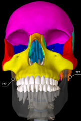
nasal bone

zygomatic bone
.

maxilla

sphenoid bone

optic canal of sphenoid bone

lacrimal bone

ethmoid bone

vomer

mandible

orbits
the depressions in which the eyeballs are located
frontal bone
frontal bone

parietal bone
parietal bone

temporal bone
temporal bone

external auditory meatus of temporal bone
external auditory meatus of temporal bone

mastoid process of temporal bone
.

zygomatic process of temporal bone

zygomatic arch
formed by the zygomatic process joining with a process of the zygomatic bone
coronal suture

squamous suture

lambdoid suture
no
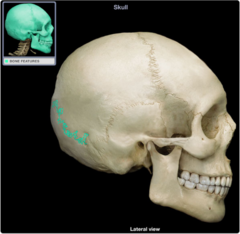
occipital bone

sphenoid bone

ethmoid bone

lacrimal bone

lacrimal fossa of lacrimal bone

nasal bone

zygomatic bone
no

maxilla

mandible

ramus of mandible

angle of mandible
no

condylar process of mandible

coronoid process of mandible

sagittal suture
joins the two parietal bones
lambdoid suture

sagittal suture
sagittal suture

coronal suture

hard palate

maxilla

palatine bone

vomer

sphenoid bone
no

temporal bone

carotid canal of temporal bone

jugular foramen of temporal bone

occipital bone

occipital condyles of occipital bone

foramen magnum of occipital bone

occipital condyles
bumps on the sides of magnus foramen
carotid canal of temporal bone
act as passageways for the internal carotid arteries
frontal bone

frontal sinus of frontal bone

ethmoid bone
.
