BIOL 319 Lab Practical 1 - KB
 Call Kai
 Learn
 Practice Test
 Spaced Repetition
 Match
 Flashcards
 Knowt Play
1/176

There's no tags or description
Looks like no tags are added yet.
177 Terms
Muscles that flex the hand
Palmaris longus,
flexors (carpi rad., uln., digitorum superfiicalis, profundus, pollicis longus)
Muscle that flexes and abducts the hand
flexor carpi radialis
muscle that flexes and adducts the hand
flexor carpi ulnaris
Muscle that flexes phalanges 2-5, proximal and middle
flexor digitorum superficialis
muscle that flexes phalanges 2-5, distal
flexor digitorum profundus
Muscles with origin - medial epicondyle of humerus
palmaris longus,
flexor carpi radialis/ulnaris (+ulna),
flexor digitorum superficialis (+ulna, radius)
pronator teres but its different and sutff
Muscles with origin - ulna
Flexor carpi ulnaris (+humerus),
flexor digitorum superficialis (+ humerus, radius),
profundus (+interosseus membrane)
Flexor pollicis longus origin - insertion
Anterior portion of radius and interosseous membrane - distal phalanx of pollex
Muscles that extend the hand
Extensor carpi radialis longus (+abduct)/brevis (+abduct)/ulnaris (+adduct),
extensor digitorum (+phalanges 2-5)
Muscle that extends thumb
extensor pollicis longus/brevis,
abductor pollicis longus (+ abducts, abducts hand also)
Muscles with humeral origins
Extensor carpi radialis longus (lat supracondylar ridge)/brevis (lat epicondyle)/ulnaris (lat epicondyle)(+ulna),
extensor digitorum (lat epicondyle)
Muscles with posterior radius/ulna, interosseous membrane origins
Abductor pollicis longus,
extensor pollicis longus/brevis
Insertions of posterior muscles acting on hand
Extensor carpi radialis longus - metacarpal 2
brevis - 3
ulanris - 5
Extensor digitorum - middle and distal phalanges of digits 2-5
Abductor pollicis longus - metacarpal 1/pollex
Extensor pollicis longus/brevis - Proximal and distal phalanges of pollex
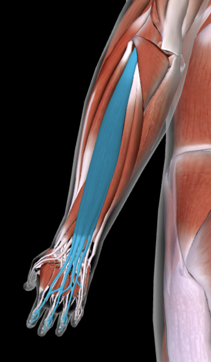
Extensor Digitorum

Extensor Carpi Ulnaris
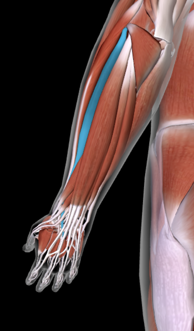
Extensor Carpi Radialis brevis
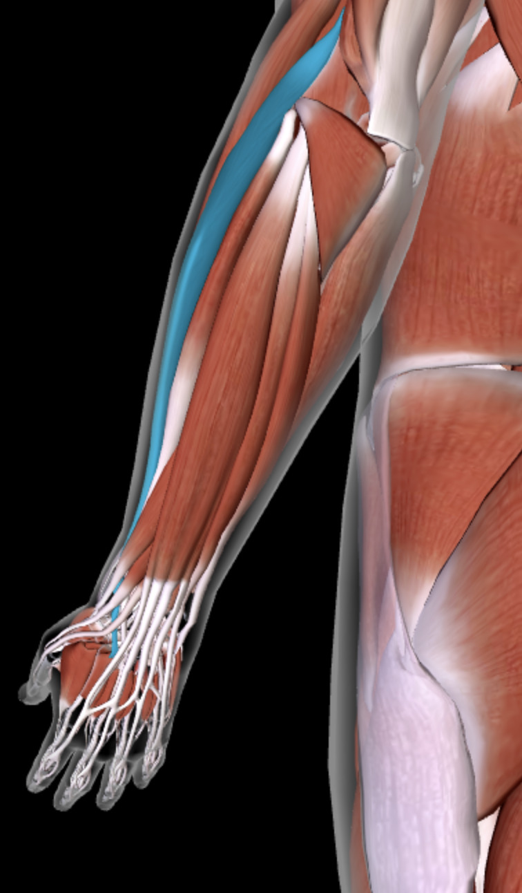
Extensor Carpi Radialis Longus
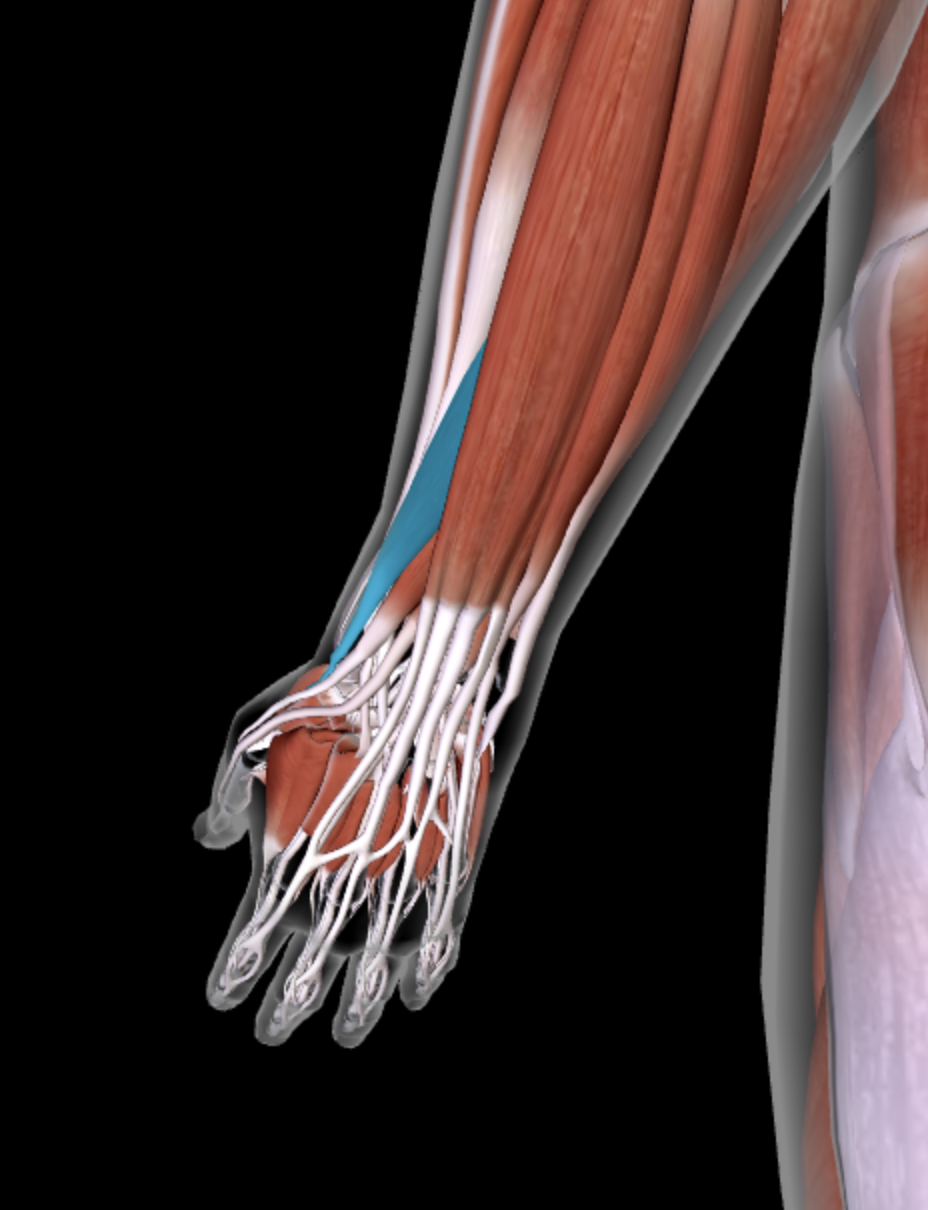
Abductor Pollicis Longus

Extensor Pollicis longus

Extensor Pollicis brevis

Palmaris Longus
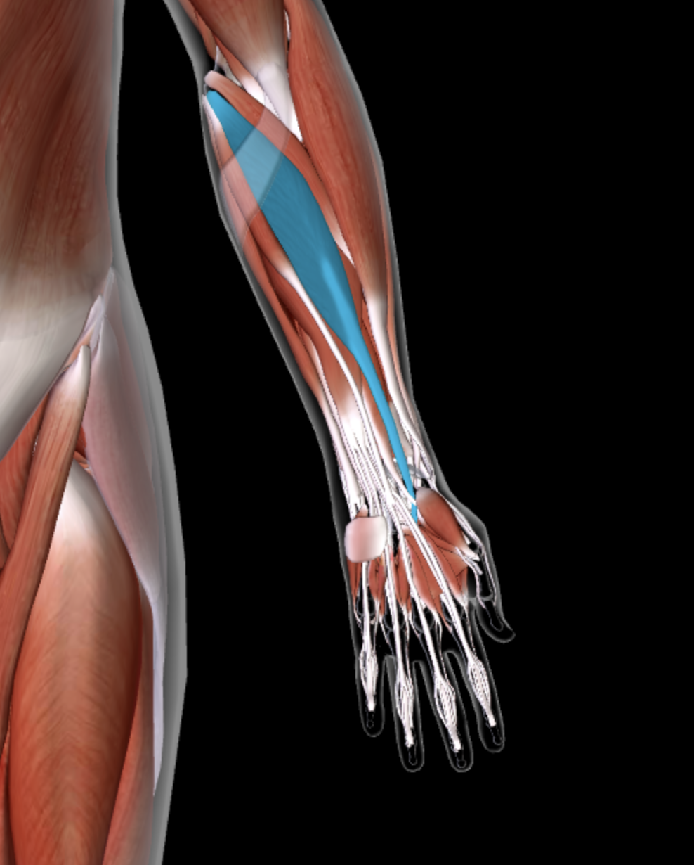
Flexor carpi radialis

Flexor carpi ulnaris

Flexor digitorum superficialis

Flexor digitorum profundus
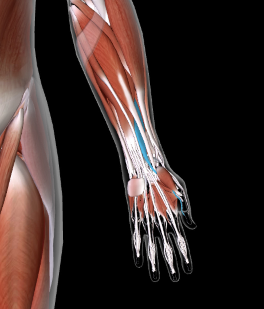
Flexor pollicis longus

Name the Muscles Pictured
Yellow - Levator Scapulae
Dark Blue - Rhomboideus Major (Minor above)
Light blue - Supraspinatus
Yellow - Infraspinatus
Magenta - Subscapularis
Pink - Teres Minor
Green - Teres Major
Flesh - Coracobrachialis

Name the anterior forearm muscles pictured
blue - brachialis
green - brachioradialis
Flesh - pronator teres
Yellow - pronator quadratus
Between radius and ulna, intrinsic - supinator

Name the muscles pictured
blue - sartorius
yellow - pectineus
orange - adductor longus
yellow - adductor brevis
gray - adductor magnus
green - gracilis

Name the muscles pictured
flesh - rectus femoris
blue - vastus lateralis
teal - vastus medius
green - semimembranosus (post.)
yellow - vasus intermedius
purple - tensor fasciae latae (post.)
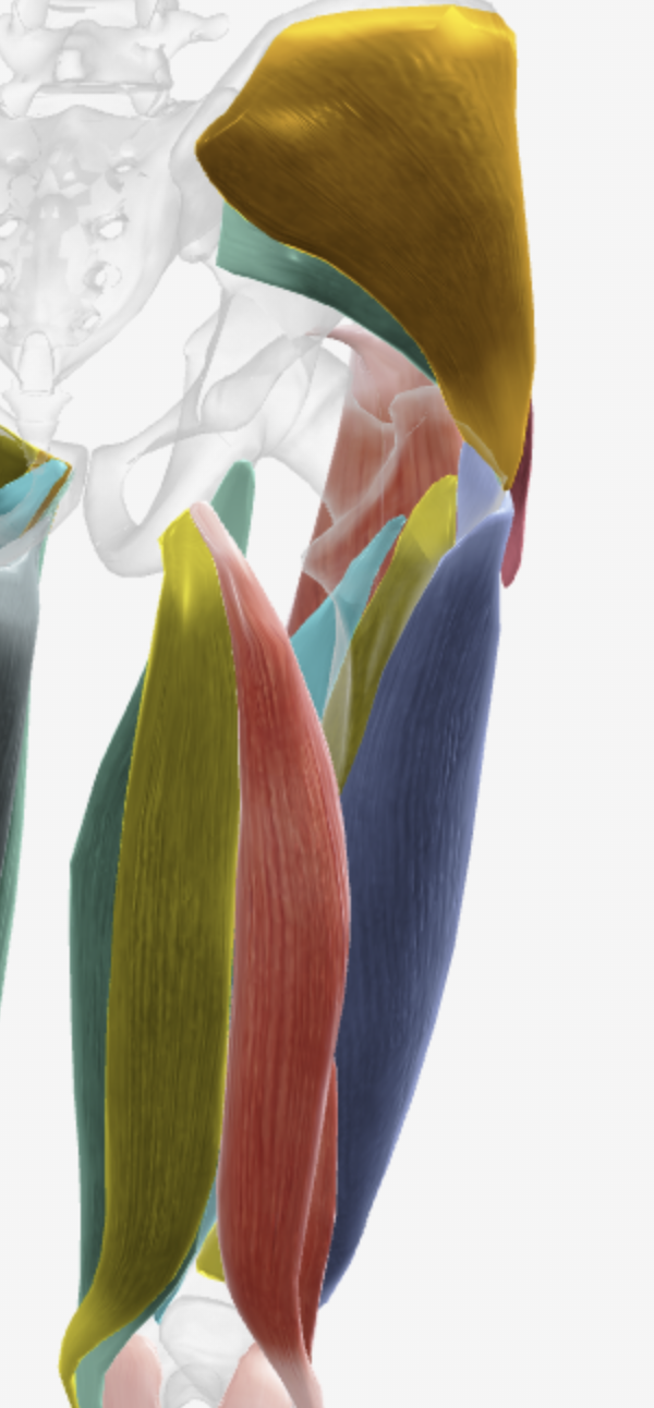
Name the muscles pictured
Orange - gluteus medius
green - gluteus minimus
purple (barely showing) - tensor fasciae latae
Blue - vastus lateralis (ant.)
flesh - biceps femoris
yellow - semitendinosus
green - semimembranosus

Name the right side muscles pictured
flesh - gastrocnemius
dark yellow - popliteus
teal - soleus
yellow - tibialis posterior (post)
pink - flexor digitorum longus (post)
purple - flexor hallucis longus (post)
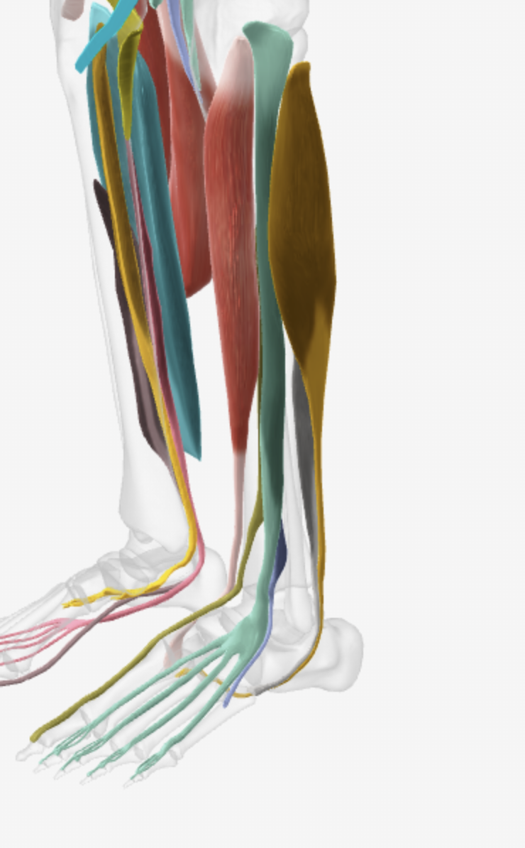
Name the left side muscles pictured (anterior)
Dark yellow - fibularis longus
green - extensor digitorum longus
gray - fibularis brevis
purple - fibularis tertius
flesh - tibialis anterior
yellow - extensor hallucis longus
Gastrocnemius, Soleus, Popliteus, Tibialis POsterior, Flexor digitorum longus, flexor hallucis longus INNERVATION
Tibial nerve
Fibularis longus, brevis, tertius INNERVATION
Superficial fibular nerve, superficial, deep
Tibialis anterior, extensor digitorum longus, hallucis longus INNERVATION
Deep fibular nerve
Biceps femoris, semintendinosus and semimembranosus INNERVATION
Tibial/fibular nerves, tibial, tibial
Tensor fasciae lata, gluteus maximus, medius, minimus INNERVATION
Superior gluteal nerve, Inferior, superior, superior
Rectus femoris, vastus lateralis, intermedius, medialis INNERVATION
femoral nerve
Iliopsoas, sartorius, gracilis, pectineus, adductor brevis, longus, magnus INNERVATION
Femoral/spinal L1-3, Femoral, Obturator, Femoral + Obturator, Obturator, Obturator, Obturator and tibial
Plantarflexers
Gastrocnemius,
soleus,
popliteus,
tibialis posterior, (invert)
flexor digitorum longus, (invert) (flex toes 2-5)
flexor hallucis longus, (invert) (flex hallux)
fibularis longus and brevis (NOT TERTIUS)
Dorsiflexers
Fibularis tertius (evert)
extensor hallucis longus, (invert) (extend hallux)
extensor digitorum longus (evert) (extend toes 2-5)
tibialis anterior (invert)
Leg Flexers
popliteus
biceps femoris (extends thigh)
seimtendinosus (extends thigh)
semimembranosus (extends thigh)
gracilis (adducts thigh)
sartorius (flex and laterally rotate thigh)
Thigh Adductors
Gracilis (flexes leg)
Pectineus
Adductor brevis (laterally rotates)
longus (laterally rotates)
magnus (laterally rotates)
Leg Extenders
Rectus femoris (flexes thigh)
Vastus lateralis
intermedius
medialis
Posterior leg OI (plantarflex foot)
Gastroc - condyles of femur, calcaneus by calcaneal tendon
Soleus - posterior prox tibia; fibula, calcaneus by calcaneal tendon
Popliteus - lateral condyle of femur, proximal tibia
Tibialis posterior - interosseus membrane/tibia/fibula, Metatarsals 2-5/ 3 tarsals
Flexor digitorum longus - posterior tibia, distal phalanges of digits 2-5
Flexor hallucis longus - midshaft of fibula, distal phalanx of hallux
Lateral leg OI (everts foot)
Fibularis longus - Head/shaft of fib/lat condyle of tibia, metatarsal 1/cuneiform 1
brevis - shaft of fib, metatarsal 5
tertius - fibular/interosseous membrane, dorsum of metatarsal 5
Anterior leg OI (dorsiflex foot)
tibialis anterior - tibia/inetrosseous membrane, metatarsal 1/cuneiform 1
extensor digitorum longus - lateral condyle of tibia/shaft of fibula, middle/distal phalanges 2-5
extensor hallucis longus - anterior shaft of fibula/ineterosseous membrane, distal phalanx of hallux
Hamstings OI (extend thigh, flex leg)
Biceps femoris - ischial tuberosity/linea aspera of femur, head of fibula/lateral condyle of tibia
Semitendinosus - ischial tuberosity, prox/med tibia
Semimembranosus - ischial tuberosity, med condyle of tib/collateral ligament
Muscle of posterior and lateral thigh OI (abduct and rotate thigh)
TFL- anterior superior iliac spine, iliotibial band of FL to lateral condyle of tibia
Gluteus maximus- outer iliac blade/iliac crest/sacrum/coccyx, gluteal tuberosity of femur/iliotibial band of FL
medius/minimus - outer iliac blade, greater trochanter of femur
quadriceps femoris /anterior thigh OI (extend leg)
Rectus femoris - anterior inferior iliac spine
Vastus lateralis. - greater trochanter of femur/linea aspera of femur
inetrmedius - proximal/anterior femur
medalis - linea aspera of femur (medial)
All - tibial tuberosity by patellar ligament
medial thigh OI (adduct thigh)
Iliopsoas - T12 (spine)/L1-5 (spine)/iliac foassa, lesser trochanter of femur
Sartorius - Anterior superior iliac spine, proximal/medial tibia
Gracilis - pubis (symphysis), proximal/medial tibia
Pectineus - pubic crest, pectineal line of femur
Adductor brevis - pubis, linea aspera @ prox portion of femur (cuz its small)
longus - pubis, linea aspera @ middle portion of femur (cuz its long)
magnus - pubis/ischium, linea aspera/adductor tubercle of distal femur (cuz its huge)
Transverse
T/B
Coronal
F/B
Sagittal
L/R
Regions of Trunk
R hypocondriac | Epigastric | L hypochondriac
_____________________________________
R lumbar | Umbilical | L Lumbar
_____________________________________
R iliac. |. hypogastric. | L iliac
Brachial, Crural, Coxal, Acromial
Arm, Leg, Hip, Shoulder
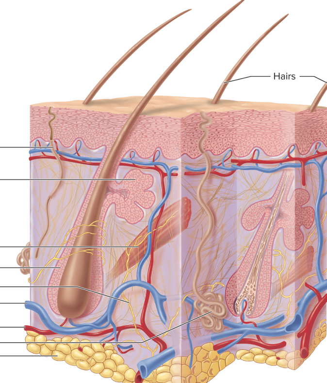
Name features top to bottom, then layers top to bottom
Sweat duct
Sebaceous gland
Arrector pili muscle
hair follicle
Nerve
Vein
Artery
Weat gland
Fat
Epidermis (skin/integument)
Dermis (skin/integument)
papillary
reticular
Hypodermis (subcutaneous)

Name strata top to bottom
Corneum (dead)
Lucidum (dead)
Granulosum (dying cells)
Spinosum
Basale (mitosis)
(+ basement membrane)
__________
Dermis

Label top to bottom
Hair shaft
Surface of skin
Arrector pili muscle
Hair root
Hair follicle (root sheath)
hair bulb
hair papilla
] Hypodermis

Label top to bottom
free edge
nail body
nail groove
nail fold
lunula
cuticle
nail root

Label top to bottom Left to Right
Cuticle
nail root
nail matrix
bone
Nail body
Free edge
Hyponychium
nail bed
] epidermis
Bone numbers?
Axial
22 skull
1 hyoid
26 vertebral column
24 ribs
1 sternum
6 auditory ossicles
Appendicular
Pectoral Girlde
2 clavicle
2 scapula
Pelvic girdle
2 hip bone
Upper limb
2 humerus
2 radius
2 ulna
16 carpals
10 metacarpals
28 phalanges
Lower limb
2 femur
2 patella
2 tibia
2 fibula
14 tarsals
10 metatarsals
28 phalanges
Tubercle, Tuberosity
Small elevation on a bone, larger elevation
Tronchanter
large process on femur
Condyle, epicondyle
smooth surface that articulates with another bone, raised area on condyle
Foramen, meatus/canal, fossa
shallow hole, deep hole, shallow surface depression

Label top to bottom
Articular cartilage
ephiphysis
ephiphyseal lines
spongy bone
compact bone
medullary cavity (yellow marrow)
periosteum
endosteum
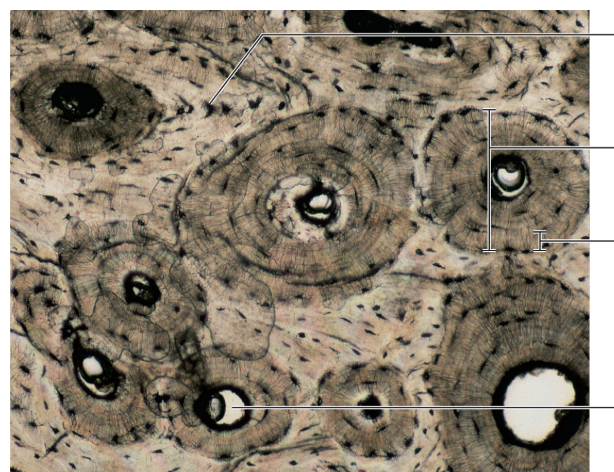
Label top to bottom
Lacuna
osteon
lamella
central canal

Label top to bottom
Canaliculi
Lacuna
Central canal
Lamella

Label top to bottom left to right
ANTERIOR
superior angle
acromion process
superior border
coracoid process
glenoid cavity
subscapular fossa
lateral (axillary) border
medial (vertebral) border
inferior angle
POSTERIOR
superior angle
supraspinous fossa
superior border
acromion process
scapular spine
glenoid cavity
medial (vertebral) border
infraspinous fossa
lateral (axillary) border
inferior angle

Label top to bottom left to right
RIGHT CLAVICLE - inferior
acromial end
conoid tubercle
sternal end

Label top to bottom left to right
RIGHT HUMERUS
ANTERIOR
greater tubercle
Lesser tubercle
intertubercular groove
deltoid tuberosity
medial supracondylar ridge
coronoid fossa
capitulum
trochlea
POSTERIOR
head
greater tubercle
anatomical neck
surgical neck
lateral supracondylar ridge
olecranon fossa
medial epicondyle

Label top to bottom left to right
RIGHT RADIUS AND ULNA
ANTERIOR
Olecranon process
Trochlear (semilunar) notch
Coronoid process
Radial notch of ulna
Head
Neck
Radial tuberoosity
Ulna
Radius
Interosseous ridges
Head of ulna
POSTERIOR
Olecranon porcess
Head of radius
radius
Ulna
styloid process of ulna
ulnar notch
styloid process of radius

Label top to bottom left to right
RIGHT HAND
POSTERIOR
Lunate
Triquetrum
Hamate
Capitate
Base
Shaft
Head
Scaphoid
Trapezium
Trapezoid
Base
shaft
Head
Pollex
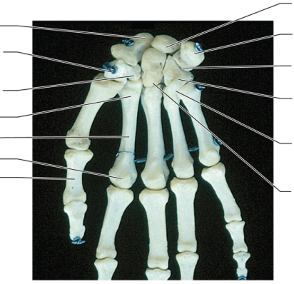
Label top to bottom left to right
RIGHT HAND
ANTERIOR
Scaphoid
Trapezium
Trapezoid
Base
shaft
Head
Pollex
Lunate
Pisiform
Triquetrum
Hamate
Hamulus of hamate
Capitate
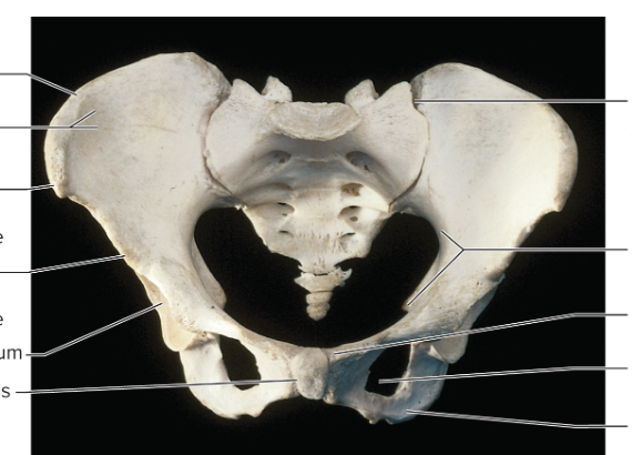
Label top to bottom left to right
ANTERIOR PELVIC GIRDLE
Iliac crest
Iliac fossa
Anterior superior iliac spine
Anterior inferior iliac spine
Acetabulum
symphysis pubis
Sacroiliac joint
Plvic brim
Pubic crest
Obturator foramen
Ischial tuberiosity
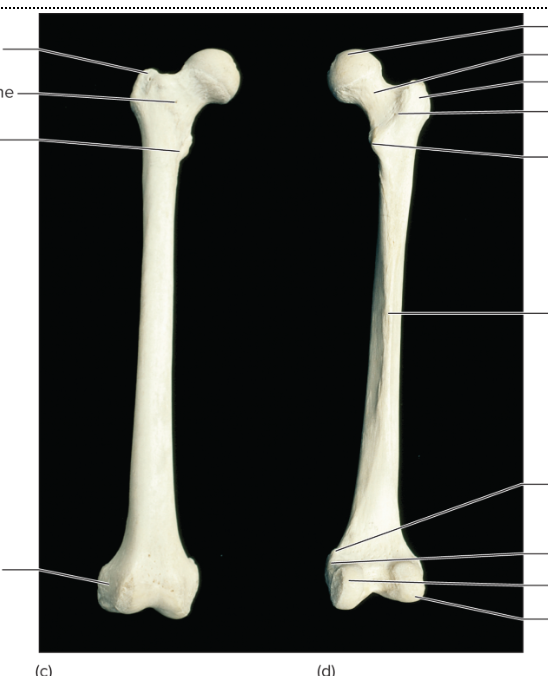
Label top to bottom left to right
RIGHT FEMUR
ANTERIOR
Greater trochanter
interochanteric line
lesser trochanter
lateral epicondyle
POSTERIOR
head of femur
neck of femur
greater trochanter
intertrochanteric crest
lesser trochanter
linea aspera
adductor tubercle
medial epicondyle
medial condyle
lateral condyle

Label top to bottom left to right
PATELLA
ANTERIOR
base
apex
POSTERIOR
base
articular facets
apex

Label top to bottom left to right
TIBIA/FIBULA
ANTERIOR
Intercondylar eminence
Medial condyle
Lateral condyle
Tibial tuberosity
Head of fibula
tibia
Anterior crest
fibula
Lateral malleoulus
Medial malleolus
POSTERIOR
Intercondylar eminence
medial condyle
head of fibula
medial malleolus
lateral malleolus

Label top to bottom left to right
RIGHT FOOT
SUPERIOR
distal phalanx
middle phalanx
proximal phalanx
first metatarsal
first cuneiform
second cuneiform
third cuneiform
navicular
cuboid talus
calcaneus
INFERIOR
Distal phalanx
proximal phalanx
third metatarsal
first cuneiform
second
third
cuboid
navicular
talus
calcaneus

Label top to bottom
TYPICAL VERTEBRA
SUPERIOR
spinous process
lamina
transverse process
pedicle
vertebral foramen
body
LATERAL
superior articular process
transverse process
body
inferior articular process
spinous process
Spine from top to bottom
Bfast @ 7 Atlas, Axis (1,2) (Cervical 1-7) Vertebra Prominens (7)
Lunch @ 12 (Thoracic 1-12)
Dinner @ 5 (Lumbar 1-5)
5 fused (sacrum)
4 fused (coccyx)

Label top to bottom
CERVICAL VERTEBRAE
SUPERIOR
Bifid spinous process
Vertebral foramen
Superior articular facet
Transverse foramen
Body
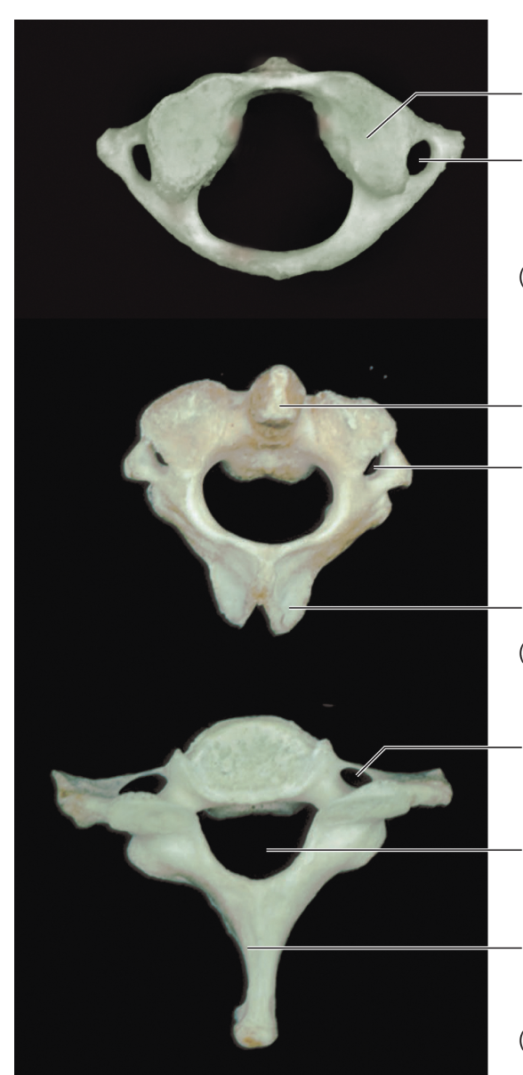
Label top to bottom
SUPERIOR
ATLAS
Superior articular facet
trasnverse foramen
AXIS
Dens
transverse foramen
bifid spinous process
VERTEBRA PROMINENS
Transverse foramen
vertebral foramen
spinous process


Label top to bottom, top to bottom left to right
THORACIC VERTEBRAE
SUPERIOR
spnous process
Vertebral foramen
trasnverse process
superior articular facet
rib facet
body
LATERAL
superior articular process
rib facet
body
superior articular facet
trasnverse process
spinous process

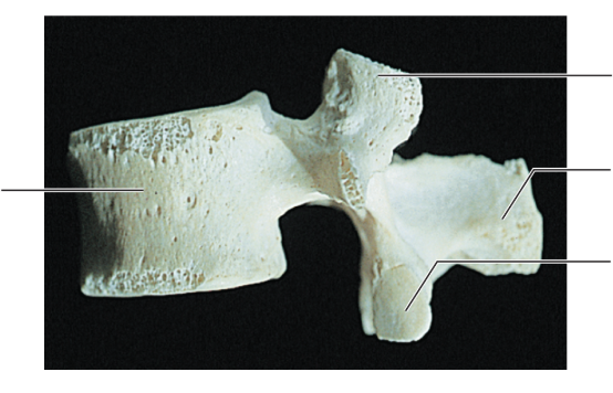
Label top to bottom left to right, top to bottom left to right
LUMBAR VERTEBRAE
SUPERIOR
spinous process
lamina
superior articular facet
pedicle
vertebral foramen
body
LATERAL
body
superior articular porcess
spinous process
inferior articular process

Label top to bottom left to right
SACRUM and COCCYX
ANTERIOR
sacral promonotory
ala
trasnverse line
anterior sacral foramen
coccyx
POSTERIOR
sacral canal
superior articular facet
articular surface
posterior sacral foramen
medial crest
lateral crest
coccyx

Label top to bottom
HYOID FLOATING BONE
Greater horn
Lesser horn
body
repeated

Label top to bottom left to right
RIB
INFERIOR
head
neck
tubercle
angle
sternal end
coastal groove
Ribs order
True ribs - 1-7
False ribs - 8-12 (8-10 attach at rib 7 to sternum) (11 and 12 are floating)

Label top to bottom left to right
THORACIC CAGE
manubrium
sternal angle
coastal notch
body
coastal cartilage
xiphoid porcess
jugular notch
clavicular notch
true ribs
false ribs

Label top to bottom
CRANIUM
INFERIOR VIEW
Mandible
Maxilla
Palatine
vomer
Sphenoid
Temporal
Occipital
Parietal

Label top to bottom
Lacrimal
Nasal
zygomatic
Inferior nasal concha
Vomer
Maxilla
Alveolus (the space)
Mandible

Label top to bottom left to right
Coronal suture
glabella
supraorbital margin
temporal bone
frontal process of the maxilla
nasal bone
infraorbital margin
zygomatic bone
perpendicular plate of ethmoid bone (nasal septum)
vomer (nasal septum)
nasal cavity (nasal septum)
maxilla
alveolar processes
body of mandible
mental foramen
frontal bone
parietal bone
supraorbital foramen
optic canal
orbital plate of frontal bone
sphenoid bone (greater wing)
superior orbital fissure
lacrimal bone
infraorbital formaen
middle nasal concha
inferior nasal concha
anterior nasal spine
oblique line of mandible
mandible
mandibular symphysis
mental protuberance

Label top to bottom left to right
optic canal
superior orbital fissure
sphenoid
ehmoid bone
inferior orbital fissure
infraorbital foramen
mandibular symphysis
frontal bone
glabella
supraorbital margin
nasal bone
lacrimal bone
zygomatic
inferior nasal concha
maxilla
alveolar process
mental foramen
mandible

Label top to bottom left to right
coronoid process
mandibular condyle
mandibular notch
mandibular foramen
alveolar process
mental foramen
condylar process
ramus
body
angle
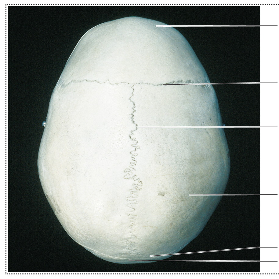
Label top to bottom left to right
Frontal bone
coronal suture
sagittal suture
parietal bone
lamboid suture
occipital bone
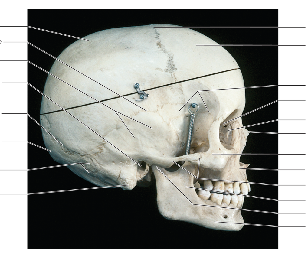
Label top to bottom left to right
parietal bone
squamous suture
temporal bone
external acoustic meatus
lambdoid suture
external occipital protuberance
occipital bone
mastoid process
coronal suture
frontal bone
sphenoid bone
ethmoid bone
lacrimal bone
nasal bone
zygomatic bone
maxilla
zygomatic arch
ramus of mandible
mandibular condyle
body of mandible
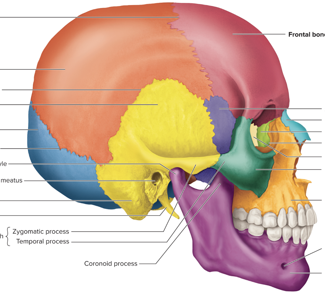
Label top to bottom left to right
coronal suture
parietal bone
squamous suture
temporal bone
occipital bone
lambdoid suture
mandibular condyle
external acoustic meatus
mastoid process
styloid process
zygomatic arch { zygomatic porcess, temporal porcess
coronoid porcess
frontal bone
sphenoid bone
nasal bone
lacrimal bone
nasolacrimal canal
ethmoid bone
zygomatic bone
maxilla
mental foramen
mandible