KNES 191 Anatomy Practical Vocabulary for Exam I
 Call Kai
 Learn
Learn Practice Test
 Spaced Repetition
 Match
Match Flashcards
 Knowt Play
1/312

Earn XP
Description and Tags
Comprehensive vocabulary flashcards covering directional terms, anatomical planes, joint movements, bone landmarks, skeletal divisions, skull features, vertebrae, bone histology, bone mineral density categories, muscle roles, and key axial muscles from the KNES 191 study guide.
Name | Mastery | Learn | Test | Matching | Spaced | Call with Kai | Chat |
|---|
No analytics yet
Send a link to your students to track their progress
313 Terms
Superior
Above or closer to the head.
Inferior
Below or toward the feet.
Anterior / ventral
Toward the front.
Posterior / dorsal
Toward the back.
Medial or internal rotation
Closer to the midline.
Lateral or external rotation
Farther from the midline.
Ipsilateral
On the same side.
Contralateral
On opposite sides.
Proximal
Closer to trunk or reference point.
Distal
Farther from trunk or reference point.
Superficial
Near the surface.
Deep
Farther inside.
Sagittal plane
Divides the body into right and left portions; midsagittal makes equal halves.
Frontal / coronal plane
Divides the body into anterior and posterior portions.
Transverse / horizontal plane
Divides the body into superior and inferior portions; appears as a cross-section.
Flexion
Joint movement where joint angle decreases.
Extension
Joint movement where joint angle increases.
Hyperextension
Extension beyond anatomical position.
Abduction
Movement away from midline.
Adduction
Movement toward midline.
Dorsiflexion
Movement where top of foot moves toward shin (up).
Plantarflexion
Movement where foot points downward.
Supination / pronation
Movement where palm faces anterior-up or posterior-down.
Process
General bony prominence or bump.
Ramus of the Mandible
Extension that creates an angle with the rest of the bone.

Condyle
Smooth rounded prominence with cartilage for articulation.
Facet
Smooth, flat articular surface.
Trochanter
Large rough rounded projection.
Tuberosity
Rough rounded projection; larger than a tubercle.
Tubercle
Small rough rounded projection.
Fossa
Depression ranging from a trench to a broad shallow basin.

Foramen
Rounded passageway through bone.

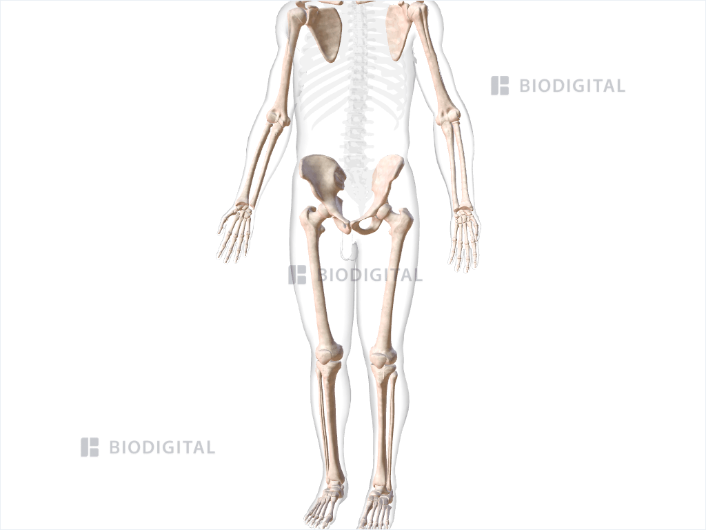
Axial skeleton
Central axis division consisting of skull, hyoid, vertebral column, ribs, and sternum/thoracic cage.
Appendicular skeleton
Division containing upper and lower limbs plus the pectoral and pelvic girdles that attach them to the axial skeleton.
Foramen magnum
Large opening on inferior occipital bone for spinal cord.

External occipital protuberance
Midline bump on posterior skull located on the occipital bone.

Zygomatic arch
Lateral cheek arch formed by the temporal process of the zygomatic bone and the zygomatic process of the temporal bone.

Mastoid process
Rounded projection posterior and inferior to ear canal on the temporal bone.

Atlas C1
Ring-shaped first cervical vertebra with no typical body; supports skull and permits 'yes' nodding at atlanto-occipital joint.

Axis C2
Second cervical vertebra having the dens projecting superiorly, forming a pivot allowing rotation for 'no'.
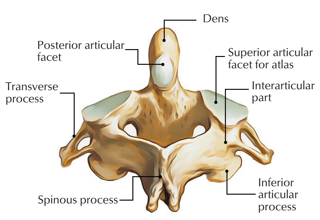
Dens
Tooth-like process of axis inside ring of atlas acting as pivot for rotation.
True ribs 1-7
Ribs that attach directly to sternum through their own costal cartilages.

False ribs
Ribs that do not attach directly to sternum; ribs 8-10 attach indirectly through cartilage above; ribs 11-12 do not attach anteriorly.

Floating ribs 11-12
Subset of false ribs with no anterior attachment to sternum.

Jugular notch
Superior midline depression of manubrium between clavicular notches.

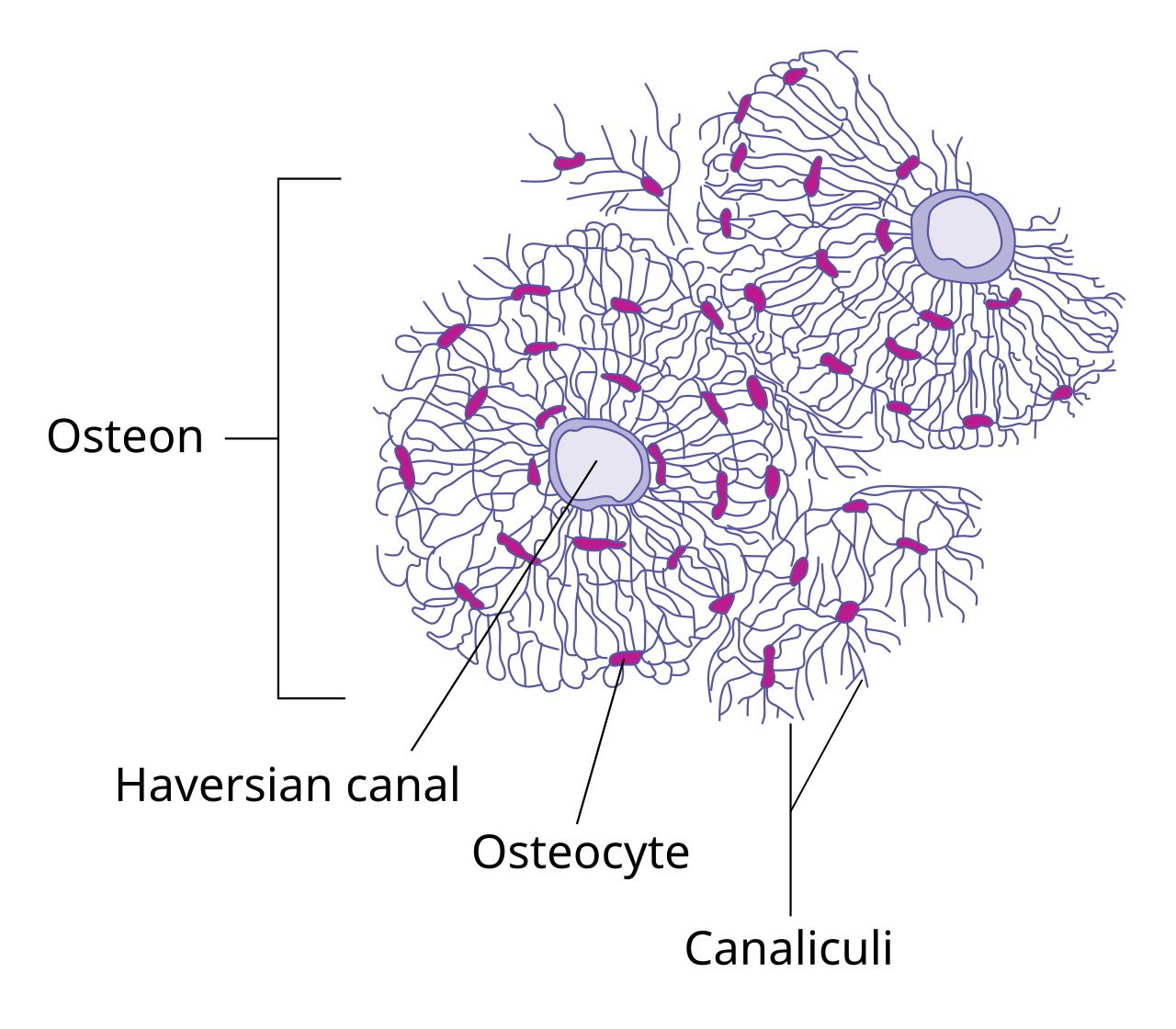
Osteon
Structural unit of compact bone consisting of concentric lamellae around a central canal.

Lamellae
Layers of calcified matrix; concentric in an osteon, arranged as trabeculae in spongy bone.

Central canal
Center of osteon containing blood vessels and nerves.

Lacuna
Small space housing an osteocyte.

Osteocyte
Mature bone cell that maintains bone matrix.

Canaliculi
Tiny channels connecting lacunae and allowing osteocyte communication/exchange.
Trabeculae
Thin plates/struts forming spongy bone.

Osteoprogenitor cell / osteogenic cell
Stem-like bone cell that can become an osteoblast.

Osteoblast
Bone cell that builds and deposits new bone matrix.

Osteoclast
Large cell that resorbs/breaks down bone.

Cortical bone
Another name for compact bone.

Cancellous bone
Another name for spongy bone.

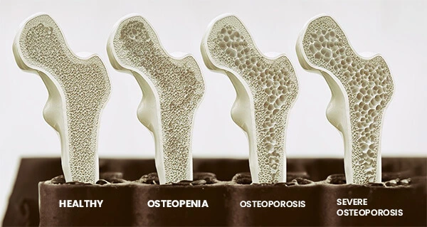
Osteopenia
Bone mass category for reduced bone mass less severe than osteoporosis, corresponding to a T-score below −1.0 through −2.49.

Osteoporosis
Fragile porous bone condition with high fracture risk, corresponding to a T-score of −2.5 or lower.
Origin
Usually the more fixed and proximal attachment of a muscle.
Insertion
Usually the more movable and distal attachment of a muscle; moves toward origin during contraction.
Agonist / prime mover
Muscle that provides most of the force for the desired movement.
Synergist
Muscle that assists the prime mover or stabilizes a joint.
Antagonist
Muscle that produces the opposite action and is lengthened as the agonist acts.
Orbicularis oris
Circular muscle around lips that constricts the mouth.

Orbicularis oculi
Circular muscle surrounding eye that closes the eye.

Masseter
Thick muscle over lateral ramus/angle of mandible that elevates mandible and closes jaw.

Buccinator
Deep cheek muscle anterior to masseter that compresses the cheeks.

Temporalis
Fan-shaped muscle over temporal bone superior to ear that elevates mandible.

Platysma
Thin superficial sheet across anterior neck that tenses skin of neck and depresses mandible.

Sternocleidomastoid
Superficial diagonal strap from sternum/clavicle to mastoid process that causes cervical flexion (bilateral) or opposite side rotation (unilateral).

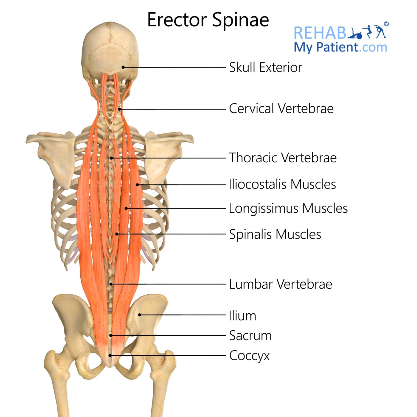
Erector spinae
Large paired longitudinal columns beside spine that extend the spine.
Rectus abdominis
Superficial vertical muscle of abdominal wall that flexes the trunk.

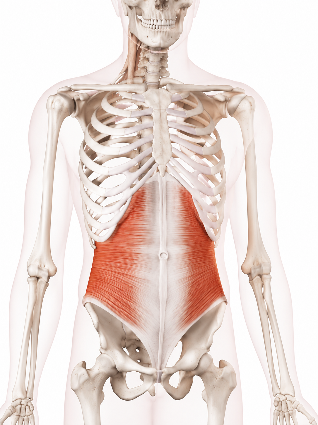
Transversus abdominis
Deepest abdominal muscle with horizontal fibers that compresses the abdomen.
Skull (22 major bones)
Cranium (8) and Facial (14)

Occipital bone
Located at the back of the skull / cranium


Frontal bone
Located in the front of the skull and forms the structure of the forehead.


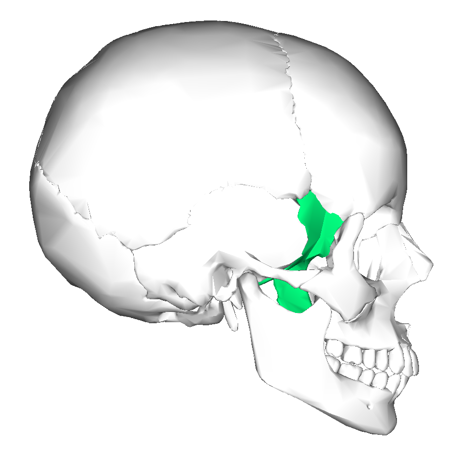
Sphenoid bone
At the middle of the central skull base, sitting just behind the eyes and wedged between other cranial bones.
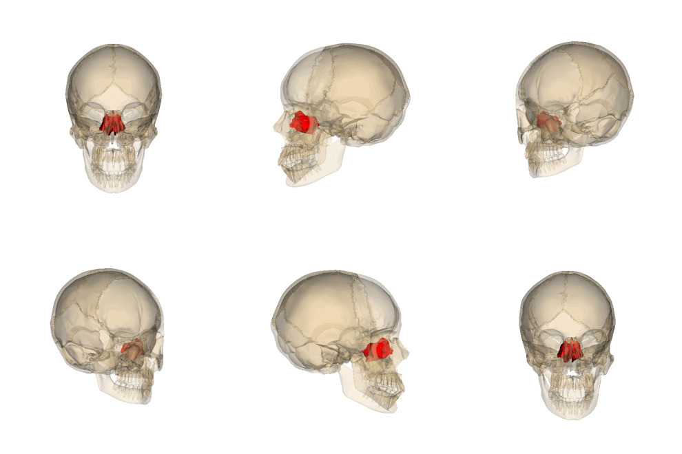
Ethmoid bone
At the anterior part of the skull base, sitting directly between the eye sockets (orbits) and forming the roof of the nasal cavity.


Parietal Bones
At the top and sides of the skull.

Temporal Bones
A pair of bones located at the lower sides and base of the skull, positioned directly beneath the parietal bones.


Maxillary bones
A pair of connected bones located in the center of the face, forming the upper jaw and the mandible third of the facial skeleton (aka maxillae)


Lacrimal Bones
A pair of small, fragile bones located in the inner wall of each eye socket (orbit)


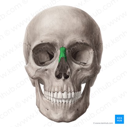
Nasal Bones
A pair of small, oblong bones located in the upper-middle part of the face, forming the bridge of the nose
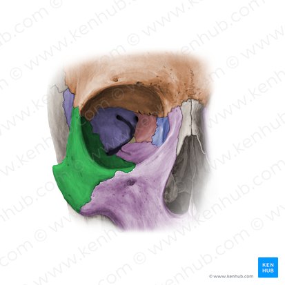
Zygomatic Bones
Commonly known as the cheekbones, this pair of diamond-shaped facial bones are located on the upper lateral sides of the face.


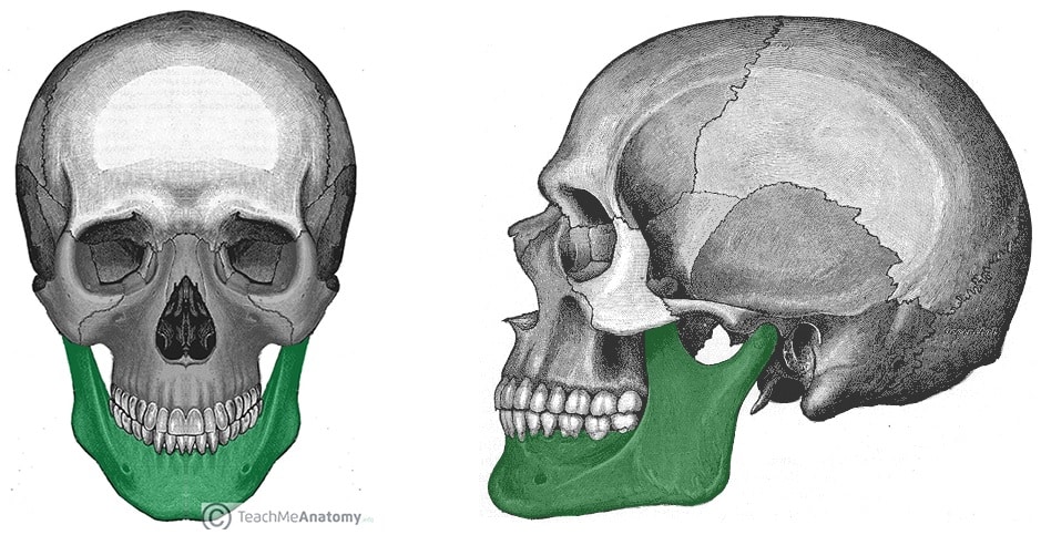
Mandible
Commonly known as the jawbone, this bone is located at the bottom of the face, forming the entire lower jaw.

Palatine Bones
A pair of L-shaped bones located deep in the back of the nasal cavity, positioned between the upper jaw (maxilla) and the roof of the mouth (hard palate).


Inferior nasal conchae bones
A pair of thin, curved, scroll-like bones located horizontally along the lower side walls of the nasal cavity.


Vomer
A small thin, plow-shaped bone located along the midline of the nasal cavity, forming the lower and back portion of the nasal septum (the wall that divides the nose into left and right nostrils).
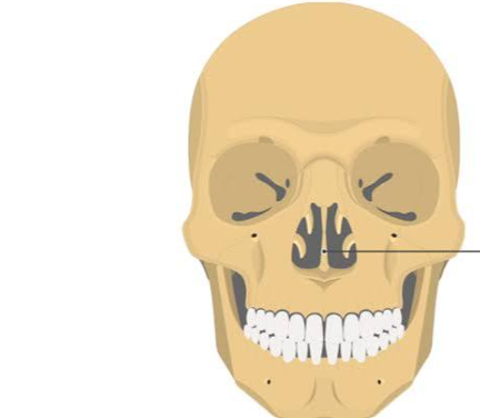

Sinuses
The skull contains four pairs, which are hollow, air-filled cavities. They are lined with mucus membranes, lighten the weight of the skull, and warm or humidify inhaled air.

Skull sutures
Rigid, immovable joints that lock the bones together. The four primary ones are the coronal, sagittal, lambdoid, and squamous.

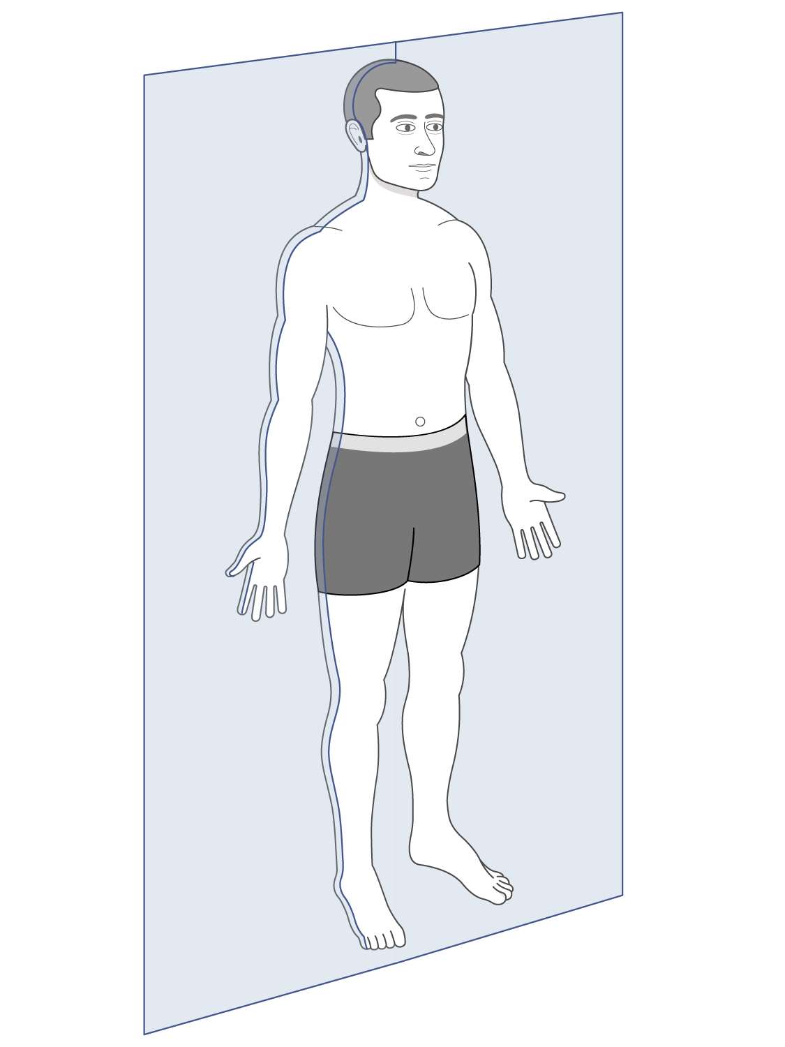
Median or anteroposterior
Another name for Sagittal plane / midsagittal plane

Lateral
Another name for Coronal / frontal
Axial skeleton
The skull, hyoid bone, vertebral column, and thorax or thoracic cage is located in the what

Appendicular skeleton
The pectoral girdle / shoulder girdle, upper limbs, hip (or pelvic) girdle / coxal bone / coxa / os coxa, and lower limbs are part of the

Hyoid bone
Located in the front of the upper neck, positioned horizontally between the lower jaw (mandible) and they thyroid cartilage (voice box)

Vertebral Column
Also known as the spine or backbone and is located along the midline of the body’s back.


Carpals
8 bones: Located at the base of the hand, forming the wrist joint.


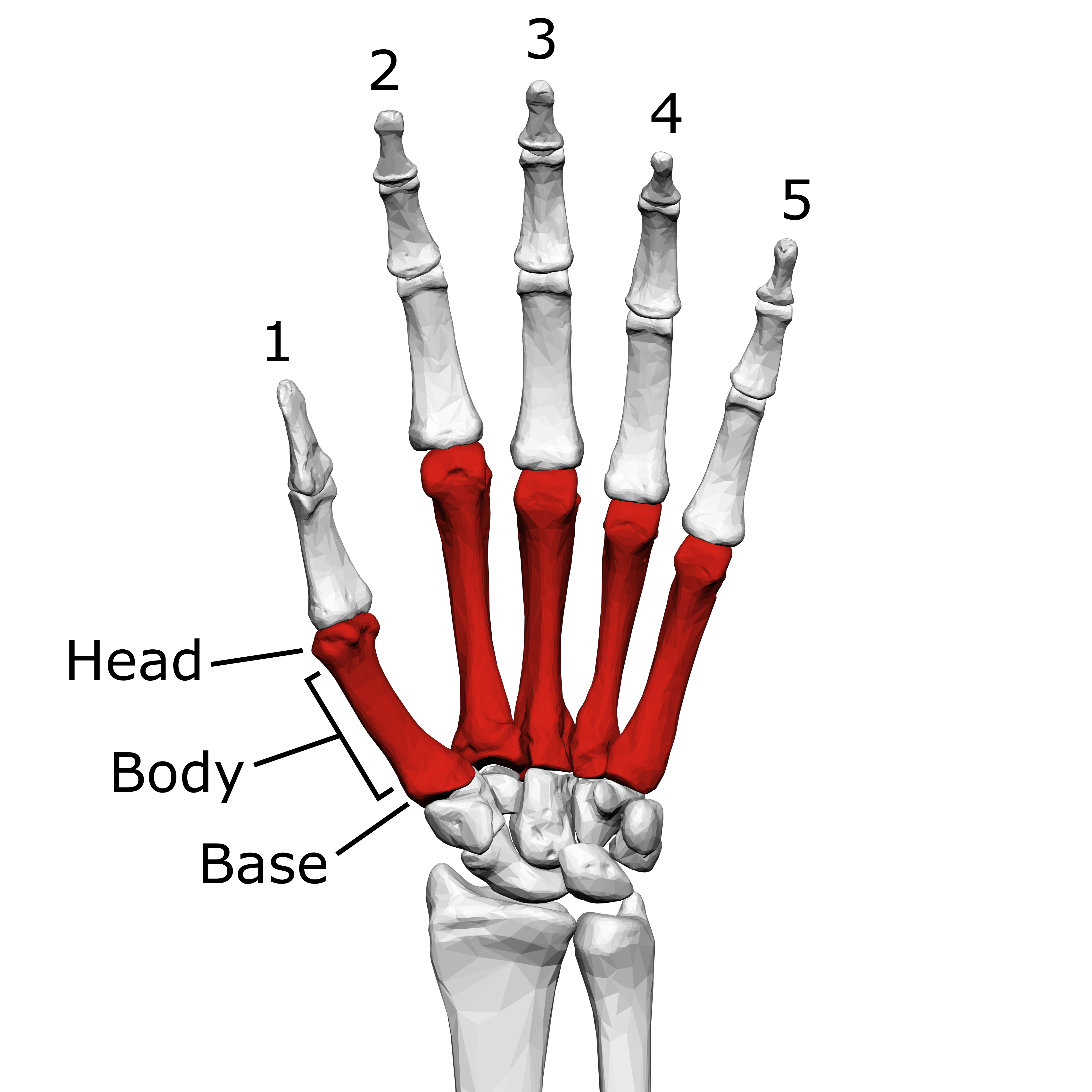
Metacarpals
5 bones: Located in the palm area of the hand.
Skull
Located at the top of the neck, housing brain and organs of special sense. Includes the cranium and face bones.
