Non-imaging Testing Part 1
 Call Kai
 Learn
 Practice Test
 Spaced Repetition
 Match
 Flashcards
 Knowt Play
1/38

Earn XP
Description and Tags
CW doppler analysis
39 Terms
how it looks, NOT measurable, waveform contour
qualitative “quality”
how it measures, velocities & ratios & indices, % diameter reduction
quantitative “quantity”
non-imaging testing includes
CW, pressure testing, and plethysmographic studies
CW doppler capabilities and limitiations
presence/absence of arterial occlusive disease
approximate level of disease, NOT exact location or severity
tech dependant
CW = requires — crystals
2; 1 transmitting/1 recieving
in CW doppler analysis there is no depth specificity aka —, unable to choose location of signal
range resolution
types of recording
audio, analog, spectral analysis
doppler shifts are within audible range
audio
zero-crossing frequency meter on strip chart recorder ESTIMATES freq shifts, not very sensitive
analog
displays TRUE or INDIVIDUAL frq shifts according to time. more sensitive
spectral analysis

analog recording

spectral analysis recording
technique for CW doppler analysis
supine
8-10 MHz CW probe
probe angle 45-60 degrees to skin surface
CW doppler analysis is a — evalustion
segmental
the segmental evaluation for CW doppler analysis
groin (CFA), thigh (SFA), pop, ankle (PYA/DP)

normal interpretation of a CW doppler analysis
triphasic or biphasic
CW doppler analysis is mainly —
qualitative
abnormal interpretation of CW doppler analysis
monophasic or monophasic, continuous
deterioration in signal contour from one level to the next =
disease between those levels
pulsatility index =
measure resistance
the pulsatile index is —
quantitative
pulsatility index equation
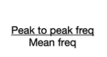
normal PI — as you go down the limb
increases
any drop in PI between levels =
disease between those levels
time from beginning of systole to peak AKA upstroke
acceleration time
increased acceleration time > —
133 msec

increased acceleration time of > 133msec means that there is
proximal (inflow) disease
when acceleration time is not affected, there is —
no proximal disease
what is the significance of a monophasic continuous waveform at the level of the groin
inflow disease
what does pulsatility index measure
resistance
resistance and pulsatility index are — related
directly
disease in which of the following vessels would cause an acceleration time of 142msec in the CFA
common iliac artery
what interpretive description is considered a qualitative assessment
triphasic contour has forward, reversed, and return to forward flow
a biphasic waveform is documented at the left CFA and a monophasic waveform is documented at the level of the popliteal. at what level is the disease located
femoral
thumping sounds would be heard —
proximal to an occlusion
inaudible would be heard —
over the occlusion
low frequency disturbances would be found when there is
turbulent flow
increased velocities mean the frequency shift would be elevated, producing a —
higher pitch sound
a bruit is an audible sound you can hear upon auscultation and is caused by —
high velocity, turbulent flow