Biol 313 Exam 3
 Call Kai
 Learn
Learn Practice Test
 Spaced Repetition
 Match
Match Flashcards
 Knowt Play
1/248

There's no tags or description
Looks like no tags are added yet.
Name | Mastery | Learn | Test | Matching | Spaced | Call with Kai | Chat |
|---|
No analytics yet
Send a link to your students to track their progress
249 Terms
3 major sub-cavities of the ventral body cavity
thoracic, abdominal and pelvic
What is the thoracic cavity defined by inferiorly
the diaphragm
is there a physical divide between the abdominal and pelvic cavities
no
what is the abdominal cavity walled by
musculoaponeurotic walls (anterolaterally), diaphragm (superiorly) and the pelvic floor muscles (inferiorly)
what are the two bony rings that support/protect the abdomen
inferior margin of the thoracic skeleton (superiorly)
pelvic girdle (inferiorly)
what is the serous membrane that covers the anterolateral abdominal wall and the visceral organs
peritoneum
what part of the peritoneum goes with the abdominal wall
parietal peritoneum
what part of the peritoneum goes with the visceral organs
visceral peritoneum

region 1
right hypochondrium

region 2
epigastric
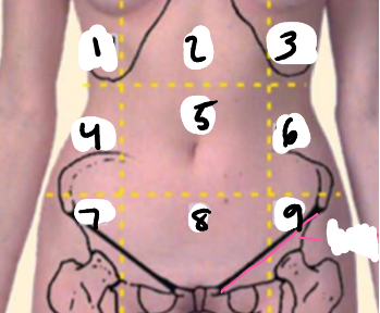
region 3
left hypochondrium

region 4
right lateral (flank)

region 5
umbilical

region 6
left lateral region
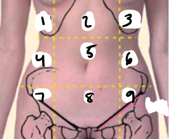
region 7
right inguinal (groin)

region 8
pubic

region 9
left inguinal (groin)

body plane 1
semilunar lines

body plane 2
midclavicular line (MCL)

body plane 3
transpyloric plane
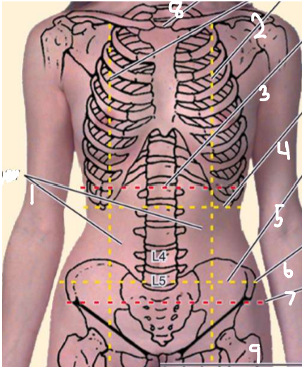
body plane 4
subcostal plane

body plane 5
transtubercular plane
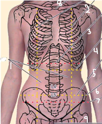
body part 6
iliac tubercle

body plane 7
interspinous plane

body plane 8
jugular line
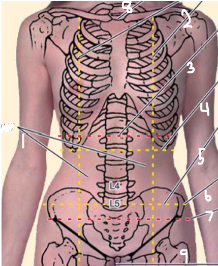
body plane 9
pubic symphysis
Right Upper Quadrant (RUQ) Organs
liver (right lobe)
gallbladder
stomach (pylorus)
duodenum (pts 1-3)
pancreas (head)
R suprarenal gland
R kidney
R colic (hepatic) flexure
Ascending colon (superior pt)
Transverse colon (R half)
Right Lower Quadrant (RLQ) Organs
cecum
appendix
most of ileum
ascending colon (inferior)
R ovary
R uterine tube
R ureter (abdominal pt)
R spermatic cord (abdominal pt)
uterus
urinary bladder
Left Upper Quadrant (LUQ) Organs
liver (L lobe)
spleen
stomach
jejunum and proximal ileum
pancreas (body and tail)
L kidney
L suprarenal gland
L colic flexure
Transverse Colon (L half)
Descending colon (superior pt)
Left Lower Quadrant (LLQ) Organs
sigmoid colon
descending colon (inferior pt)
L ovary
L uterine tube
L ureter (abdominal pt)
L spermatic cord (abdominal pt)
Uterus (if enlarged)
Urinary bladder (if full)
what does the hypodermis of the anterolateral abdominal wall contain
variable amounts of adipose tissue
3 muscle layers of the deep hypodermis
superficial fascia, muscle then peritoneum
five muscles of the anterolateral abdominal wall
external and internal oblique
transversus abdominis
rectus abdominis
pyramidalis (not found on everyone)
external oblique
fibers = inferomedial
external surfaces of lower ribs (5-12)
aponeurosis inserts on linea alba
linea alba = place of intramuscular exchange of superficial and deep fibers
internal oblique
fibers run superomedially
origin at thoracolumbar fascia and iliac crests
insertion of aponeurosis on linea alba
intermuscular exchange of fibers with contralateral external oblique muscles
transversus abdominus
deepest muscle layer
origin at internal surfaces of lower ribs, thoracolumbar fascia
insertion: aponeurosis inserts on linea alba
rectus sheath
formed by merger of aponeuroses of all three flat muscles
inferior border thickens = forming inguinal ligament
rectus abdominus embedded in this
pierced by inguinal canal
where do all layers of the anterolateral wall fuse
the umbilicus
what does the rectus sheath converge inferiorly to create in males
fundiform ligament
rectus abdominus
divided vertically by linea alba
divided horizontally by tendinous intersection
eight bellies
origin at pubic crest
insertion at xiphoid process and inferior costal cartilages
pyramidalis muscle
absent in 20% of people
origin at pubic crest
insertion at linea alba
no real function
Causes of protuberance of the abdomen
6 F’s
food, fluid, fat, feces, flatus (gas), fetus
venous flow reversal
happens when flow through superior or inferior vena cava is obstructed
blows through anastomoses collateral pathways
blood is basically flowing backwards because of a blockage and finds alternate routes to get back

what are the gutters of the posterior abdominal wall occupied by
kidneys, perirenal fat and pararenal fat
what is the most powerful muscle in the body called and what is it composed of
iliopsoas muscle
composed of iliacus and psoas muscles
what muscle causes hip flexion
iliacus

Iliacus muscle
large, triangular muscle
major and minor
originates on iliac crest and inserts on lesser trochanter of femur
Quadratus lumborum
originates on iliac crest
inserts on tips of transverse processes of L1-L4 and inferior margin of rib 12
flexes vertebral column and fix ribcage during inspiration
abdominal aorta
13cm (2.5cm in diameter)
begins at aortic hiatus and ends by dividing into common iliac arteries at L4 level
can sometimes see/feel pulsations when wall is relaxed
Inferior vena cava
begins at level L5 with merging of common iliac veins
leaves abdomen through caval opening at level T8
20cm in length
largest vein in body
no valves
to the right of the aorta
where does the Inferior vena cava drain blood from
abdominal wall, lower limbs, portal blood and non-portal blood from abdomen and pelvis
most blood from digestive tract goes through hepatic portal system then drains into IVC
where does the thoracic duct drain into
junction of left subclavian and internal jugular veins
lymphatic drainage
cisterna chyli → thoracic duct → Left subclavian vein
cisterna chyli
where all lymphatics from abdomen and lower limbs converge, and where everything drains through
Rupture of Diaphragm
frequently from car accidents
95% on left side (because liver protects the right hemidiaphragm)
stomach, small intestine, mesentery, transverse colon or spleen may enter thorax
Congenital Diaphragmatic Hernia
1 in 2200 newborns
almost always affects left side owing to liver on right
left lung usually fails to inflate at birth
high mortality rate (76%)
Posterior Abdominal Pain
if kidneys, ureters, cecum, appendix, sigmoid colon, lumbar lymph nodes and lumbar nerves are diseased movement of iliopsoas can be painful
test = lying patients on unaffected side and asking them to extend thigh, pain means positive sign
Abdominal Aortic Aneurysm
can be congenital or acquired weakness in arterial wall
deep palpations of mid-abdomen can detect it
severe pain and high mortality (90%) due to blood loss
peritoneum
mesothelium lining abdominopelvic cavity
moist with peritoneal fluid, allows movement without friction
cavity is completely closed in males, but open to exterior in females (through uterine tubes, uterus and vagina)
three compartments of the peritoneum
supracolic (greater sac)
omental bursa (lesser sac)
infracolic (greater sac)

Peritoneal formations
mesentery
mesocolon
omentum (greater and lesser)
greater omentum
attached to greater curvature of the stomach
lesser omentum
attached to lesser curvature of the stomach
Peritonitis
infection and inflammation in the peritoneum
often caused by rupture of the GI tract (ulcer perforation or appendix rupture)
widespread can be fatal due to rapid absorption of bacterial toxins
ascites
excess fluid in the peritoneum
Peritoneal adhesions
peritoneum is damaged and surfaces become inflamed and sticky with fibrin
when healing, fibrin can be replaced with connective scar tissue binding visceral peritoneum to parietal peritoneum (sticks organs together)
Adhesiotomy
surgical separation of adhesions
how can the peritoneum be used during renal failure
a ‘dialysis machine’ which by injecting saline into the peritoneal cavity and then draining them a few hours later will, by using the fluids, absorb toxins by concentration gradient diffusion
falciform ligament
divides the liver into right/left lobes
attaches to diaphragm and anterior abdominal wall to the level of the umbilicus
keeps the liver in place
what are the intestines largely covered by
the greater omentum
celiac trunk
serves stomach, spleen and liver
goes to the phrenic artery, gastric artery and hepatic artery
superior mesenteric artery
serves small intestine, cecum, ascending colon and transverse colon
interior mesenteric artery
serves descending colon, sigmoid colon and rectum
inferior mesenteric vein
drains rectum, sigmoid colon and descending colon
superior mesenteric vein (right and middle colic veins)
drains small intestine, cecum, ascending colon and transverse colon
splenic vein (left colic vein)
drains spleen and inferior mesenteric vein
where do all veins drain
hepatic portal vein to the liver
esophagus
10 in long
surrounded by smooth muscle (2/3rds) and skeletal muscle (1/3)
follows curvature of vertebral column
terminates at z-line inferior to GI sphincter
stomach
between esophagus and duodenum of small intestine
food blender and reservoir
mechanical, chemical and enzymatic digestion
mixes bolus with gastric juice producing chyme
can hold 2-3L of food, and is able to change size
only place that does mechanical, chemical and enzymatic digestion
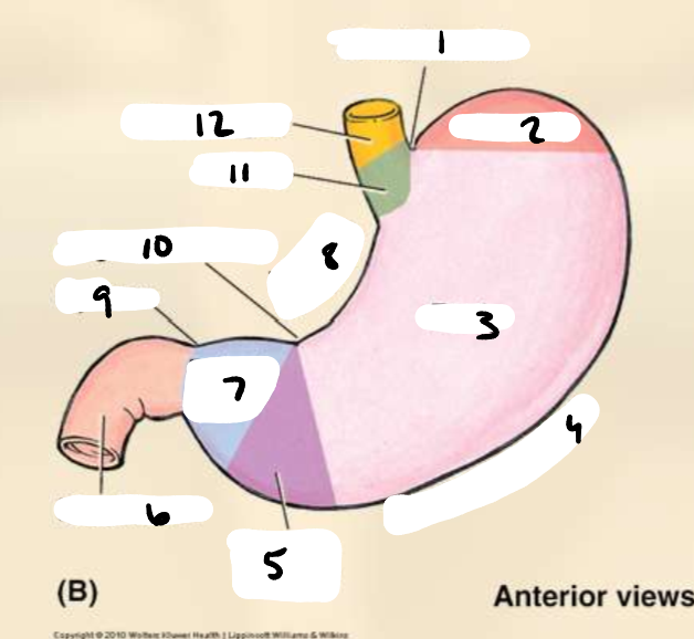
point 1
cardiac notch

point 2
fundus

point 3
body

point 4
greater curvature

point 5
pyloric antrum

point 6
duodenum

point 7
pyloric canal
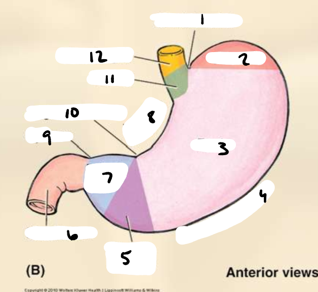
point 8
lesser curvature

point 9
pylorus

point 10
angular incisure
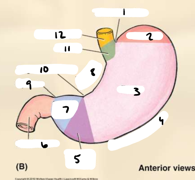
point 11
cardia

point 12
esophagus
what is the stomach closed by
pyloric sphincter and GI sphincter
What is the interior of the stomach lined with
rugae (folds)
not always there if a person is full


point 1
Left gastric artery

point 2
celiac trunk
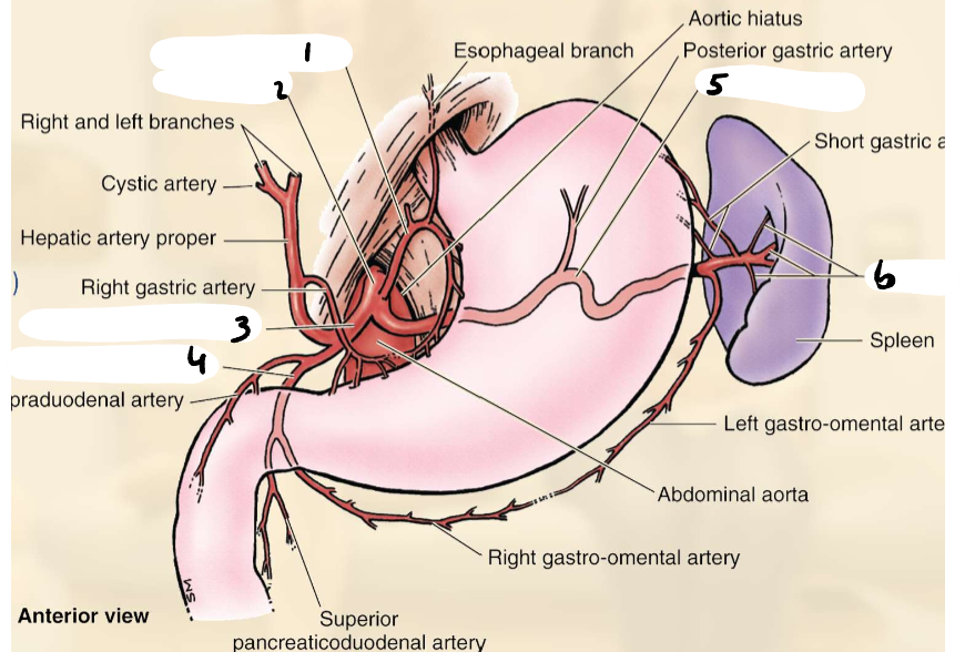
point 3
common hepatic artery

point 4
gastroduodenal artery

point 5
splenic artery

point 6
splenic branches
small intestine
primary digestion and nutrient absorption site
divided into three regions (duodenum, jejunum and ileum)
begins are pyloric sphincter
ends at ileocecal valve (opening to large intestine)