Anatomy Lab Practical 2 (Fall Semester)
 Call Kai
 Learn
 Practice Test
 Spaced Repetition
 Match
 Flashcards
 Knowt Play
1/214

There's no tags or description
Looks like no tags are added yet.
215 Terms
Incus
Transmits vibratory motion from the malleus to the stapes in the middle ear.

Malleus
Receives sound vibrations from the tympanic membrane and transmits them to the incus.

Stapes
Transmits vibrations from the incus to the oval window of the inner ear.

Ethmoid sinus
Lightens skull weight and humidifies/warms inhaled air.

Cribriform plate
Supports the olfactory bulb and allows passage of olfactory nerve fibers.

Crista galli
Serves as an attachment point for the falx cerebri (dura mater).

Middle nasal concha
Increases air turbulence in the nasal cavity to warm, moisten, and filter air.

Perpendicular plate of ethmoid bone
Forms the superior part of the bony nasal septum.

Ethmoid bone
Contributes to the anterior cranial fossa, nasal septum, and orbital walls.

Anterior cranial fossa
Houses and protects the frontal lobes of the brain.

Middle cranial fossa
Houses and protects the temporal lobes and pituitary gland.

Posterior cranial fossa
Houses and protects the cerebellum, pons, and medulla oblongata.

Frontal sinus
Lightens skull weight and acts as a voice resonating chamber.

Glabella
Smooth area between the eyebrows on the frontal bone.
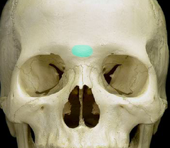
Supraorbital margin / Superciliary arch
Supraorbital margin: Forms the superior protective boundary of the eye socket. Superciliary arch: Bony ridge above the eye socket supporting the eyebrow.
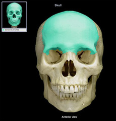
Frontal bone
Forms the forehead and the superior walls/orbits of the eyes.

Hyoid
Unattached anchor bone supporting the tongue and larynx muscles.

Inferior nasal concha
Increases surface area and turbulence in the nasal cavity to filter, heat, and humidify air.

Vomer
Forms the inferior and posterior part of the nasal septum.

Lacrimal bone
Houses the lacrimal sac/fossa for tear drainage.

Alveolar process of mandible
Supports the lower teeth.

Condylar process of mandible
Articulates with the temporal bone to form the temporomandibular joint (TMJ).

Coronoid process of mandible
Insertion point for the temporalis muscle (jaw closure).

Genial tubercle
Attachment site for tongue and suprahyoid muscles (genioglossus/geniohyoid).

Angle of mandible
Corner junction point between the body and ramus of the mandible.

Mandibular foramen
Passageway for the inferior alveolar nerve and vessels supplying lower teeth.

Mandibular notch
U-shaped notch between coronoid and condylar processes.

Ramus of mandible
Vertical posterior portion of the mandible.

Mental foramen
Passageway for the mental nerve and vessels to the chin and lower lip.

Mental protuberance
Bony prominence forming the chin.

Alveolar process of maxilla
Supports the upper teeth.

Maxillary sinus
Largest paranasal sinus; lightens skull and humidifies air.

Palatine process of maxilla
Forms the anterior hard palate.

Nasal bone
Forms the bridge of the nose.

Foramen magnum
Passageway connecting the cranial cavity with the spinal canal (brainstem/spinal cord transition).
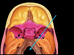
Inion / External occipital protuberance
External occipital protuberance: Attachment site for the nuchal ligament and trapezius muscle. Inion: Most prominent point of the external occipital protuberance.

Occipital condyle
Articulates with the atlas (C1 vertebra) to allow nodding of the head.

Occipital bone
Forms the posterior and base wall of the cranium.
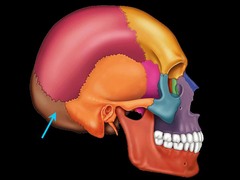
Palatine bone
Forms the posterior part of the hard palate and nasal cavity wall.

Parietal bone
Forms the sides and roof of the cranial cavity.

Foramen ovale
Passageway for the mandibular nerve (V3 of cranial nerve V).

Foramen rotundum
Passageway for the maxillary nerve (V2).

Foramen spinosum
Passageway for the middle meningeal artery.

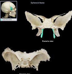
Lateral pterygoid plate
Attachment site for pterygoid muscles aiding jaw movement.

Medial pterygoid plate
Attachment site for tensor veli palatini and pterygoid muscles.
Sella turcica
Bony depression housing and protecting the pituitary gland.
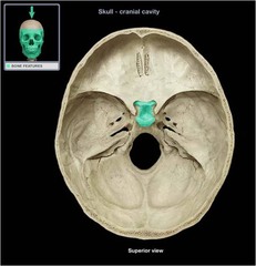
Sphenoid sinus
Paranasal sinus lightening the sphenoid bone.

Superior orbital fissure
Passageway for cranial nerves controlling eye movements (III, IV, V1, VI).

Sphenoid bone
Keystone bone of the cranial floor articulating with all other cranial bones.

Coronal suture
Immovable fibrous joint uniting frontal and parietal bones.

Lambdoid suture
Immovable fibrous joint uniting occipital and parietal bones.

Sagittal suture
Immovable fibrous joint uniting left and right parietal bones.

Squamous suture
Immovable fibrous joint uniting temporal and parietal bones.

External acoustic meatus
Canal conducting sound waves to the tympanic membrane.

Foramen lacerum
Passage sealed by cartilage in life, transmitting minor nerves/vessels.

Internal acoustic meatus
Passageway for facial (CN VII) and vestibulocochlear (CN VIII) nerves.

Jugular foramen
Passageway for internal jugular vein and cranial nerves IX, X, XI.

Mastoid process
Attachment site for neck muscles (e.g., sternocleidomastoid).

Styloid process of temporal bone
Attachment site for tongue and pharynx muscles/ligaments.

Temporal bone
Forms the inferolateral walls of the skull and houses middle/inner ear structures.

Zygomatic arch
Structural bridge providing attachment for the masseter muscle.

Zygomatic bone
Forms the prominence of the cheek and part of the lateral orbital wall.

Temporal process of zygomatic bone
Joins the zygomatic process of the temporal bone to form the zygomatic arch.

Buccinator
Eating / Chewing: Compresses the cheek against the teeth to keep food centered on the grinding surface during mastication.

Depressor anguli oris
Communication / Expression: Depresses the corner of the mouth for non-verbal expression and emotional communication.

Depressor labii inferioris
Eating / Drinking: Pulls the lower lip downward (used during drinking from a cup or speaking).

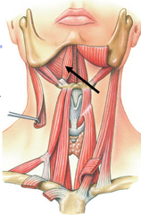
Anterior belly of digastric
Swallowing / Eating: Elevates the hyoid bone and depresses the mandible to initiate the first phase of swallowing food.
Levator labii superioris
Eating / Facial Expression: Elevates the upper lip (used during eating, smiling, or facial hygiene).

Masseter
Eating / Mastication: Powerful elevation of the mandible to bite and chew food solids during meals.

Nasalis
Respiration / Hygiene: Flares or compresses the nasal passages during heavy breathing or nose blowing.

Frontalis
Grooming / Sensory Awareness: Raises eyebrows and wrinkles the forehead when looking upward during personal grooming or scanning the environment.

Occipitalis
Postural Adjustment: Fixes and retracts the scalp to stabilize head posture during movement.

Orbicularis oculi
Hygiene / Protection: Closes the eyelids tightly to protect the eyes during face washing, showering, or bright sunlight.

Orbicularis oris
Eating / Drinking / Speaking: Closes and protrudes the lips (essential for sealing lips around a spoon, drinking from a straw, and speech articulation).

Risorius
Communication: Pulls the corners of the mouth laterally during smiling and social interaction.

Sternocleidomastoid
Mobility / Scanning: Rotates and flexes the neck, allowing a patient to scan their surroundings, look both ways while walking, or turn to talk.

Temporalis
Eating / Mastication: Elevates and retracts the mandible to firmly close the jaw and grind food during eating.

Zygomaticus major
Social Communication: Elevates the corners of the mouth upward and outward during smiling and laughing.
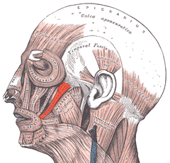
Zygomaticus minor
Facial Expression: Elevates the upper lip during speaking and expression.

Atlanto-occipital
Articulation allowing flexion, extension, and lateral bending of the head ("yes" movement).

Tempromandibular
Articulation enabling jaw movement during chewing and speaking.

Arbor vitae
Branching white matter inside the cerebellum bringing information to and from the cortex.

Cerebellar hemispheres
Coordinate complex voluntary movements, balance, and posture.

Falx cerebelli
Small dural fold separating the two cerebellar hemispheres.

Folia
Parallel folds/ridges on the cerebellar cortex that increase surface area.

Tentorium cerebelli
Horizontal dural fold separating the cerebrum from the cerebellum.

Vermis
Narrow worm-like midline structure connecting cerebellar hemispheres; helps maintain balance and posture.

Central sulcus
Prominent groove separating the frontal lobe from the parietal lobe.

Corpus callosum
Large white matter tract bundle connecting left and right hemispheres for interhemispheric communication.

Falx cerebri
Vertical dural fold in the longitudinal fissure separating the left and right cerebral hemispheres.

Fornix
C-shaped fiber bundle that outputs signals from the hippocampus to the mammillary bodies.

Frontal lobe / Pre-frontal cortex
Frontal lobe: Executive functions, voluntary motor control, reasoning, decision-making, and personality. Pre-frontal cortex: Complex cognitive behavior, personality expression, planning, and social moderation.

Lateral sulcus
Deep groove separating the temporal lobe from the frontal and parietal lobes.

Occipital lobe
Processing primary visual information received from the eyes.

Parietal lobe
Processing somatic sensory information (touch, temperature, pain, proprioception).

Parieto-occipital sulcus
Deep cleft separating the parietal lobe from the occipital lobe.

Postcentral gyrus
Primary somatosensory cortex; receives tactile and proprioceptive sensory input.

Precentral gyrus
Primary motor cortex; initiates voluntary skeletal muscle movements.

Temporal lobe
Processing auditory information, language comprehension (Wernicke's area), memory, and emotion.

Hypothalamus
Master regulator of autonomic nervous system and endocrine functions (temperature, hunger, thirst, sleep).
