patho exam 2 combined
 Call Kai
 Learn
 Practice Test
 Spaced Repetition
 Match
 Flashcards
 Knowt Play
1/342

There's no tags or description
Looks like no tags are added yet.
343 Terms
renal and urinary systems functions
maintaining fluid electrolyte balance
maintain acid-base balance
excrete hydrogen
regulate BP
release erythropoietin to stimulate RBC production
when there isn’t enough oxygen in the blood, bone marrow cells get signaled to make RBC
activation of vitamin D
the nephron
functional unit of kidney
glomerulus: bundle of capillaries that sit within Bowman’s capsule
site of filtration → filters out sodium (water follows), potassium, hydrogen, phosphate
urine flows from Bowman’s capsule → proximal convoluted tubule → loop of Henle → distal convoluted tubule → collecting duct
blood vessels that surround the nephron are involved in reabsorption of solutes and water
at the end, you will have some sort of concentrated urine depending on what your body needs
could be concentrated or diluted depending on what your body reabsorbs

regulation of urine output, fluid balance and BP
several factors involved
juxtaglomerular cells (like stretch receptors that gauge what the blood pressure is)
release renin in response to low BP
renin activates RAAS to increase BP (retain fluid and water)
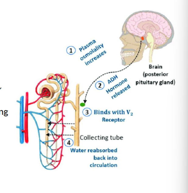
antidiuretic hormone (ADH)
released by posterior pituitary in response to decreased BP or increased serum osmolality
ADH promotes fluid reabsorption from collecting duct
just tells collecting ducts to reabsorb water (not sodium, just water)
ANP and BNP
released by heart in response to stretching
promotes excretion of Na and H2O
decreases fluid overload
when there is too much fluid in your blood, it will overload the ventricles and atria and cause them to stretch
renin-angiotensin-aldosterone system
renin is released from juxtaglomerular cells in response to low BP or sympathetic stimulation
renin converts angiotensinogen (produced by liver) into angiotensin I
angiotensin converting enzyme (ACE - produced by lungs) converts angiotensin I into angiotensin II
angiotensin II has 2 main functions
vasoconstriction
stimulates release of aldosterone from adrenal cortex

regulation of acid base balance
during acidotic states, kidney promotes:
excretion of H+
reabsorption of HCO3
synthesizes of new HCO3
during alkalotic states, kidney promotes:
retention of H+
simple cysts
common
single or multiple
unilateral or bilateral
usually very small
< 1 cm
usually don’t produce signs or symptoms
may cause flank pain
may cause hematuria
may become infected

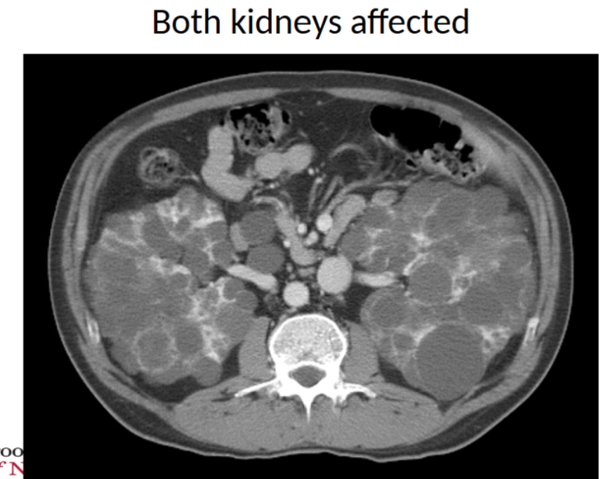
autosomal dominant polycystic kidney disease (ADPKD)
most common renal cystic disease
etiology
two gene mutations that lead to excessive growth and differentiation of epithelial cells
result is destructive, fluid-filled cysts in the kidney and other organs
can lead to fibrosis
cysts may detach and continue to grow
damage to renal tissue believed to be due to
inflammatory mediators
apoptosis of renal tubule cells
autosomal dominant polycystic kidney disease: clinical manifestations
may be asymptomatic for decades
flank pain
cysts can bleed or become infected
can also lead to issues elsewhere
cysts in liver and pancreas
diverticuli
aneurysms in cerebral arteries
heart valve issues
renal calculi (nephrolithiasis)
“kidney stones” → masses of crystals, proteins or other substances that obstruct the flow of urine
most develop in kidney, but may start anywhere in urinary tract
composed of particles normally excreted in urine
many factors involved in development
high concentration of these particles in bloodstream
start precipitating out and coalescing into stones
hydration status
like it’ll be a normal amount of solution, but there’s not enough water for it to dissolve so it’ll form stones instead
anatomy of urinary tract
diet and intestinal absorption
metabolism
UTIs
renal calculi: risk factors
requires supersaturated urine
depends on pH of urine and concentration of ions
alkaline urine usually more likely to develop renal calculi
also requires a nucleus that facilitates stone formation
small cluster of crystals held together by ionic force
organic materials produced by epithelium

renal calculi: calcium stones
most common (70-80%)
conditions that increase serum Ca or decrease fluid volume will increase risk of calcium stones
hyperparathyroidism (parathyroid is for the regulation of calcium)
the parathyroid is taking too much calcium from the bones and putting it in the blood
can lead to multiple myeloma
increased reabsorption of bone
renal calculi: struvite stones
magnesium ammonium phosphate (struvite) stones
often associated with bacterial infection / UTI
more common in people AFAB
bacteria secrete urease
urease converts urea → ammonia
ammonia makes urine alkaline
alkaline urine promotes formation of stone
can grow into staghorn calculi
renal calculi: uric acid stones
more common in individuals who excrete large amounts of uric acid in their blood (those with gout or abnormal metabolism of purines)
also associated with high amount of purines in diet (meat, beer)
not visible on x-ray
renal calculi: cysteine stones
more common in children
an amino acid that precipitates more readily in acidic urine
conditions that increase cysteine in blood (genetic predisposition) will increase cysteine in urine and increased risk of cysteine stones
renal calculi: clinical manifestations
flank pain radiating to the groin
may be accompanied by nausea and vomiting
renal calculi: summarized
calcium
most common (70-805)
any condition that increases calcium concentration in blood (hyperparathyroidism, conditions that remove Ca from bone, etc.)
also common with low urine volume (precipitation)
magnesium ammonium phosphate (struvite)
can only form in alkaline urine
requires bacterial infection
can result in staghorn calculus (requires lithotripsy)
uric acid
caused by conditions that produce high uric acid in blood (gout)
not visible on x-ray
cysteine
more common in children
genetic problem with renal transport of cysteine
similar to struvite, but no UTI
urinary tract infections (UTi)
inflammation of urinary epithelium (can occur anywhere)
most often caused by bacterial infection
most are GI bacteria that enter through urethra due to poor hygiene
some can enter from bloodstream (more common among the immunocompromised)
most common pathogen is Escherichia coli
klebsiella, proteus, Enterobacter, pseudomonas also can cause UTI
urine from bladder normally washes out bacteria in urethra
increased risk when this is disrupted
uncomplicated → bladder infection in someone with no other comorbidities
complicated → when the infection goes up and infects the kidneys or when the person is pregnant or if they have comorbidities
urinary tract infection: normal host defenses
washout phenomenon
washes pathogens out of urethra with urination
protective mucin layer
lines bladder - prevents attachment and infection
ureteral peristalsis
moves urine and pathogens down from kidneys
low pH of urine inhibits bacterial growth
like if you drink cranberry or graprefruit juice
normal flora of periurethral area
lactobacillus → defends against uropathogens
urinary tract infection: contributing factors
virulence of pathogens
presence of pili or fimbriae that help pathogens adhere
fimbriae → finger like projections that come out of the pathogen that help create anchor points (so they aren’t able to get washed out when the person urinates)
decreased urinary flow, ureteral obstruction or reflux
catheterization
allows the bacteria to climb up
pregnancy
baby presses on ureter and has the potential to push the urine backwards
use of antibiotics that kill normal flora
diabetes mellitus
aren’t able to metabolize glucose → all the excess glucose in the urine serves as food for the bacteria
acute cystitis
inflammation of bladder
most common site of UTI
patho
often due to retrograde movement of bacteria up urethra
e. coli is most common
some strains possess fimbriae that that help them attach to epithelium and flagella that help them move up urinary tract
many strains of e. coli release toxins that induce inflammation
acute cystitis: clinical manifestations
inflammation → edema in bladder → stimulates stretch receptors that create a sensation of fullness
frequency
urgency
dysuria (pain when urination)
suprapubic / low back pain
hematuria (hemorrhagic cystitis → bladder becomes so inflamed that it starts to bleed)
blood in urine
cloudy / foul smelling urine
older adults often present with mental status change
acute pyelonephritis
infection of upper urinary tracts (kidneys and ureters) - more common in people AFAB
patho
usually the result of ascending infection from bladder
also can gain access from bloodstream (less common)
inflammation usually affects renal tubules in the renal pelvis, calyces and medulla
BUN (blood urea nitrogen) and creatinine tests for kidney function
also glomerular filtration rate (GFR)
acute pyelonephritis: clinical manifestations
fever
malaise
chills
flank pain
symptoms of UTI
urgency, frequency, etc. due to stretching of bladder
glomerular disease
glomerulus is a net of capillaries within Bowman’s capsule
filtration takes place
glomerulonephritis → inflammation of glomerular structures
2nd leading cause of kidney failure in US
broken down into 2 broad categories based on the symptoms that the person experiences
nephritic and nephrotic
glomerular disease: causes
immunologic → most common
antibodies reacting with glomerular antigens (type II HSR)
IgG or IgM binds to a receptor on a cell and leads to inflammation
antigen-antibody complexes trapped in glomerulus (type III HSR)
antibody creates a complex around a cell (bind to a structure in circulation) then binds to a nephron or blood vessel and causes inflammation there
non-immunologic
diabetic nephropathy
create molecules that harm the nephron
hypertensive nephropathy
toxic medications, etc.
hereditary
glomerular disease: pathologic changes
three main changes associated with glomerular disease
proliferation: increase in cell number and formation of “crescents”
thickening of basement membrane
sclerosis/fibrosis: accumulation of proteins and collagen that obstruct glomerulus
two broad categories of primary glomerular diseases based on presentation
nephritic syndrome
nephrotic syndrome
nephritic syndrome
glomerular diseases characterized by INFLAMMATION of glomeruli
clinical presentation
oliguria
because of the inflammation, you aren’t going to be able to filter things out (less urine)
hypertension
since you can’t excrete the water, BP increases = hypertension
azotemia (nitrogenous wastes in blood)→ proteinuria not as pronounced as in nephrotic syndrome
you can’t excrete BUN and creatinine
hematuria → blood in urine
specific conditions
post infectious glomerulonephritis
rapidly progressive (crescentic) glomerulonephritis
ITIS = ITIC
nephrotic syndrome
glomerular diseases characterized by increased glomerular PERMEABILITY
clinical presentation
profound proteinuria
large molecules are going to get dumped into urine (albumin is most common protein in blood)
hypoalbuminemia → edema
since all the albumin is in the urine, there won’t be a lot in the blood vessels (albumin attracts water into vessel)
water then stays in the tissue and leads to edema
lipiduria with compensatory hyperlipidemia
lipids in urine because of permeability
however the liver overcompensates for the loss of lipids in urine so there will be an increase of lipids in blood
specific conditions
minimal change disease
membranous nephropathy
focal segmental glomerulosclerosis
collagen in glomerulus and hardens it
O in OTIC = HOLE
nephritic syndrome key points
major abnormality → inflammation
characteristics
decreased glomerular filtration → oliguria
increased nitrogenous wastes → azotemia
poor kidney function leads to HTN
damaged capillaries allow RBCs to escape → hematuria
some proteinuria, but not significant

nephrotic syndrome key points
main abnormality → increased glomerular permeability
characteristics
massive proteinuria
hypoalbuminemia → edema
lipiduria → overcompensation of LDL by liver
post-infectious glomerulonephritis
most common in children
nephritic syndrome (ITIS = ITIC)
post infectious = after an infection
Like if you had strep throat, then you have kidney malfunction, you probably have this (it usually follows strep throat)
patho
most often follows a group A beta hemolytic streptococcal infection
antibodies produced against group A beta hemolytic streptococcal antigen
antibodies bind to antigens in circulation and form immune complexes
immune complexes aggregate in glomerulus and promote inflammation (type III hypersensitivity)
proliferation of glomerular cells and infiltration by leukocytes follows

post infectious glomerulonephritis: clinical manifestations
nephritic syndrome 1-2 weeks after strep throat infection
oliguria
hypertension
azotemia
hematuria
fever
malaise
rapidly progressive (crescentic) glomerulonephritis
glomerular disease characterized by development of cellular crescents in the glomerulus and rapid loss of renal function
nephritic
patho
initiated by either
immune complexes that deposit in glomerulus and promote inflammation (type III hypersensitivity)
antibodies (IgG) against glomerular basement membrane that stimulate inflammation (type II hypersensitivity)
inflammation promotes proliferation of Bowman capsule epithelial cells and infiltration by leukocytes to create crescent that obliterates Bowman’s space
sometimes IgG cross reacts with alveolar basement membrane to produce pulmonary hemorrhage in addition to renal failure (Goodpasture syndrome)
if it affects kidney and lung = goodpasture
rapidly progressive (crescentic) glomerulonephritis: clinical manifestations
nephritic syndrome
hypertension
azotemia
hematuria
oliguria
nephritic syndrome summary
examples
post infectious glomerulonephritis
follows group A beta-hemolytic strep throat
immune complexes in glomerulus stimulate inflammation
rapidly progressive glomerulonephritis
rapid proliferation of glomerular cells
recruitment of monocytes and macrophages that obliterate Bowman’s space
creates a “crescent” shaped blockage
manifestations
decreased glomerular filtration → oliguria
increased nitrogenous wastes → azotemia
poor kidney function leads to HTN
damaged capillaries allow RBCs to escape → hematuria
some proteinuria, but not significant
minimal change disease (lipid nephrosis)
most frequent cause of nephrotic syndrome in children
relatively benign
patho
believed to be due to immune reaction that results in epithelial cell damage
result is increased permeability and proteinuria
often associated with respiratory illness
minimal change disease (lipoid nephrosis): clinical manifestations
nephrotic syndrome
profound proteinuria / hypoalbuminemia
edema
lipiduria with compensatory increase in lipids in the blood
renal function remains good, however
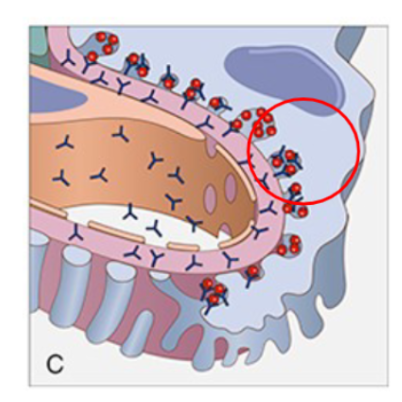
membranous nephropathy
glomerular disease associated with thickening of glomerular basement membrane
one of the most common causes of glomerular disease
comparable to a type III HSR
patho
deposits of antigens and IgG antibodies form between basement membrane of glomerular capillaries and Bowman’s capsule
activation of inflammation
increased capillary permeability
thickening of basement membrane
membranous nephropathy: clinical manifestations
nephrotic syndrome
proteinuria
edema
lipiduria
focal segmental glomerulosclerosis
glomerular disease characterized by sclerosis of some glomeruli (not all)
more common in persons of African-American and Hispanic heritage
patho
believed that a circulating cytokine induces epithelial cell injury
plasma proteins and extracellular matric proteins (collagen) become deposited
scar tissue forms → sclerosis
focal segmental glomerulosclerosis: clinical manifestations
nephrotic syndrome
proteinuria
edema
lipiduria
nephrotic syndrome: key points
examples
minimal change disease
most common in children
may be related to infection
no HTN and rare loss of kidney function
responds well to corticosteroids
membranous nephropathy
most common in adults
immune complexes deposit on glomerular basement membrane
basement membrane thickens
stimulates release of enzymes that destroy capillary walls
focal segmental glomerulosclerosis
deposit of collagen in glomeruli
damages glomerular tissue
more common in people of Hispanic and African-American / Caribbean-American heritage
manifestations
massive proteinuria
hypoalbuminemia → edema
lipiduria → overcompensation of LDL by liver
glomerular disease diagram

Wilms tumor
one of the most common primary neoplasms of children
tumor composed of elements resembling fetal tissue
due to genetic mutation
associated with several congenital abnormalities
aniridia
they don’t have an iris (colored portion of eye)
hemihypertrophy
one half of the body is bigger than the other side (like one arm is bigger than the other)
clinical manifestations
large, asymptomatic abdominal mass
HTN
releases epinephrine which activates the fight or flight response and increases HTN
managed with surgery and chemotherapy
good prognosis - 5 year survival 90%
renal cell carcinoma
cancer kidneys
accounts for 80% of renal neoplasms
risk factors
smoking
obesity
hypertension
NSAIDs
exposure to industrial solvents, asbestos, heavy metals
renal cell carcinoma: clinical manifestations
hematuria
dull flank pain
palpable flank mass in think patients
weight loss, fatigue, fever, anemia if cancer is advanced
acute kidney injury (AKI)
sudden decline in renal function
affects 5% of hospitalized patients
mortality of 50-50%
decreased in glomerular filtration with decreased urine output and increased retention of nitrogenous wastes, K and PO4
three possible etiologies
pre-renal: a disorder characterized by decreased renal perfusion
intra-renal: a disorder characterized by poorly functioning renal tissue
post-renal: a disorder characterized by obstruction of the urinary tract
pre-renal etiology
most common cause of AKI
problem getting blood to kidney
patho
decreased renal perfusion results in decreased filtration
reduced filtration promotes retention of fluids and nitrogenous wastes
may be due to
hypovolemia
sepsis
decreased cardiac output (MI or HF)
renal artery constriction / stenosis
artery is stiff
intra-renal etiology
kidney can’t make urine
patho
often the result of acute tubular necrosis (ATN)
also can be the result of glomerulonephritis, vascular disease (HTN, DIC), or disease of the interstitial tissue (cancer, infection)
two categories of ATN
ischemic ATN
damage to renal cells is due to prolonged ischemia and hypoxia
nephrotoxic ATN
damage of renal cells is due to a toxic (IV contrast, antibiotics (aminoglycosides → nephrotoxic), heavy metals (mercury, arsenic), bacterial toxins)
acute tubular necrosis (ATN)
destruction of renal tubule epithelial cells with suppression of renal function
damage leads to necrosis of epithelial cells
epithelial cells obstruct lumen of renal tubule
increased pressure in lumen causes decreased glomerular filtration
fluid leaks from nephron into interstitial tissue
usually reversible → depends on:
recovery of injured cells
clearance of necrotic cells
post-renal etiology
uncommon
can’t get urine out of the body
patho
obstruction of urinary tract bilaterally
obstruction causes increase pressure in lumen of urinary tract (ureters, etc)
GRF gradually declines
several hours of anuria and flank pain → polyuria
acute kidney injury (AKI): clinical manifestations
progressive
onset/initiating phase
time from precipitating event to injury
oliguric phase
sudden, significant decline in GFR
retention of nitrogenous wastes (BUN, creatinine, etc)
low urine output
fluid retention (edema, pulmonary congestion, HTN)
neurologic symptoms may develop due to buildup of nitrogenous wastes)
diuretic phase
kidneys heal
urine output improves → diuresis
nitrogenous wastes don’t improve yet
recovery phase
filtration returns to normal
nitrogenous wastes improve
tubular edema resolves
Oo, Dr
onset / initiating
oliguric
diuretic
recovery
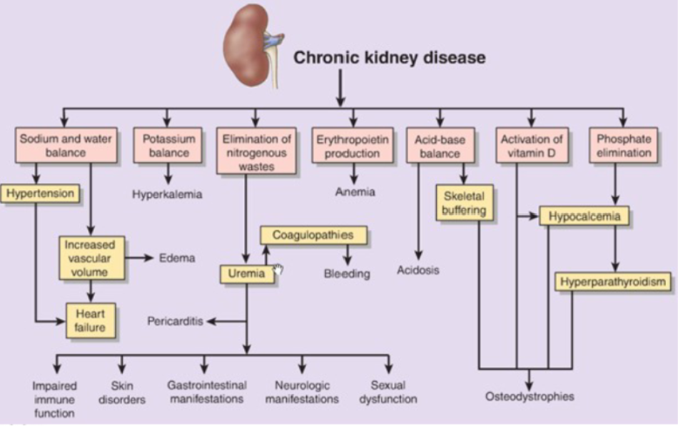
chronic kidney disease (CKD)
progressive loss of kidney function due to permanent loss of nephrons
often due to systemic disease (HTN, DM, SLE)
progressive loss of renal function
symptoms won’t appear until renal function is below 25% normal
patho
nephron loss begins in response to secrete or prolonged injury
persistent injury → fibroblast proliferation → focal segmental glomerulosclerosis and interstitial fibrosis
chronic kidney disease: clinical manifesations
azotemia (retention of urea, creatinine, and other nitrogenous wastes)
creatinine is released by muscle and is excreted by kidney
can lead to uremic syndrome (nausea, vomiting, diarrhea/constipation, pruritis → itching)
how the body reacts to elevated levels of nitrogenous wastes
metabolic acidosis
H+ is not excreted
may require dialysis
fluid and electrolyte imbalances
potassium
K+ cannot be excreted - levels can rise and become life threatening
helps with function of heart → too high/too low can develop arrhythmias
normal levels of potassium = 3.5 - 5
normal albumin also 3.5 - 5
Na+ and H2O
Na and H2O retention can occur → HTN
Ca and PO4
abnormalities will be noticed late
kidneys cannot excrete PO4 → serum PO4 will be increased
PO4 binds to Ca in circulation → hypocalcemia
If you have a lot of phosphate in blood, then it will bind with calcium and make the levels of the blood go down (bc they’re binding together to make phosphate-calcium)
normal calcium levels is 9 - 11
hypocalcemia may stimulate release of PTH from parathyroid glands → increased bone reabsorption
Parathyroid gland will see that calcium levels are down and then will pull calcium out of the bone → osteoporosis and risk for fractures
kidneys cannot activate vitamin D → gut cannot absorb Ca well
proteinuria
due to renal damage
anemia (HGB < 12 for people AFAB / < 13.5 for people AMAB)
kidney cannot sense low oxygen tension or secrete erythropoietin
Kidney secretes erythropoietin to signal the bone marrow to make more RBC (But if kidneys fail, they can’t send out that signal = anemia)
neurologic complications
due to uremia and hyperkalemia
nitrogenous wastes and K+ damage neurons
increased K+ associated with increased neuromuscular excitability
hiccups
muscle cramps
muscle twitching
change in mental status (impaired memory, concentration and judgement, confusion, etc.)
bladder cancer
most frequent form of urinary tract cancer in the US
cause is unknown
may be linked to carcinogens excreted into urine and stored in bladder
occupational exposure to dyes, rubber, textiles, paint, petroleum
cigarette smoking accounts for 30-50%
clinical manifestations
painless hematuria
frequency, urgency, dysuria
managed with surgery - chemotherapy may be attempted for advanced cancer
alveolar anatomy
location of gas exchange - large surface area
several cell types are present in alveolus
Type I pneumocyte
type II pneumocyte
alveolar macrophage
type I pneumocyte
make up 95% of alveolar epithelium
provide structure
easily damage
where gas exchange occurs
type II pneumocyte
secrete surfactant - prevents alveolar collapse (atelectasis)
repair the epithelium by producing type I pneumocytes
what helps the alveoli from not sticking together
alveolar macrophage
phagocytic cells responsible for innate immunity
protect against infections
measures of oxygenation: PaO2
partial pressure of oxygen in arterial blood (normal > 80 mmHg)
a measure of how much pressure your oxygen exerts within your blood vessels
evaluated on the ABG
as PaO2 increases, more O2 is forced into HGB
O2 continues to bind to HGB until the HGB is saturated
measures of oxygenation: SaO2
oxygen saturation
describes the % of HGB that is bound to O2
measured by pulse ox
ventilation / perfusion (V/Q)
diffusion of O2 from alveolus into blood also is dependent on:
ventilation (V): the amount of air getting to the alveoli
perfusion (Q): the amount of blood making it to the alveoli
a V/Q mismatch is the most common cause of hypoxemia

pulmonary function tests (FEV1)
forced expiratory volume in one second
measure amount of air expelled in one second following full inspiration
the person takes a big deep breath in and then blows out forcefully
measurement to figure out how much is blown out in one second
should be pretty high
pulmonary function tests (FVC)
forced vital capacity
the total amount of air that can be expired following full inspiration (not limited to 1 second)
person takes a big deep breath in and then blows out
see how much air they can empty from their lungs after a full inspiration
seeing how much total air volume exists in the lungs
FEV1 / FVC ratio
< 0.7 is indicative of obstructive lung disease
the common cold
most common respiratory tract infection
etiology: viruses
rhinoviruses are most common
parainfluenza virus
respiratory syncytial virus (RSV)
coronaviruses
time of year determines specific virus
most often spready by direct contact
less common by aerosol
common cold: clinical manifestations
begins with feeling of dryness/stuffiness in nasopharynx
clear, watery nasal secretions follow
tears
swollen mucous membranes
post-nasal drip → sore throat
2 day incubation period where you don’t see many symptoms
virus promotes inflammation
increased vascular permeability → that’s what leads to nasal secretions
vascular dilation
management
self limiting
rest and treat symptoms
vitamin c
chicken soup
rhinosinusitis
inflammation of nasal sinuses
patho
upper respiratory infection (usually viral) promotes mucosal swelling
swelling blocks flow out of sinuses
fluid, mucous, pressure increase
nasal polyps also can impede flow
can be acute or chronic
chronic lasts > 12 weeks
usually due to bacterial or fungal infection
rhinosinusitis: clinical manifestations
facial pain
the pt will have pressure wherever their paranasal sinuses are
if you were to palpate those areas, it would feel warm and it would hurt
headache
purulent nasal discharge
if it was a bacterial infection
fever
epiglottitis
dramatic, potentially fatal inflammation of the epiglottis and pharynx
etiology
often due to bacterial infection (S. pyogenes, S. pneumonia and S. aureus are most common)
epiglottitis: clinical manifestations
pale
lethargic
sitting with mouth open and chin thrust forward
muffled voice
stridor
drooling → they can’t swallow the saliva they are producing
anxiety
inspiratory retractions (muscles)
flaring of nares
epiglottitis: nursing considerations
DO NOT examine throat or attempt culture
complete obstruction of airway could be imminent → death
medical emergency - establish airway immediately

influenza
viral respiratory infection
three types: A, B and C
influenza A and B cause seasonal flu epidemics
subtypes of influenza A are categorized by proteins on the surface
hemagglutinin (H): helps virus attach to respiratory epithelium
neuraminidase (N): helps newly formed virus release from epithelium
many possible combos: H1N1, H5N1, etc.
influenza: virulence
more contagious than bacterial infection
spready by inhaling infected droplets or touching infected surfaces
incubation 1-4 days
infectious 1 day before symptom onset
airborne transmission
influenza: patho
virus infects upper airway
virus attaches to respiratory epithelial cells (hemagglutinin)
virus enters respiratory epithelial cells to replicate
epithelial cells are damaged during viral replication
fluids and inflammatory mediators leak out of epithelial cells
inflammatory mediators promote inflammation (redness and swelling of respiratory tract)
damage to goblet cells = reduced mucous secretion
damage to ciliated columnar cells → reduced ciliary motion
underlying basal cells are exposed
the only thing that will be left are the basal cells and basement membrane
holes between them allow extracellular fluid to escape (runny nose)
influenza: virus spread to lower airway
bronchial and alveolar epithelial cells are shed
single layer of basal cells is left
increased likelihood of bacterial attachment and infection
pneumonia can develop from viral or bacterial infection
as air moves lower into your airway, it will start killing more cells which can lead to pneumonia (viral pneumonia)
since many of the cells of the epithelium are stripped away, bacteria can come down and attach to the free spots (bacterial pneumonia)
influenza: clinical manifestation
abrupt onset of fever and chills
rapid onset of malaise (characteristic of flu)
muscle aches
headaches
nasal discharge
non-productive cough
sore throat
viral pneumonia may occur → rapid progression of symptoms
secondary complications → bacterial pneumonia, croup (in children)
influenza prevention
annual vaccine to reduce risk in all people over 6 years of age
if a vaccine is good against 3 strains = trivalent
4 strains = quadvalent
particularly important in
clients with chronic respiratory illness (emphysema, asthma, etc)
clients who are immunosuppressed
older adults
young children
pneumonia
inflammation of lower respiratory tract
commonly caused by infectious agents
can be caused by non infectious agents (inhaled toxins, hot air, etc.)
8th leading cause of death in the US
risk factors for pneumonia
older age
immunosuppression
chronic respiratory disease (COPD)
smoking
altered level of consciousness
usually when you aspirate something, you are able to cough it up
but with ALC, you lose that ability
endotracheal tubes
pneumonia: classifications
setting: community acquired vs hospital acquired
type of agent: typical (bacterial) vs atypical (viral or mycoplasma pneumonia)
pattern of distribution: lobar (consolidation of one lobe) vs bronchopneumonia (patchy)
pneumonia: community acquired (CAP)
occurs outside hospital or within 48 hours of admission
streptococcus pneumoniae is most common
haemophilus influenza
legionella pneumophila
influenza
easier to treat compared to HAP
pneumonia: hospital acquried (HAP)
occurs 48 hours after admission or more
many have antibiotic resistance
pseudomonas aeruginosa
very distinct smell when it’s in the mucus
klebsiella pneumonia
acute bacterial pneumonia
aspiration of secretions from oropharynx is most common
streptococcus pnumoniae is the most common organism
some bacteria gain access through inhalation (Legionella)
endotracheal tubes bypass upper airway defenses (cough, mucociliary blanket, etc.) and increase risk
normally microorganisms are destroyed by alveolar macrophages or can be coughed out
loss of cough reflex, reduced function of cilia, damage to endothelium and suppression of the immune system increase risk of colonization
pneumococcal pneumonia
model bacterial pneumonia
aspiration of streptococcus pneumoniae into lower airway
alveolar macrophages ingest S. pneumoniae
macrophages release inflammatory mediators → inflammation
steps in pneumococcal pneumonia
stage 1: congestion
alveoli and surrounding tissue fill with protein rich fluid (edema) → because of increased capillary permeability from inflammation
destroys the type II pneumocytes (the ones that keep the alveoli open and provide surfactant)
bacteria not ingested by macrophages multiply in edema and spread to adjacent alveoli
stage 2: red hepatization
RBCs, neutrophils and fibrin accumulate in the alveoli and interstitial tissue → consolidation
neutrophils will show up (the first responders) → recruit fibroblasts that produce collagen
more red cells will show up because of the vasodilation
stage 3: grey hepatization
RBCs dissipate and leave neutrophils and fibroblasts
collagen from the fibroblasts aren’t an elastic molecule which will make your lungs stiff
neutrophils and macrophages continue to fight the infection
stage 4: resolution
enzymes or macrophages remove fluid and debris
bacterial pneumonia
clinical manifestations
cough, dyspnea, fever
crackles
signs of consolidation (tactile fremitus, egophony, whispered pectoriloquy)
prevention
prevent aspiration
remove ETTs when possible
vaccination
viral pneumonia
influenza is the most common cause
usually milder, self-limiting but can set the stage for bacterial infection
doesn’t create the same consolidation (not as noticeable on x-ray)
viral infections damage ciliated epithelial cells and goblet cells
virus attaches to respiratory epithelial cells
virus enters respiratory epithelial cells to replicate
epithelial cells are damaged during viral replication
underlying basal cells are exposed
bronchial walls become edematous and infiltrated by leukocytes → pneumonia
no fluid accumulation / consolidation → not visible on x-ray
tuberculosis
infection caused by Mycobacterium tuberculosis
possesses waxy capsule
protects itself from immune system
affects lungs mainly but can affect other organs
risk factors
crowded living environments (barracks, dorms, prisons, nursing homes, etc.)
people without a fixed residence
people with substance use disorders
lack of screening
tuberculosis: spread
spread by respiratory droplets (coughing, sneezing, etc.)
droplet evaporates
bacteria remains suspended in air and inhaled
very contagious
tuberculosis: strong immunity
approx. 10% of those exposed will develop disease
in someone with an intact immune system, the bacteria is usually contained in a granuloma
95% of persons

primary tuberculosis: the initial stages
first 3 weeks of initial infection (previously unexposed person)
bacilli enter the airways and land in alveoli
upper airways most common
alveolar macrophages ingest bacilli
cellular enzymes cannot destroy the bacillus at this point because of the waxy capsule
bacilli replicate within macrophage
if client has intact immune system, infected macrophages initiate cell-mediated immunity
primary tuberculosis: cell mediated immunity (someone with strong immune system)
after first 3 weeks
infected macrophages (APCs) attach TB antigen to MHC II → presentation to CD4 cells → activation of T helper cells
T helper cells recruit more macrophages and lymphocytes
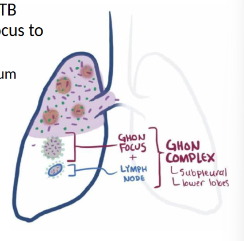
formation of granuloma (tubercle) to isolate mycobacterium
also called a Ghon focus
macrophages in middle and lymphocytes on outside
T helper cells also reprogram infected macrophages to release stronger enzymes to destroy TB
enzymes destroy bacteria and lung tissue (caseous necrosis)
PPD is positive at this point
if there is a delay forming the tubercle, TB might multiply and spread from ghon focus to the regional lymph nodes
causes necrosis (caseous necrosis) in the hilum
called a ghon complex
ghon complex will ultimately “heal”
can become fibrotic or calcified
visible on x-ray
latent tuberculosis
in most patients, the bacteria will go into a latent stage
bacteria are still viable but dormant
person is not infectious at this time
can reactivate with immunosuppression or re-exposure
secondary tuberculosis
disease that develops in previously sensitized host due to
new exposure (memory cells)
reactivation of latent TB when immune system is down
infection spreads to upper lobes of lung
immune response is rapid due to previous sensitivity
multiple areas of caseous necrosis
lesions can erode into airways → expel contents into airways
empty, calcified / fibrotic space left behind (cavitation)
after the lesions clear out and are exposed by you coughing, it leaves behind a hold inside your lung
patient develops bronchopneumonia
TB can erode into blood vessels and spread to other organs (kidneys, meninges, liver, etc)
this is called miliary TB
tuberculosis clinical manifestations
primary tuberculosis with an intact immune system
asymptomatic or flu-like symptoms
secondary tuberculosis
fatigue
anorexia
low grade fever
night sweats
hemoptysis
pleuritic pain
diagnostics
acid-fast bacillus
tuberculosis: weak immunity
in someone who is immunocompromised, the bacteria can replicate and cause illness
5% of persons

primary progressive tuberculosis (someone with WEAK immune system)
the macrophages cannot contain the bacteria
clients progress from primary TB to clinically significant (progressive) disease immediately
resembles bacterial pneumonia
elderly and immunosuppressed are highest risk
clinical presentation
fever
fatigue
weight loss
night sweats
bacillus gains access to sputum - client is infectious
bacillus can enter bloodstream and spread to other organs (miliary TB)

lung cancer
malignancies that arise from epithelium of respiratory tract (bronchogenic carcinomas)
2nd most common cause of death in the US → we don’t have a good screening available
most common cause of cancer-related death
risk factors
smoking (10-15% of smokers will develop lung cancer)
occupational exposure to certain substances (asbestos fibers, diesel fuel, radon gas, silica, etc.)
genetics
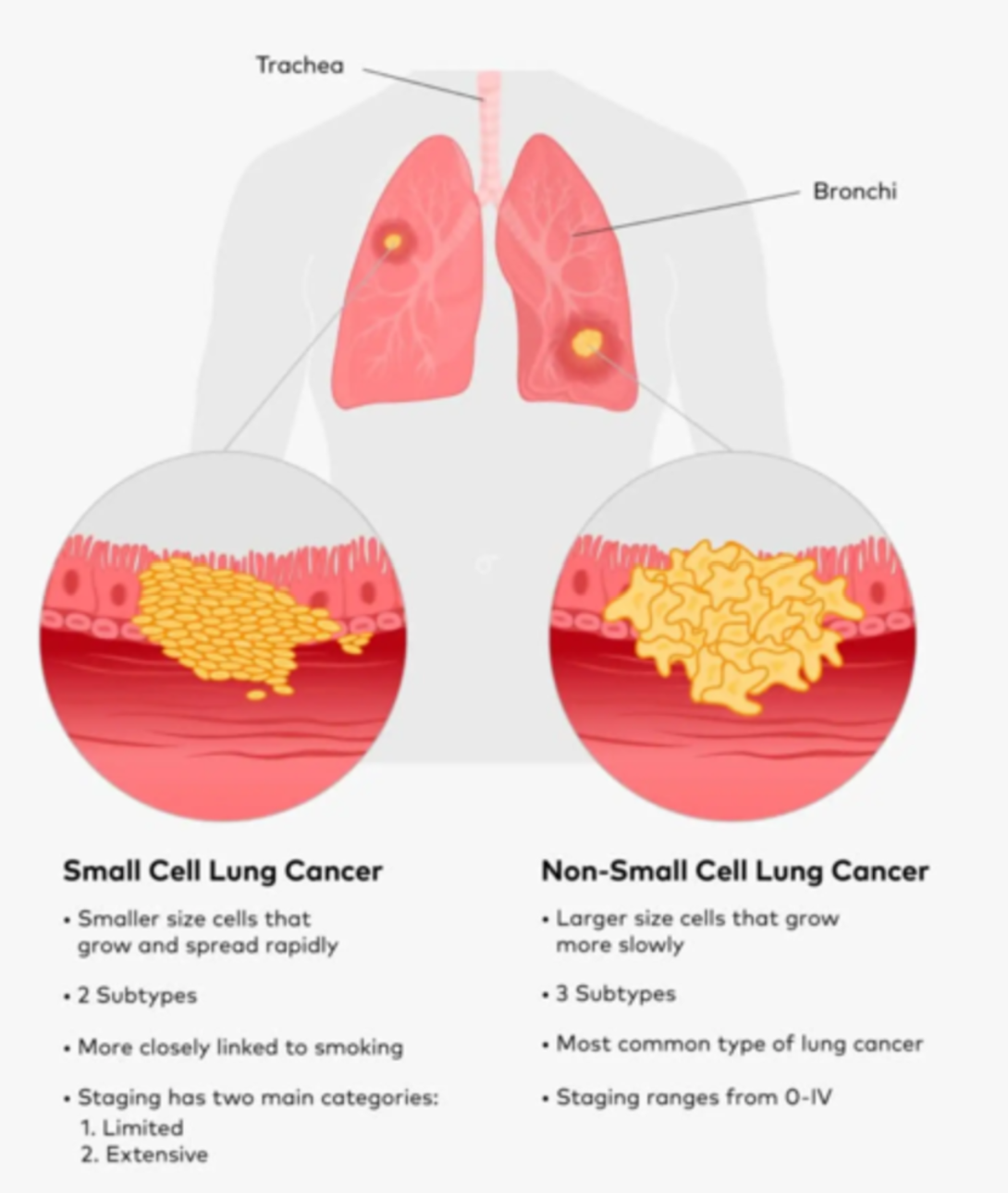
lung cancer: types
two major categories based on biopsy
small cell lung cancer (harder to detect)
non-small cell lung carcinoma (85%)
squamous cell carcinoma
adenocarcinoma
large cell undifferentiated carcinoma