bio 225 exam 3: reproduction
 Call Kai
 Learn
 Practice Test
 Spaced Repetition
 Match
 Flashcards
 Knowt Play
1/64

There's no tags or description
Looks like no tags are added yet.
65 Terms
reproduction
necessary for maintenance of the species
driving force of evolution
2 main types: asexual and sexual
asexual reproduction
a single parent creating genetically identical (or nearly identical) offspring
genetic variation is not necessary in relatively constant environments
budding and fragmentation
asexual reproduction: budding
new organism grows off of the parent and detaches fully-formed
asexual reproduction: fragmentation
parent splits, and each piece regrows into a full organism
pathogenesis
asexual reproduction in which an egg develops without fertilization
ex: whiptail lizards
2 females will simulate copulation with one another which induces oogenesis
female behavior is based on estradiol (female-like) and progesterone (male-like)
sexual reproduction
2 parent contribute nearly equal amounts of genetic material to generate genetically new offspring
usually haploid + haploid → diploid

asexual vs sexual reproduction
asexual
parent 1 splits into identical versions of itself
in the case of a drought, no more species
sexual
consecutive generations with varying genetics
in the case of a drought, reproduction is still possible

advantages of sexual reproduction
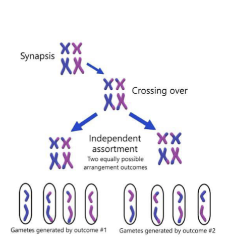
generates genomic variation at 3 levels:
random reorganization of chromosomes during meiosis (independent assortment)
hybrid chromosomes by recombination/crossing over
diploid offspring carry unique genetic combinations from both parents (multiplied x2)
creates population of distinct genotypes
hermaphrodites
individuals with the capacity to produce both egg and sperm
simultaneous hermaphrodites
produce egg and sperm at the same time
some can fertilize
serial hermaphrodites
change sex in response to environmental cues
protogynous: egg-producing becomes sperm-producing
protandrous: sperm-producing becomes egg-producing

gametogensis
production of gametes
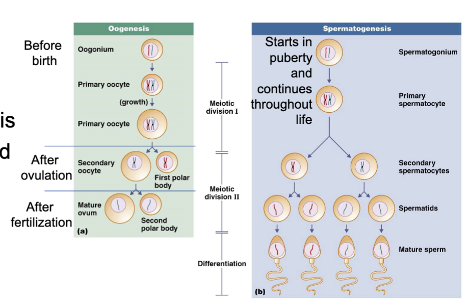
chromosomal segregation is the same for oogensis and spermatogenesis
both result on haploid gametes (ova and sperm)
regulated by hormones
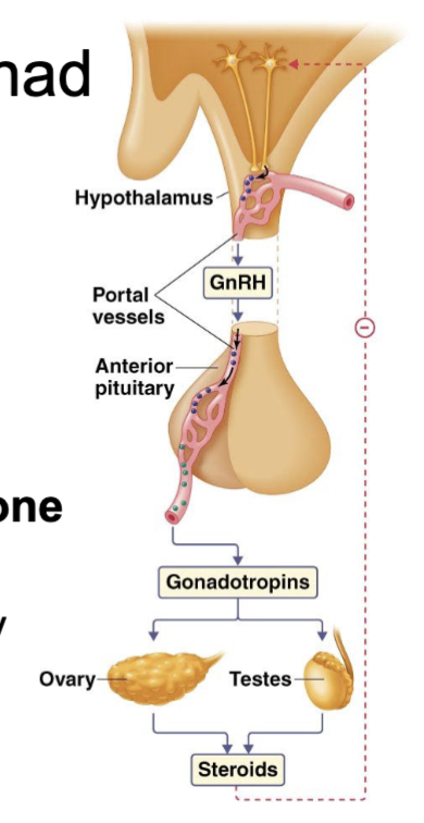
hypothalamic-pituitary-gonad (HPG) axis
gonadotropin-releasing hormone (GnRH)
synthesized and released from the hypothalamus
delivered to anterior pituitary gland
regulates FSH/LH synthesis and release
gonadotropins: follicle-stimulating hormone and luteinizing hormone (FSH and LH)
tropic hormones produced by anterior pituitary
control steroid hormone synthesis in gonads
reproductive steroid hormones
reproductive steroid hormnes
bind to nuclear hormone receptors in target cells → regulate gene expression
produced in gonads for vertebrates
essential for MANY systems out of reproduction
regulate metabolism, bone health, immune function, and cardiovascular health
lots of neuronal functions including memory and mood
separating sex, gender, and gonads in reproductive physiology
reproductive physiology simplifies systems to describe a sex binary → although humans are a sexually dimorphic species, this does not wholly reflect the spectrum of human sexes
testicular reproductive tract
in most species, sperm production begins at puberty and continues throughout life
takes about 70 days from start to finish of spermatogenesis
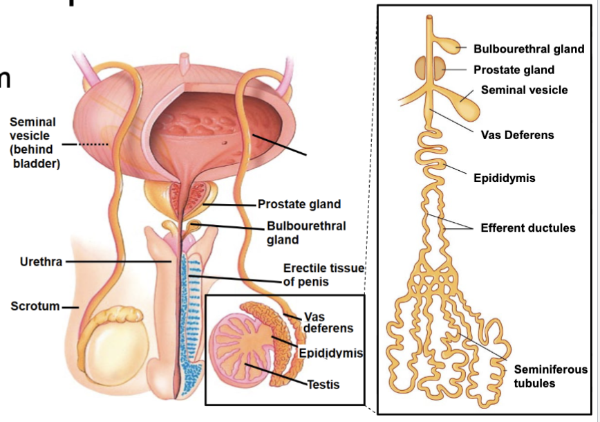
seminiferous tubules are coiled within the testes
testicular cell anatomy
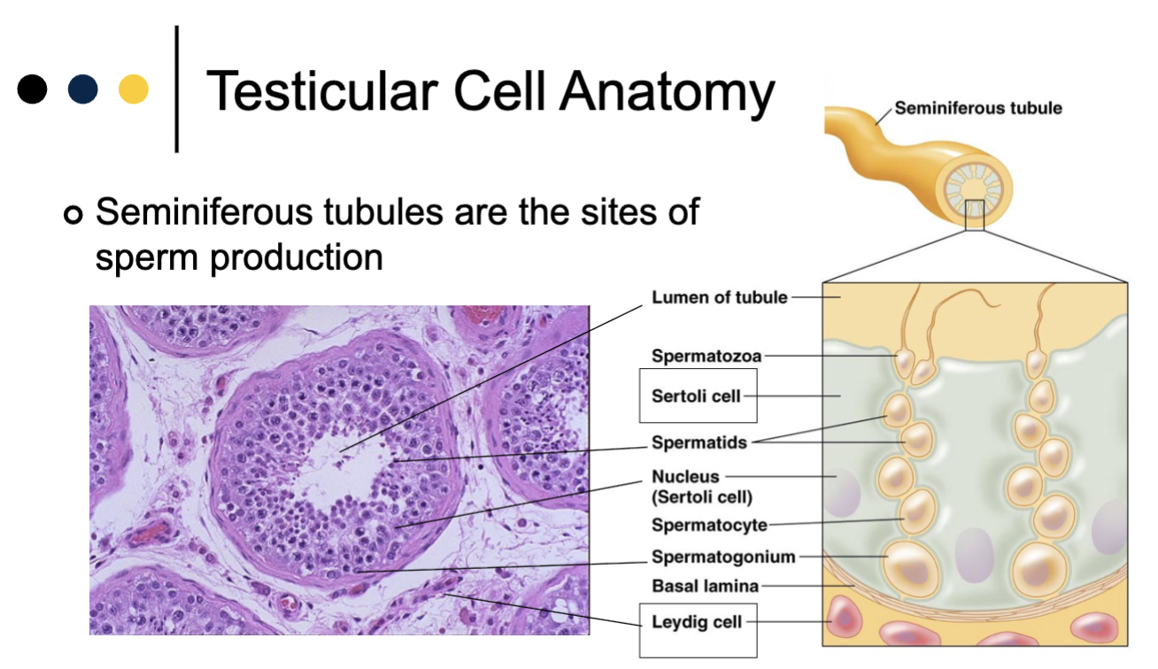
seminiferous tubules are the sites of sperm production
spermatogenesis
seminiferous tubules are the sites of sperm production
leydig cells: produce testosterone in response to LH
sertoli cells: fill gaps between cells and support germ cell growth in response to FSH
spermatogenic cells: germ cells on their way to becoming sperm
spermatogensis = maturation of germ cells into sperm
spermatogonium → spermatocyte → spermatid → spermatozoon

spermiogenesis
spermiogenesis: sperm maturation after meiosis
spermatids change shape:
lose cytoplasm
develop flagellum
condense DNA and form acrosome
nearly mature spermatozoa that cannot swim are released into the lumen of the tubule
finish maturation in epididymis to develop into motile sperm

sperm release
during ejaculation, sperm are propelled by cilia and smooth muscle contractions along the vas deferens which connects to the urethra
seminal fluid added to sperm by exogrine glands
seminal vesciles: nutrients and fluids
prostate: nutrients, many citrate
bulbourethral gland: mucus as a lubricant

regulation of testicular reproduction
slide 25

ova-uterine reproductive tract
ovaries
oocyte (egg) maturation
hormone production
fallopian tubes
location of fertilization
cilia sweep embryo to uterus
uterus
endometrium: implantation/gestation
myometrium: labor/menstruation
ova-uterine reproductive cycles: estrous cycle
present in most mammals
sexual receptivity coincides with specific phase of cycle
little uterine tissue loss
fertile females exhibit behavioral cues and pheromones called “estrus” or “in heat”
ova-uterine reproductive cycles: menstrual cycle
present only in some primates (and a few other exceptions)
sexual receptivity occurs at many phases of the cycle
substantial uterine tissue loss
ova-uterine reproductive cycle
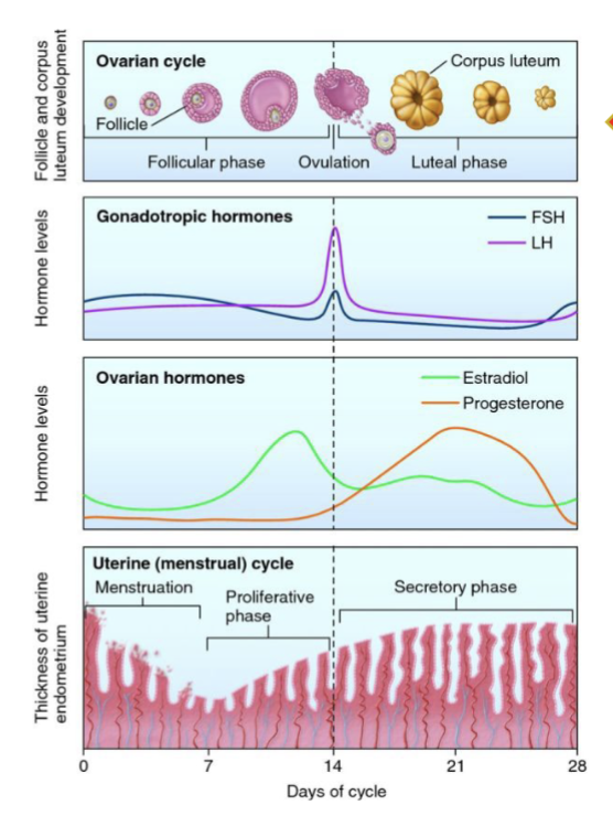
interconnection of hormone levels, ovarian function, and uterine response
begins at puberty, continues until pregnancy or menopause
ovary anatomy
cortex:
outer 1mm
stiff
contains mainly primordial follicles
medulla:
inner portion
les rigid
blood vessels
growing follicles

ovarian follicles
follicle: the functional unit of the ovary
oocyte:
granulosa cells: provide nutrients to germ cell and produce estradiol
theca cells: produce testosterone

folliculogensis
the process of ovarian follicle development
steps: slide 32

hormone regulation of folliculogenesis
changes over time
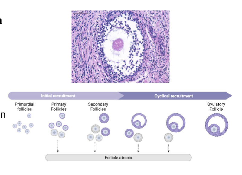
initial recruitment (random) → cyclic recruitment
slide 34

the ovarian reserve
follicles are not generated or replaced after brith
ovarian reserve: the inactive primordial follicles which represent future fertility
depletion of the ovarian reserve → menopause
follicular atresia (death)
follicles can undergo atresia throughout folliculogenesis
characterized by:
reduction in number of granulosa cells
oocyte fragmentation
connective tissue invasion into follicular fluid
collapse of follicle
steroidgenesis
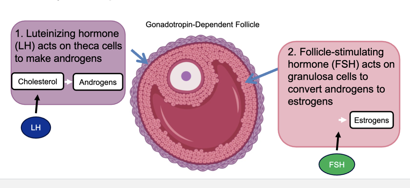
two-cell-two-gonadotropin theory
LH acts on theca cells to make androgens (LH binds to cholesterol → androgens)
FSH acts on granulosa cells to convert androgens to estrogens
follicular steroidogenesis
slide 39

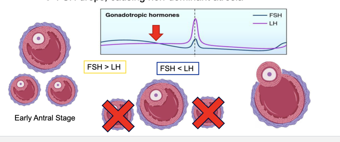
selection of dominant follicle by FSH
high FSH stimulates growth of a cohort of antral follicles
in monovular species, a single follicle will become dominant
FSH drops, causing non-dominant atresia
ovulation
a strong burst of LH triggers ovulation in the dominant follicle
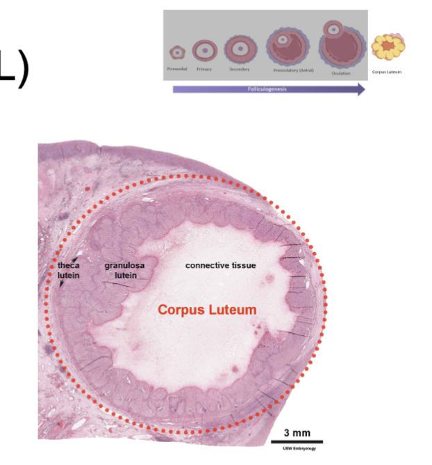
corpus luteum (CL)
following ovulation, granulosa/theca cells luteinize (yellow)
becomes highly vascularized
CL becomes progesterone until placenta takes over
if no pregnancy, CL degrades into corpus albicans (scar)
uterus is highly responsive to ovarian hormones
slide 45

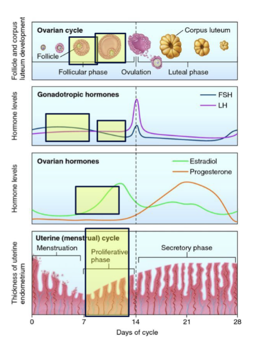
ova-uterine cycle: follicular phase
high FSH → growing antral follicles
growing antral follicles begin to produce estradiol
estradiol stimulates proliferation of the uterus endometrium
as FSH drops, one dominant follicle is selected for ovulation
ova-uterine cycle: ovulation
very high estradiol switches from a negative feedback to positive feedback, causing a “surge” of GnRH
high GnRH causes peaks in levels of FSH and LH
huge peak of LH triggers ovulation 10-12 hours later
oocyte completes meiosis I

ova-uterine cycle: luteal phase
without the oocyte, what remains of the follicle differentiates into the corpus luteum
luteinized granulosa cells produce more progesterone than estradiol
progesterone sustains the endometrium in case of the embryo implantation
without implantation, corpus luteum degenerates and progesterone levels drop
with low estradiol and progesterone, endometrium also degenerates, causing menstruation

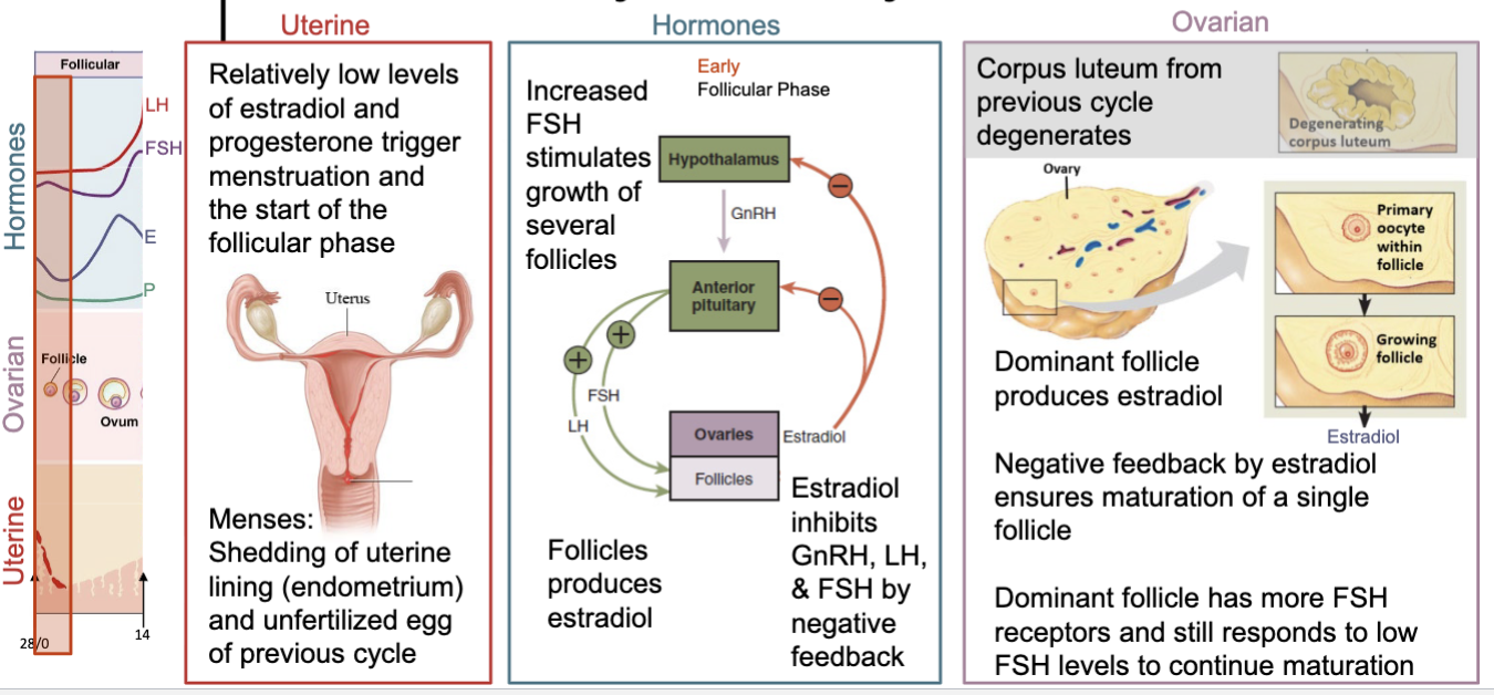
menstrual cycle: early follicular phase
uterine:
relatively low levels of estradiol and progesterone trigger menstruation and the start of the follicular phase
menses: shedding of uterine lining (endometrium) and unfertilized egg of previous cycle
hormones:
increased FSH stimulates growth of several follicles
follicles produce estradiol
estradiol inhibits GnRH, LH, and FSH by negative feedback
ovarian:
corpus luteum from previous cycle degenerates
dominant follicle produces estradiol
negative feedback by estradiol ensures maturation of a single follicle
dominant follicle has more FSH receptors and still responds to low FSH levels to continue maturation
menstrual cycle: late follicular phase
uterine:
proliferative phase: increasing estradiol stimulates thickening of the uterine lining
endometrial growth requires rebuilding the epithelium and underlying stromal layers by cell devision
hormones:
high estradiol for 48 hours switches it from inhibiting to stimulating GnRH (and LH and FSH)
FSH and LH levels increase sharply
ovarian:
a single dominant follicle emerges
this maturing follicle releases estradiol, which adds more estradiol-producing cells to the maturing follicle → therefore raising estradiol further
as FSH and LH levels rise, the mature follicle stops producing estradiol and prepares for the next step

menstrual cycle: ovulation
uterine:
endometrium has been significantly replenished and thickened
cervical mucus altered tos upport sperm motility
hormones:
massive surge in LH levels drives ovulation
ovarian:
about 10-12 hours after the peak in LH, the dominant follicle ruptures and releases the “secondary” oocyte (now in metaphase of meiosis II)
the empty follicle immediately begins transforming into corpus luteum
rupture is accompanied by a “gush” of follicular fluid that washes the oocyte out of the follicle and toward the opening of the fallopian tube
the oocyte can then be fertilized by sperm that are already present (sperm survive up to 7 days in the fallopian tube) or that arrive within 24 hours (unfertilized oocytes only survive 12-24 hours after ovulation)

menstrual cycle: luteal phase
uterine:
secretory phase: driven by increased progesterone
endometrium becomes plush, spongy, and dense with blood, making it structurally and functionally optimized for implantation
hormones:
progesterone inhibits GnRH, FSH and LH to suppress new follicle maturation
corpus luteum secretes progesterone
progesterone maintains a healthy endometrium
ovarian:
corpus luteum is maintained and secretes progesterone
if no implantation occurs, the corpus luteum degenerates during the next follicular phase

pregnancy hormones
upon implantation, the embryonic cells produce human chorionic gonadotropin (hCG)
hCG maintains the CL so that it keeps producing progesterone
once the placenta develops, it makes progesterone and the CL degrades

pregnancy testing
commercial pregnancy tests detect hCG in urine
hCG has a common structure with LH → stimulates follicle growth and luteinization
first scientific test for pregnancy: inject patient urine into a female animal and observe whether the ovaries increased in size
embyonic development is highly evoluationarily conserved
slide 57

fertilization
gametes require an aqueous environment to fuse (evolutionary challenge)
species have evolved different ways to overcome this: external and internal fertilization
external fertilization
most common in aquatic animals
huge gametes numbers are released
egg and sperm release are synchronized
more cells = more change for successful fertilization
internal fertilization
most common in terrestrial animals
usually associated with mating behavior and accessory sex organs
copulation - permits sperm to move directly from male reproductive system to female
mammalian fertilization
a single spermatozoon reaches the fallopian tube first and touches the egg
zona pellucida = thick glycoprotein layer surrounding the oocyte
acrosomal reaction: the acrosome of the sperm bursts, releasing enzymes to degrade the zona pellucida
mammalian fertilization steps
the sperm nucleus enters the egg
sperm releases PLC
PLC → IP3 → release of Ca2+ from ER
waves of Ca2+ trigger egg activation
completion of meiosis
exocytosis of cortical granules to block further sperm entry

zygote stage (1 cell)
2 parental pronuclei
polar body visible
very low transcriptional activity
highly regulated by maternally derived mRNA and proteins
fusion of the pronuclei triggers the first mitotic division
first few mitotic divisions occur without cell growth (cleavage)
resulting cells are called blastomeres

cleavage stage (2 cells to 8 cells)
up through the 8-cell stage, blastomeres are totipotent
each blastomere retains the capacity to form a whole new organism
first cell fate decision (16 cells)
after the 4th cell division, some cells become internalized
mechanical cues trigger gene expression changes to set cell fate (no longer totipotent)
inner cells → inner cell mass (ICM) → fetus
outer cells → trophectoderm (TE) → placenta
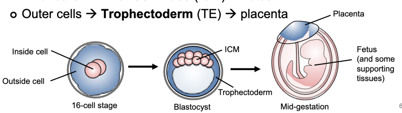
blastocyst formation (~60-300 cells)
tight junctions form between the outside cells, allowing the structure to become water-tight
aqueous cavity forms and inflates the embryo into a blastocyst

embryonic hatching
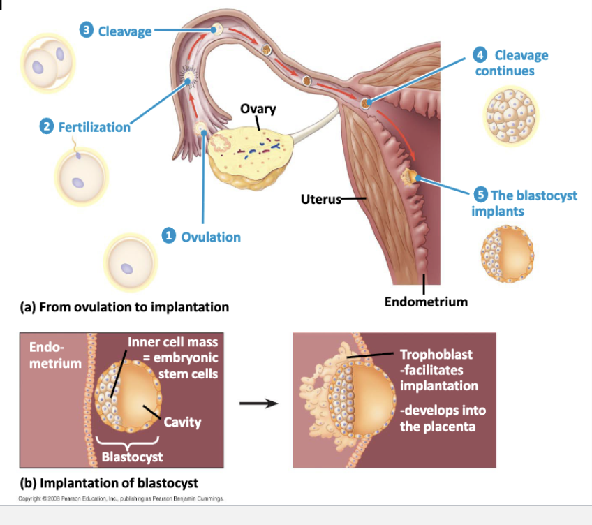
mature blastocysts “hatch” out of the zona pellucida to interact with the uterine endometrium for implantation
implantation
once hatched, the trophectoderm cells can attach to the endometrium
begin producing hCG, which feeds back to the CL in the ovary
most potential pregnancies go undetected
based on (very rough) estimates, upt o 50% of fertilized embryos do not survive past implantation
placenta formation
only organ that develops from 2 genomes
essential for pregnancy success
nutrient/waste exchange
hormone production: progesterone, hCG, and estradiol
invading embryonic trophectoderm cells interface with maternal arteries
fully developed by end of first trimester (before that, CL produces hormones)
nutrients and oxygen enter fetal veins that converge into the umbilical cord
arteries in umbilical cord draws waste away from the fetus into the maternal blood
whole structure delivered during labor
human embryonic and fetal development
organs are vulnerable during critical periods of their development
greatest sensitivity for birth defects occurs around the time of pregnancy detection
sex determination
primary sex determination during embryonic development
bipotential gonads differentiate into ovary or testes
secondary sex differentiation during growth and later development
sexually dimorphic characteristics: reproductive structures (except gonads), coat color/patterns, behaviors, etc
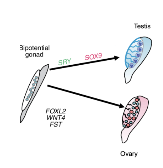
genetic/chromosomal sex determination
slide 75

environmental sex determination
temperature-dependent
based on environmental temperature during early development
other determinants include social group of pH

partuirition (birth)
progesterone levels decrease, allowing uterine muscle (myometrium) to contract
fetal cells produce oxytocin that causes the placenta to release prostaglandins
posterior pituitary releases more oxytocin
both induce further contractions
