CT PROTOCOL PRACTICE EXAM 3
 Call Kai
 Learn
 Practice Test
 Spaced Repetition
 Match
 Flashcards
 Knowt Play
1/14

There's no tags or description
Looks like no tags are added yet.
15 Terms
It requires contrast.
Which of the following is incorrect about a High-Resolution Chest CT.
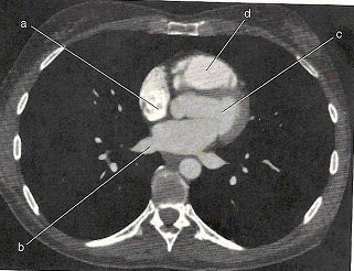
Left ventricle
What is labeled as 'C' on the image?
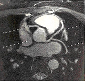
CTA for Coronary Arteries
Based on the image, what thoracic exam was performed?
Prospective ECG gating
Which method of ECG tracing is depicted in the image?

Pulmonary nodule
What thoracic pathology is shown on the image?

R-R interval
One complete cardiac cycle and is sometimes referred to as the ____________.
To reduce respiratory motion in the bases of the lungs
It is recommended for protocols to detect PE scan are scanned in a caudal to cranial direction. Why is this recommended?
Aortic dissection
What thoracic pathology is shown on the image?

D-dimer
This lab test is considered valuable when diagnosing a patient with PE. It is an inexpensive screening tool. If this lab test is within normal limits, a PE is highly unlikely. However, if the lab test is positive, it doesn't necessarily indicate that the patient has a PE. What lab test is this?
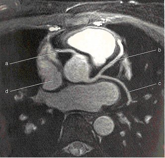
Right coronary artery
What anatomy is displayed by the letter 'A' on the image?
Inspiratory prone
High resolution CT protocols may include three series of scans: inspiratory supine, expiratory supine, and ______________.
Pulmonary nodule
What thoracic pathology is shown on the image?

Lower extremities
Most pulmonary emboli are caused from thrombi originating in the ____________ known as deep vein thrombosis (DVT).
High-Resolution Chest CT
______________ protocol of the chest is used for the assessment of lung parenchyma in patients with diffuse lung disease such as fibrosis and emphysema.
CT Chest with contrast
A patient comes in a CT of the chest. The reason for the exam on the requisition states: abnormality seen on chest x-ray". What exam protocol is the most appropriate?