Flashcards PLTW Human Body Systems 2.1 test review
 Call Kai
 Learn
 Practice Test
 Spaced Repetition
 Match
 Flashcards
 Knowt Play
1/56

There's no tags or description
Looks like no tags are added yet.
57 Terms
action potential
A brief electrical impulse that travels along the axon of a neuron.

electricity is created by
the sudden reversal of the overall charge
action potential steps 1&2
1. The resting membrane potential of a neuron is usually around -70mV
2.The open potassium channels cause neurons to overshoot their resting potential and the cell becomes hyperpolarized, meaning the membrane potential has dropped below its resting potential
action potential steps 3&4
3. A stimulus is applied to the cell, causing the membrane potential to rise
4. After stimulation, the neuron reaches what is known as its threshold membrane potential. This is generally around -55 mV
action potential steps 5&6
5. The action potential reaches its peak. Sodium channels close and potassium channels open, which allows potassium to flow out of the cell
6. The potassium channels close and the sodium-potassium pump allows the membrane to return to resting membrane potential, ready to be activated again.
action potential steps 7&8
7. With potassium channels open, the relative voltage inside the cell falls in a process called repolarization, and the neuron's membrane potential drops back toward resting potential.
8. When the threshold is reached, a large number of sodium channels open, allowing positively charged sodium ions into the cell. This causes depolarization of the neuron as the membrane potential rises to 0 and then becomes positive
Ion Movement
●Potassium channel moves potassium ions out of the cell.
●Sodium channel moves sodium ions into the cell.
●Sodium-potassium pump moves 3 sodium out and 2 potassium in the cell
Neuronal Communication
•Neurons communicate with each other through synapses.
•The presynaptic cell is the sending cell, and the postsynaptic cell is the receiving cell.
•The synapse is the space between them.
•The vesicle contains neurotransmitters at the axon terminal.
•Calcium ions move into the cell, causing neurotransmitters to be released into the synaptic cleft and bind to receptor proteins on the receiving cell.

Explain how neurons convey information using both electrical and chemical signals.
The electrical signals are action potentials, which transmit the information from one neuron to the other; the chemical signals are neurotransmitters, which transmit the information from one neuron to the next. The electrical signal travels down the axon to the axon terminals where it tells the vesicles to release the neurotransmitters into the synaptic cleft which travel to the receptors of the receiving cell which releases the second messengers
Neurotransmitters
the body's chemical messengers used by the nervous system to transmit messages between neurons, or from neurons to muscles

Axon
where electrical impulses from the neuron travel away to be received by other neurons.

Axon Terminal
-any of the button-like endings of axons that change electrical impulses or action potentials within a neuron into chemical messages in the form of neurotransmitters.

Dendrites
appendages that are designed to receive communications from other cells by bringing electrical signals to the cell body. They often resemble a tree like structure.

Vesicles
structures in neurons that store neurotransmitter molecules before releasing them into the synapse in response to electrical signaling within the cell.

Receptors
In neurons, specialized protein molecules on the postsynaptic membrane; neurotransmitters bind to these molecules after passing across the synaptic cleft.

Central Nervous System (CNS)
The part of the nervous system which, in vertebrates, consists of the brain and spinal cord, to which sensory impulses are transmitted and from which motor impulses pass out, and which supervises and coordinates the activity of the entire nervous system.

Peripheral Nervous System (PNS)
The part of the nervous system that is outside the central nervous system and comprises the cranial nerves excepting the optic nerve, the spinal nerves, and the autonomic nervous system.

Cerebrum
Integrating center for memory, learning, emotions
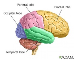
Cerebellum
Coordinates muscles and maintains bodily equilibrium

Brain Stem
The brainstem is composed of three parts: the midbrain, pons, and medulla oblongata. Together, these three parts are involved in vision, hearing, motor control, alertness, temperature regulation, breathing, heart rate, and blood pressure.

Gyri
Convoluted ridges on the brain's surface

Sulci
Shallow furrows separating adjacent gyri

Frontal Lobe
- It helps control voluntary movements, expression and language, cognition, planning, and is the control center for personality , behavior, and emotion.

Temporal Lobe
help with processing pain and auditory stimuli, emotions, and memories. It allows you to understand what you hear, retain visual memories, and process and remember emotions.

Parietal Lobe
responsible for sensory perception and integration. It interprets input from other areas of the body such as taste, hearing, sight, touch, and smell.

Occipital Lobe
responsible for perceiving, processing, and remembering visual stimuli. It allows you to see color, size, distance, depth, and recognize faces and objects.

Neurons
-Primary signaling cells of the nervous system. Neurons send and receive electrical and chemical signals to communicate with each other in the nervous system and with other types of cells in the body.

Glial Cells
Provide protection and maintain homeostasis for neurons

Cell Body
Stores genetic material and produces energy

Synapse
the portion of the neuron where information is received. The synapse is composed of small receivers, called dendrites

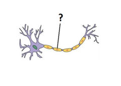
Nodes of Ranvier
periodic gaps in the insulating sheath (myelin) on the axon of certain neurons. These nodes allow ions to diffuse in and out of the neuron, pushing the electrical signal down the axon

Myelin Sheath
Insulating layer around nerves for fast transmission
Nucleus
contains genetic material (chromosomes) for cell development and the synthesis of proteins necessary for cell maintenance and survival.

Astrocytes
- Abundant, star-shaped cells
- Anchor & brace neurons
- Form barrier & make exchanges between capillaries & neurons
- Control the chemical environment of the brain by cleaning up potassium ions & neurotransmitters

Microglia
-Small ovoid cells with thorny processes
- Spider-like phagocytes
-Diposed of debris

Ependymal Cells
- line central cavities of the broca & spinal cord
- Cells are squamous & columnar shaped and ciliated
- Circulate cerebrospinal fluid

Oligodendrocytes
Produce myelin sheath around nerve fibers
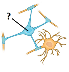
Satellite Cells
- Surround neuron cell bodies located in PNS
-Protection & cushioning neurons
-Similar to astrocytes in CNS

Schwann Cells
-Surrounds & form myelin sheath in the PNS
-Help with the regeneration of damaged peripheral nerve fibers

Sensory Neurons
get information about what's going on inside and outside of the body and bring that information into the CNS so it can be processed. Example: unipolar neuron
Motor Neurons
get information from other neurons and convey commands to your muscles, organs, and glands. Example: multipolar neuron
Interneurons
connect one neuron to another. They receive information from other neurons (either sensory neurons or interneurons) and transmit information to other neurons (either motor neurons or interneurons).
Acetylcholine
Involved in muscle contraction, learning, memory
Dopamine
Plays a role in pleasure, motivation, mood, attention
Gamma-aminobutyric acid (GABA)
Primary inhibitory neurotransmitter in the CNS
Glutamate
Primary excitatory neurotransmitter in the brain
Epinephrine & Norepinephrine
Excitatory neurotransmitters in fight or flight response
Serotonin
Regulates mood, sleep, and aids in digestion
Agonist
Substance that stimulates an action
Antagonist
Substance that blocks an action
Inverse Agonist
a molecule or chemical compound that can bind to a receptor but rather than producing an activation, it will lead to a deactivation that means decrease the baseline activity of the receptor.
Reuptake Inhibitor
Chemical that prevents reuptake of neurotransmitters
Reflex
Automatic response to a stimulus
Reaction
Thoughtful response to external stimuli
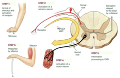
Reflex Arc
1.The patellar reflex is initiated by a tap on the patella tendon.
2.The tap causes a slight stretching of the quadriceps muscle activating the stretch receptor.
3.This begins a nerve impulse that travels to the spinal cord.
4.The motor neurons in the spinal cord activate without any signal going to the brain for processing.
5.These motor neurons then bring the signal to the muscle, causing it to contract.
Support Cells(Neuroglia) in Central Nervous System
- Astrocytes
-Microglia
-Ependymal cells
Support Cells (Neuroglia) in Peripheral Nervous System
- Satellite cells
-Schwann cells