ANATOMY- Lower Extremity Muscles (CADAVER IMAGES), Nerves and Vessels of the Lower Extremity, and some bones Diagram
 Call Kai
 Learn
Learn Practice Test
 Spaced Repetition
 Match
Match Flashcards
 Knowt Play
1/112

There's no tags or description
Looks like no tags are added yet.
Name | Mastery | Learn | Test | Matching | Spaced | Call with Kai | Chat |
|---|
No analytics yet
Send a link to your students to track their progress
113 Terms
Sartorius

Adductor Longus

Adductor Brevis

Gracilis

Adductor Magnus

Gluteus Maximus

Gluteus Medius

Gluteus Minimus

Piriformis

Semitendinosus

Semimembranosus

Extensor Hallucis Longus

Tibialis Anterior

Fibularis Longus

Fibularis Brevis

Gastrocnemius

Soleus
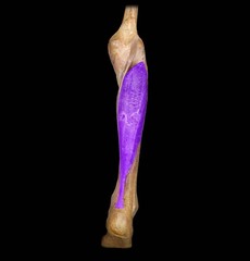
Flexor Digitorum Longus

Flexor Hallucis Longus

Tibialis Posterior

Gluteus Medius
Abducts and medially rotates thigh; , Description: Above and partially under Gluteus Maximus

Rectus Femoris
A- Extends leg at the knee and flexes thigh at the hip; part of the quadriceps group, Description: Superficial, runs straight down, middle of thigh

Vastus Intermedius
ANTERIOR THIGH
Description: Underneath Rectus Femoris, between Vastus Lateralis and Vastus Mediali; , Extends knee

Vastus Medialis
B- Description: Medial aspect of thigh, under Sartorius

Long Head of the Biceps Femoris
Description: Outside of leg, under Gluteus Maximus; Flexes leg at the knee and extends thigh at the hip; belongs to the hamstring group

Semitendinosus
POSTERIOR THIGH- pointing medially
Description: Inside of leg, next to Biceps Femoris

Sartorius
ANTERIOR THIGH-
Flexes, abducts, and laterally rotates thigh at the hip; flexes knee; A long, narrow muscle running obliquely across the front of each thigh from the hipbone to the inside of the leg below the knee.

Gracilis
ANTERIOR THIGH. (pointing MEDIALLY)
Adducts thigh at the hip and flexes leg at the knee; assists in medial rotation; groin muscle

Extensor Digitorum Longus
ANTERIOR SHIN
Extends the lateral four toes; dorsiflexion of ankle and eversion of foot; Femur to Phalange

Fibularis Longus
LATERAL SHIN
ISOLATED FUNCTION: concentrically plantarflexes and everts the foot INTEGRATED FUNCTION: decelerates ankle dorsiflexion, isometrically stabilizes the foot and ankle complex

Gastrocnemius
Aids in flexion of the knee/primary mvmt is the ankle plantar flexion; toe dancer muscle, 2 bellied, calf muscles
Tibialis Posterior
POSTERIOR Tib/Fib
Is a deep muscle that lies deep to the soleus; Inverts foot; may assist in strong plantar flexion or control pronation of foot during walking

Gluteus Maximus
1 (Posterior Hip/Femur)

Gluteus Minimus
2 (Posterior Hip/Femur)

Semitendinosus
4 (Posterior Hip/Femur)

Semimembranosus
3 (Posterior Hip/Femur)

Adductor Magnus
2 (Posterior Hip/Femur)

Soleus
13

Vastus Lateralis
10

Rectus Femoris
11

Vastus Medialis
9

Vastus Intermedius
12

Tibialis Anterior
16
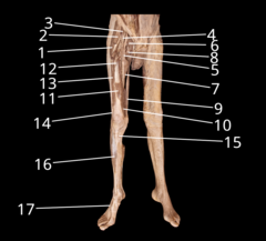
anterior inferior iliac spine
Name this structure:

greater sciatic notch
Name this structure:

lesser sciatic notch
Name this structure:

Ischial spine
Name this structure:

Ischial tuberosity
Name this structure:

superior pubic ramus
Name this structure:

inferior pubic ramus
Name this structure:

pubic symphysis
Name this structure:

pubic tubercle
Name this structure:

obturator foramen
Name this structure:

Head
Name this structure

Neck
Name this structure

greater trochanter
Name This structure

lesser trochanter

Patellar Surface
Patellar Surface

linea aspera

Lateral condyle
Name this structure:

MEdial condyle
Name this structure:

medial epicondyle
Name this Structure:

lateral epicondyle
Name this Structure:

lateral condyle of tibia
Name this structure:

tibial tuberosity
Name this structure:

medial malleolus
Name this structure:

fibular head
Name this structure:

lateral malleolus
Name this structure:

talus
Name this structure:

calcaneus
Name this structure:

cuboid
Name this structure:

medial cuneiform
Name this structure:

intermediate cuneiform
Name this structure:

lateral cuneiform
Name this structure:

navicular
Name this structure:

Metatarsal bones
Name these structures

Philanges
Name these structures

gluteus medius
Name this muscle:

gluteus minimus
Name this muscle:

piriformis
Name this muscle:

rectus femoris
Name this muscle:

vastus medialis
Name this muscle:

vastus intermedius
Name this muscle (posterior to rectus femoris):

vastus lateralis
Name this muscle:

pectineus
Name this muscle:

adductor brevis
Name this muscle:

adductor magnus
Name this muscle:

Obturator nerve
Branches to the adductor longus, adductor brevis, and gracilis

Sciatic nerve
Composed of the tibial and common fibular nerves within a fibrous sheath. Does not innervate any muscles itself, but its branches do. TIbial division innervates semimembranosus, semitendonosis, and the long head of the biceps femoris. Common fibular division innervates the short head of the biceps femoris. Splits into the tibial nerve and the common fibular nerve in the popliteal fossa.

fibular nerve
Wraps around the head of the fibular and then splits into the superficial fibular nerve and deep fibular nerve.

Deep fibular nerve
Innervates muscles of anterior compartment of leg.

Superficial fibular nerve
Innervates muscles of the lateral compartment of the leg.
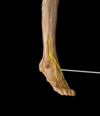
Tibial nerve
Travels between superficial and deep posterior compartments of leg, innervating muscles in both compartments. Wraps around the medial malleolus and branches into the lateral and medial plantar nerves.

Femoral artery
Passes through adductor magnus. Extension of the external iliac artery. Supplies anterior structures of the thigh.
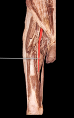
Deep femoral artery
Runs posteriorly and supplies the muscles of the posterior and medial regions of the thigh

Anterior tibial artery
Travels in anterior compartment of leg along with the deep fibular nerve to the tibialis anterior muscle. Becomes dorsal pedis artery as it crosses the ankle.

Dorsalis pedis artery
Runs on the dorsal aspect of the foot to supply muscles and nerves of this region.
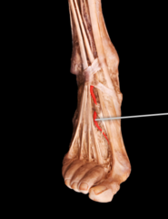
Posterior tibial artery
Goes around medial malleolus and divides into the medial and lateral plantar arteries. May appear as a continuation of the popliteal artery with the anterior tibial artery branching off of it. Also travels between superficial and deep posterior compartments with the tibial nerve, supplying both compartments.

Fibular artery
Supplies blood to the lateral compartment of the leg.

Popliteal vein
Travels with popliteal artery, continuous with femoral vein.
