i love u! good luck baby
 Call Kai
 Learn
Learn Practice Test
 Spaced Repetition
 Match
Match Flashcards
 Knowt Play
1/61

Earn XP
Description and Tags
from taiger
Name | Mastery | Learn | Test | Matching | Spaced | Call with Kai | Chat |
|---|
No analytics yet
Send a link to your students to track their progress
62 Terms
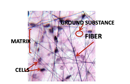
Photomicrograph of Connective Tissue

Connective Tissue

Overview of connective tissue and fibers

Fibroblast (Left), Fibrocyte (Right)

Active Fibroblast
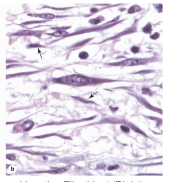
Inactive Fibroblast

Connective cell lineage (under Mesenchymal cells section)

Macrophage under (a) light microscope and (b) electron
microscope

Appearance of Adipocytes (Fat Cells)

Appearance of Plasma Cells, “Cartwheel” appearance of
the nucleus

Appearance of “cartwheel”/“spokewheel” nucleus under EM

Appearance of Mast Cells under EM

Appearance of Mast Cells under Light Microscopy with 40x
magnification (left) and 400x magnification (right)

Assembly of collagen fiber bundles
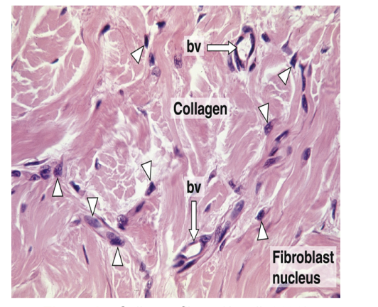
Collagen fibers
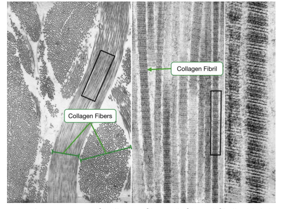
Appearance of collagen fibers (left) and fibrils (right) under
EM
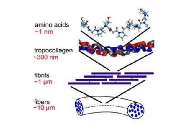
Simplified assembly order of collagen fiber bundles

Collagen synthesis
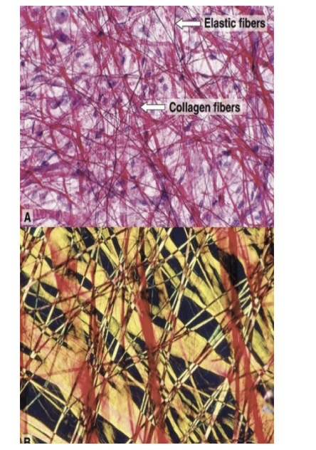
Elastic Fibers under the microscope

Reticular Fibers

Stretch and Recoil of Elastin Molecules

Mesenchymal Lineage for CT Cells and HSC Differentiated

Histological examples of loose connective tissue, Lamina propria of the small intestine (left), Mammary gland interlobular (right)

Histological example of dense irregular connective tissue
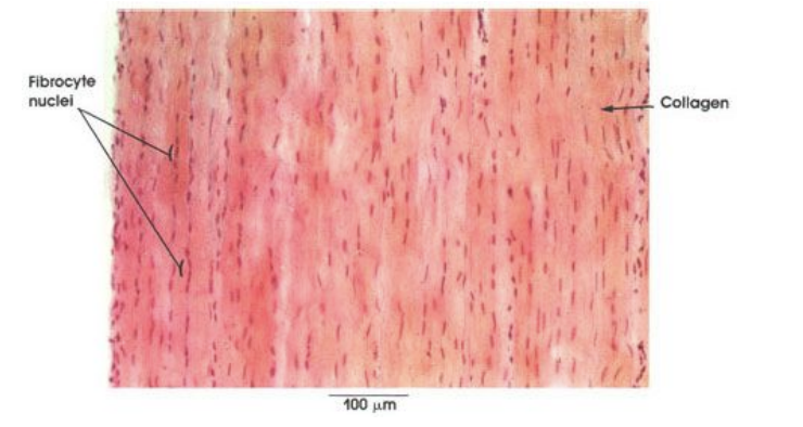
Histological example of dense regular connective tissue

Yellow adipose tissue and dense irregular connective tissue

Brown adipose tissue

Embryonic tissue (Wharton’s jelly)

Spongy bone histology

Bone canaliculus (under compat bone section)

Osteon (under compat bone section)

Composition of bone (under compat bone section)

Periphreal blood cells (left) and the embryonic origins of the blood (right)

Extracellular or Ground Matrix
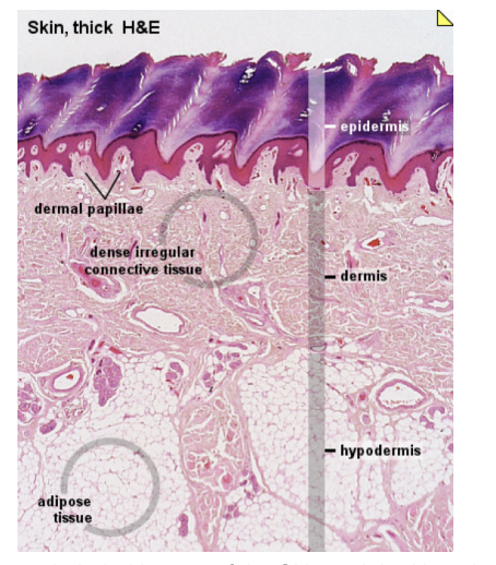
Labeled layers of the Skin and the Hypodermis
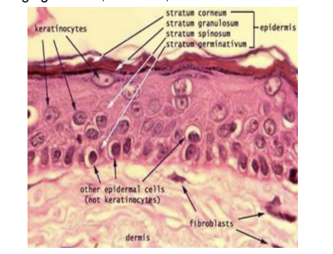
Keratinocytes

Thick vs. thin layers of the epidermis

Melanocytes in the Stratum Basale

Langerhans cells

Layers of the epidermis

Non-keratinocyte cells of the epidermis

Histological slide of the papillary and reticular dermis

Layers of the dermis

Histological slide showing (a) Meissner tactile corpuscles (TC), dermal papillae (DP), and epidermis (E); (b) Pacinian corpuscles (PC)

Adult hair histology

Histological slide of the hypodermis

Lateral view of the parts of the nail

Anteriro view of parts of the nail
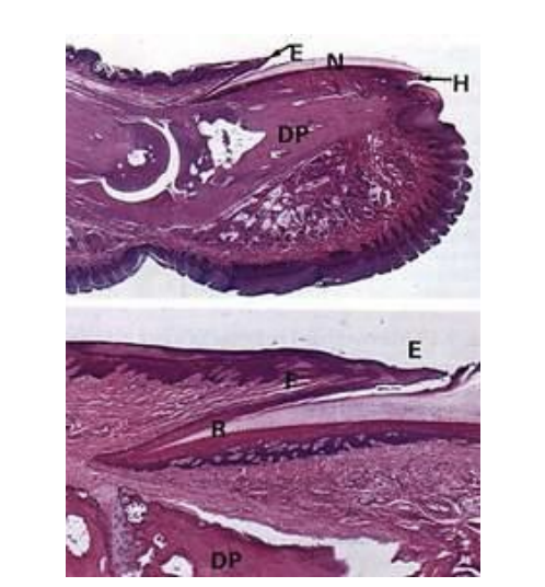
Nail histology

Sebaceous glands

Eccrine and apocrine sweat glands

Fibrocytes Collagen-I

Fibrocytes Collagen-IV

Fibrocytes P-4-H

Fibrocytes rabbit lgG
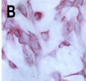
Fibroblasts Collagen-I
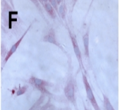
Fibroblasts P-4-H

Fibroblasts Collagen-IV

Fibroblasts rabbit lgG

Hyaline cartilage

Elastic cartilage

Fibrocartilage