CHEM 220 Midterm 3
 Call Kai
 Learn
 Practice Test
 Spaced Repetition
 Match
 Flashcards
 Knowt Play
1/26

There's no tags or description
Looks like no tags are added yet.
27 Terms
Spectroscopy
Spectroscopy is the interaction b/w EM radiation & matter
Spectrophotometry (subset of spectroscopy) describes the quantitative study of EM spectra, dealing w/ radiation in the visible, UV, and infrared regions.
In this method, sample absorbs EM radiation (light) from appropriate source & amount of light absorbed is related to [ ] of analyte in sol/sample
EM Spectrum
EM radiation possesses wave and particle particles. it consists of an oscillating electric field component (E) and an oscillating magnetic field component (M). E and M are perpendicular to each other and are perpendicular to the direction of propagation
Wave Motion
Wave motion of radiation vibrate perpendicular to the direction of propagation
wavelength (λ): distance of one complete cycle
frequency (v): the # of cycles passing a fixed point per unit of time
wavenumber (v -): reciprocal of λ & # of waves in a unit length or distance per cycle
λ = c / v and v dash - v / c
Absorption vs Emission
Absorption - photons give their energy to e-
causes excitation of valence e-
promotes molecule to higher energy level
an unstable state & e- will fall to ground state
Emission - excited molecule ejects a photon of energy
decrease in energy and molecule demoted to lower energy level
Transition
Transition is when a molecular is promoted from one energy level to another. There are 3 possible types of transitions
Rotational < Vibrational < Electronic
Spectrophotometer
Used to measure an adsorption or emission spectra. Requires the following:
Light source → need a light source that has detectable output of λ radiation in region specific instrument
no light source can emit a single λ of light but rather multiple λ (polychromic)
Wavelength sector → restricts the radiation (or light) being measured to a narrow band that is absorbed or emitted by the analyte
enhances selectivity and sensitivity of SPM
Sample cell → sample is usually liquid and contained in cuvette that is transparent, visually and also to the λ region being used
Detector → detects how much light has been emitted
Readout device
Components of wavelength selector
For good analysis, compounds should be analyzed at a λ for max absorbance. Wavelength selector has a few components:
Lenses or mirrors → focus on radiation
Entrance & exit slits → limits unwanted radiation
Dispersing element → separates λ from polychromic radiation
2 types: prism & diffraction grating
Prisms vs Diffraction Gratings
Prisms: when light passes through a prism, it is refracted — bending of light. Index of refraction depends on λ, short λ are refracted more than longer λ.
Prism monochromator
Focusing lens: source light is directed to entrance slit
Collimating lens: makes light parallel
Diffraction Gratings are highly polished surfaces w/ a large # of parallel lines, or grooves, in it
Incident light hits the groove/gratings & id dispersed
Resolving power is typically better than prisms
Optical Filters
Optical filters isolate specific λs of light w/ 3 types:
Narrow-bandpass filters → absorb all radiation except desired λ
Sharp-cutoff filters → absorbs all radiation up to a specific λ, but passes all radiation at longer λ
Interference filters → filter designed to have destructive to have destructive interference for all radiation striking it, other than for a narrow band of radiation that it transmits
Band
No λ selector can produce a single λ of radiation as a group of λ or band
The nominal λ is selected by the instrument; is the λ within the max intensity passed by the exit slit
The bandwidth is composed of the λ that passed at half the intensity of the normal λ
Suggestions for Sample Reading
Always check cuvette to make sure you have clear sides and correct path length in light pass
Do not transfer sample to cuvette w/ glass Pasteur pipette - can scratch walls
Before inserting cuvette into SPM, wipe down to remove dust
Use ‘matched’ cuvettes or put a small dot → ensures optical density of cuvette wall is the same each time
Read samples in random order
Detector
A photon detector produces an electrical signal when struck by photons. Types:
Phototubes
Photomultiplier tubes
Silicon photodiodes
Photovoltaic cells
Photo-multipler tube
Photons enter an evacuated tube
Photons strike the cathode
E- are emitted & accelerated towards first dynode
After striking, more e- are emitted & accelerated
Continues until amplified signal reached anode
Creates cascade effect w/ 106-7 e- collected for each photon entering the tube
Transmittance vs Absorbance
Transmittance: fraction of light than passes through a sample
T = P / P0
P = power of transmitted radiation
P0 = power of incident radiation
T is b/w 0-1 or %
Absorbance: amount of light not passed through a sample
A = - log (T)
Is the opposite of transmission but not a linear relationship
Most accurate spectroscopy will have reading b/w 0.4-0.9
Beers Law
A = ε b c
A = absorbance (unitless)
ε = molar absorptivity (L / mol cm)b = path length (cm)
c = [ ] (M)
Limitations: only accurate for dilute sol.
Can be applied to mixtures that have more than 1 compound that absorbs light, but there can be no interactions b/w components.
SPM Titration
titration where endpoint is found by monitoring the absorption or emission of light
can plot absorbance vs V of titrant
as titration is added, A increases
once all analyte reacted, A plateaus
should plot corrected A to account for any dilution effect:
Acorrected = (Vt / Vi) x Aobserved
Molecular absorption
at room temp, molecule is in ground state S0 (e- paired + opposite spin)
when photon is absorbed, it gets excited and promoted to higher energy singlet excited state (e.g. S1, S2)
e- spins are antiparallel
or e- can go to triplet excited state (e.g. T1); spins are parallel
to return to ground state, fluorophone can take nonradiative or radiative pathways

Nonradiative Pathways
Vibrational relaxation: molecule in an excited vibrational energy level loses energy & goes to lower vibration energy level within the SAME electronic state
same electronic state + same spin
Internal conversion: molecule moves from a higher electronic energy level to a lower energy level
diff states + same spin (e.g. S2 → S1)
Intersystem crossing: e- moves from higher electronic energy level to lower energy e- state that has a diff spin
diff electronic state + diff spin
External conversion: when absorbed energy from photon is transferred to the solv or part of the sample matrix
enegery transferred to surr
Note: combo of VR + ICV can convert entire energy of photon to heat, meaning no radiation given off on path block to ground state
Radiative Pathways
Fluorescence: emission of a photon as the fluorophore goes from the lowest vibration energy level of an excited state to a lower energy state
both states have the same spin (e.g. S1 → S0)
radiation stops as soon as light source is removed/shut off
Phosphorescence: emission of a photon as the fluorophore goes from lowest vibrational energy of an excited state to a lower energy state
Excitation vs Emission Spectra
Excitation Spectra: fluorescent intensity measured w/ constant emission λ selected & varying excitations λs
Emission Spectra: fluorescent intensity measured w/ constant excitation λ selected & varying excitations λs
Atomic Spectroscopy
Basic concept: atomize samples by heating to high temps, and then observe either the light that is emitted or absorbed
elemental e- promoted from the ground state, but ONLY electronic transitions
Energy absorbed is quantified via λ and intensity monitoring to produce a spectrum - either emission or absorption of radiation
Atomic Emission Spectroscopy (AES)
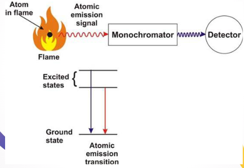
Sample is heated enough to break molecular bands and create free atoms
Valence e- are promoted to hgiehr, unstable energy levels
Excited e- fall back to lower, or ground, energy states, releasing the absorbed energy in the form of light
Emitted light passes through a monochromator
Qualitative Analysis = elements have unique “fingerprints” of specific λ of emitted
Quantitative analysis = intensity of light corresponds to how much of the element is present
Atomic Absorption Spectroscopy (AAS)
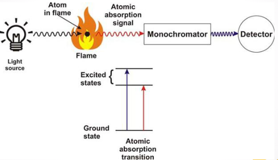
Sample is atomized to break chemical bonds and create unexcited, ground-state atoms
Need to install a specific ligth source, such as hollow cathode lamp, that matches the exact element to be measured
While lamp is on, it will measure that compound and how much light is passed through to detector
If element is found in 2+ diff forms in sample, AAS will only tell the total amount of compound, not how much was from each species
Can quantify by using calibration standards
Atomic SPM Components
Light source (for AAS)
Atomizer
Monochromator
Detector
Flame Atomizer
Uses a premix burner to combine sample, oxidant, and fuel before going to flame
Sample drawn into nebulizer by high flow of oxidant (e.g. air)
leaves tips as fine mist
Spray is directed towards glass bead
small droplets hit it and become smaller
Aerosol travels past baffles which promote mixing and blocks large droplets
Only ~5% of initial sample actually reaches flame
Furnace Atomizer
Steps of Furnace Atomizer
Drying → sample heating to ~125C for ~20s to remove any solv
Charring → furnace heated to ~1400C for about 60s to destroy any organic matter; known as pyrolysis
Atomizations → furnace heated to ~2100C for less than 10s; samples atomized and absorbance measured
Vaporization → furnace heated to ~2500C for a few secs to vaporize any remainig residue