Companion animal medicine 2
 Call Kai
 Learn
Learn Practice Test
 Spaced Repetition
 Match
Match Flashcards
 Knowt Play
1/546

There's no tags or description
Looks like no tags are added yet.
Name | Mastery | Learn | Test | Matching | Spaced | Call with Kai | Chat |
|---|
No analytics yet
Send a link to your students to track their progress
547 Terms
Jaundice
Yellow staining of serum/ tissue caused by excess bilirubin
Large, sustained increase in bilirubin production or a major impairment of bile excretion
Normal bilirubin metabolism
Old RBCs are phagocytosed
Haem > biliverdin > unconjugated (fat-soluble) bilirubin
Unconjugated bilirubin binds albumin and travels to the liver
Hepatocytes conjugate bilirubin, making it water-soluble
Bilirubin is excreted into the bile - this step is rate-limiting
Bile is stored in the gallbladder or passes directly to the duodenum
Bacteria deconjugate bilirubin to urobilinogen - most is reabsorbed, some is excreted
Pre-hepatic (haemolytic) jaundice mechanism
Massive red cell destruction (intravascular or extravascular) overwhelms the liver’s substantial capacity to take up, conjugate and excrete bilirubin
Produces predominantly unconjugated hyperbilirubinaemia
Implies a marked, ongoing haemolysis
Pre-hepatic (haemolytic) jaundice causes
IMHA, babesiosis, cytauxzoonosis, oxidant toxicity, zinc toxicity
Pre-hepatic (haemolytic) jaundice clinically
Moderate to severe regenerative anaemia
Normal to high plasma protein
Minimally deranged liver enzymes
Normal to enlarged spleen/ liver
Intra-hepatic (hepatocellular) jaundice mechanism
Biliary disease impairs uptake, intracellular processing or canalicular excretion of bilirubin (true cholestasis with regurgitation of bile into sinusoidal blood)
Jaundice is more likely when disease primarily affect periportal hepatocytes
Intra-hepatic (hepatocellular) jaundice causes
Chronic hepatitis, hepatic lipidosis, cirrhosis/ fibrosis, acute hepatotoxicity, neoplasia (primary or infiltrative), cholangitis
Sepsis/ endotoxaemia can interfere with bile flow
Intra-hepatic (hepatocellular) jaundice clinicaly
High ALT (hepatocellular damage) with high ALP/ GGT (cholestasis)
Hypoalbuminaemia
Coagulopathy
Post-hepatic (obstructive) jaundice mechanism
Physical obstruction of bile flow distal to the liver, causing conjugated hyperbilirubinaemia
Post-hepatic (obstructive) jaundice causes
Extrahepatic bile duct obstruction
Intraluminal - choleliths, biliary mucocele, neoplasia, parasites, foreign body
Extraluminal - pancreatitis (most common)
Biliary rupture (trauma/ necrotising cholecystitis) causing bile to leak into peritoneal cavity
Post-hepatic (obstructive) jaundice clinically
Markedly elevated ALP/ GGT
Pale
Dilated bile duct/ gallbladder and possible obstructing mass/ choleliths on imaging
Primary haemostasis
Platelet plug formation through platelet adhesion via vWF causing aggregation
Forms a short-lived unstable plug
Secondary haemostasis
The coagulation cascade stabilising the initial plug with fibrin, producing a durable clot
Primary haemostatic disorders
Behave like a garden hose punctured with many small holes
Either thrombocytopenia or platelet dysfunction (where platelet numbers are normal)
Primary haemostatic disorders clinical signs
Petechiae and ecchymoses (multiple, small, short-lived bleeds)
Mucosal/ surface bleeding - epistaxis, melena, haematochezia, haematuria
Bleeding begins immediately after venipuncture or minor trauma
Thrombocytopenia aetiology
Decreased production
Increased destruction
Increased consumption
Increased sequestration
Decreased production of platelets causes
Immune-mediated megakaryocytic hypoplasia, marrow aplasia, myelophthisis, drugs (e.g. oestrogens), retroviral infection (cats)
Increased destruction of platelets causes
Immune-mediated thrombocytopenia, drug-induced, infectious, vaccine-associated
Increased consumption of platelets causes
DIC, microangiopathy, vasculitis
Increased sequestration of platelets causes
Splenomegaly, splenic torsion
Platelet dysfunction (thrombocytopathia) aetiology
Congenital
von Willebrand disease (most common in dogs)
Breed-specific thrombopathias
Scott syndrome
Acquired: NSAIDs, uraemia, monoclonal gammopathies, vector-borne disease
Secondary haemostatic disorders
Behave like a hose with one large hole
Either congenital clotting factor deficiencies or vitamin K deficiency/ antagonism
Secondary haemostatic disorders clinical signs
Petechiae/ ecchymoses rare
Haematoma formation, bleeding into muscles, joints (lameness) and body cavities
Haemothorax/ hemiabomen leading to collapse, dyspnoea, abdominal distension
Bleeding after venipuncture is typically delayed, not immediate
Congenital clotting factor deficiencies aetiology
Haemophilia A and B (rare in cats, slightly more common in dogs)
Vitamin K deficiency/ antagonism aetiology
Rodenticide (anticoagulant) toxicity - inhibits vitamin K epoxide reductase
Cholestatic/ EBDO disease - fat malabsorption limits vitamin K uptake
Severe hepatic disease - most coagulation factors are hepatically synthesised
Diagnostic approach to haemostatic disorders
History
Physical examination - classify the bleeding pattern
Further testing
Causes of regenerative anaemia
Blood loss
Haemolysis (extravascular or intravascular)
Causes on non-regenerative anaemia
Anaemia of chronic disease
Anaemia of renal disease
Bone marrow disorders
Anaemia of endocrine disease
Peracute blood loss/ haemolysis
Blood loss anaemia
Acute or chronic external/ internal haemorrhage - trauma, GI ulceration/ neoplasia, coagulopathy, parasitism
Typically normal to low plasma protein
Progresses to iron deficiency anaemia
Extravascular haemolytic anaemia
RBCs phagocytosed by the mononuclear-phagocytic system in spleen/ liver/ marrow
Most common mechanism in small animals
IMHA, RBC parasites, Heinz bodies
Spherocytes are the hallmark finding
Intravascular haemolytic anaemia
Direct lysis in circulation
Caused by complement-fixing antibody, infectious agents, oxidative/ zinc toxicity, hypophosphataemia or mechanical shearing
Immune-mediated haemolytic anaemia
Primary/ idiopathic or secondary to drugs, infection, neoplasia
Diagnosed by spherocytosis, saline autoagglutination and/ or a positive direct Coombs’ test
Can be peracute and life-threatening
Anaemia of chronic disease
Most common non-regenerative anaemia
Mild, normocytic, normochromic, secondary to chronic inflammation/ infection/ neoplasia
Sequestration of iron making it unavailable for erythropoiesis
Usually resolves with treatment of underlying disease
Anaemia of renal disease
Due to reduced renal erythropoietin production
Normocytic, normochromic, poorly/ non-regenerative
Bone marrow disorders
Aplasia/ hypoplasia (e.g. FeLV)
Myelophthisis - marrow ‘crowding out’ by neoplastic cells
Myelodysplastic syndrome
Myelofibrosis/ osteosclerosis
Anaemia of endocrine disease
Mild and often incidental - hypothyroidism and hypoadrenocorticism
Iron-deficiency anaemia
Mild-moderate regeneration, becomes non-regenerative
Microcytic and hypochromic
In dogs - usually due to chronic GI blood loss (neoplasia, ulceration) or severe flea burden
In cats - rare in adults, seen mainly in kittens or with chronic GI blood loss
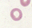
Torocytes
RBC with increased central pallor and a normal cell wall - looks punched out
Torocytes causes
Artefact with no clinical significance

Poikilocytosis
Increase in the number of abnormal RBCs of any shape
Poikilocytosis causes
Any condition disrupting normal RBC membrane integrity or production

Spherocytes
Small, spherical, densely-staining RBCs with little/ no central pallor
Spherocytes causes
Removal of membrane by macrophages - IMHA, oxidative damage, microangiopathic disease, toxicoses

Schistocytes
Small, irregularly shaped RBC fragments with sharp angles or points
Schistocytes causes
Shearing of RBC in circulation due to vasculature abnormalities
MAHA, DIC, haemangiosarcoma, iron deficiency, vasculitis, heartworm
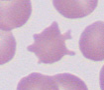
Acanthocytes
RBCs with irregular, unevenly spaced, blunt membrane projections of varying length
Acanthocytes causes
Potentially fragmentation injury
Hepatic disease, haemangiosarcoma, DIC, glomerulonephritis, lipid disorders

Anisocytosis
Variation in RBC size within the same smear
Anisocytosis causes
Regenerative anaemia (macrocytes from reticulocytosis), any active/ compensating erythropoiesis, some breed variation

Microcytes
RBCs smaller than the normal reference size
Microcytes causes
Iron deficiency anaemia, portosystemic shunts, some breeds

Polychromatophils
Immature RBCs staining blue-grey/ purple due to residual ribosomal RNA - usually larger
Polychromatophils causes
Regenerative anaemia and release of reticulocytes

Hypochromia
RBCs with an enlarged area of central pallor and a thin rim of haemoglobin, giving a pale appearance
Hypochromia causes
Decreased Hb production - iron deficiency anaemia, chronic external blood loss

Ghost cells
RBC membranes with little to no visible haemoglobin - appear as pale, empty outlines
Ghost cells causes
Intravascular haemolysis (IMHA, oxidative injury, other haemolytic disease)
Can also be an artefact

Howell-Jolly bodies
Small, round, dark purple nuclear remnants within RBCs, usually single
Howell-Jolly bodies causes
Increased RBC production or decreased removal by splenic macrophages
Regenerative anaemia, splenectomy, glucocorticoids
Normal in cats

Metarubicytes
Most mature form of a nucleated RBC. Condensed, pyknotic nucleus still present within the cell, cytoplasm ranging from basophilic to orange depending on maturity
Metarubicytes causes
Strong regenerative anaemia, bone marrow disease, lead toxicity, splenic disease, extramedullary haematopoiesis, sepsis/ endotoxaemia
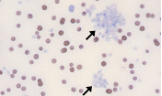
Platelet clumping
Can cause a falsely low platelet count
Platelet clumping causes
Occurs during routine blood collection and indicates normal numbers of platelets

Macroplatelets
Larger than normal, immature platelets
Macroplatelets causes
Produced in response to peripheral thrombocytopenia
Fresh whole blood contents
RBC, WBC, platelets, all coagulation factors, albumin, globulin
Indications for fresh whole blood
Anaemia with coagulopathy/ platelet disorder
Severe haemorrhage requiring massive transfusion
Stored whole blood contents
RBC, WBC, albumin, globulin, non-viable platelets, coagulation factors
Indications for stored whole blood
Blood loss anaemia
Packed RBCs contents
RBC, WBC, non-viable platelets
Indications for packed RBCs
Symptomatic anaemia of any aetiology, especially those at risk of overload
Fresh frozen plasma contents
All coagulation factors, albumin, globulin
Indications for fresh frozen plasma
Coagulopathy
Benefits of component therapy
Allows for longer storage
>1 patient can benefit from one donation
Decreased risk of volume overload + transfusion reactions
Downsides of component therapy
Requires specialised equipment
Commercial availability can be inconsistent
Hard to get a stable source of platelets
Febrile non-haemolytic transfusion reaction
An increase of at least 1ºC from pre-transfusion values within 4 hours of the end of transfusion
Infection, acute haemolysis, TRALI and bacterial contamination have been ruled out
Possibly caused by transfer of inflammatory mediators by stored RBCs
Febrile non-haemolytic transfusion reaction treatment
Generally self-limiting and not life-threatening so no need for antipyretics
Continue transfusion at a slower rate?
Transfusion associated cardiac overload
Circulatory overload due to an increase in blood volume
Acute respiratory distress and pulmonary oedema within 6 hours of transfusion
Diagnosed with radiographic or ultrasonographic evidence
Allergic transfusion reaction
Type I hypersensitivity reaction to antigen within the blood product
Occurs within 4 hours of transfusion
Can be transient/ self-limiting or life-threatening
Allergic transfusion reaction clinical signs
Dogs: erythema, urticaria, pruritic, peripheral angioedema, GI signs (vomiting)
Cats: respiratory distress, GI signs (vomiting)
Allergic transfusion reaction treatment
Non-anaphylactic - antihistamine therapy, stop transfusion and assess patient for severity
Anaphylactic - adrenaline
Acute haemolytic transfusion reaction
Onset of haemolysis within 24 hours of transfusion
Inadequate increase in PCV post-transfusion
With or without new onset of fever, tachycardia or hypotension
Causes of acute haemolytic transfusion reaction
Immunologic - secondary to incompatibility of transfused product to recipient
Non-immunologic - damage to RBCs due to thermal, osmotic, mechanical or chemical factors
Delayed haemolytic transfusion reaction
Unexplained decrease in PCV/ Hb >24h - 28d post transfusion
Delayed onset of two indicators of RBC destruction
Causes of delayed haemolytic transfusion reaction
Xenotransfusion - usually within median 2 days, happens regardless of compatibility
Type A cats receiving type B blood - usually within 2 days
Auto-transfusion
Blood taken from a patient and re-transfused into the same patient
Reduces the risk of transfusion reactions, risk of contamination/ haemolysis
Macroscopic agglutination test
Fresh anticoagulated blood on a glass side - examine for visible clumping
Mix a drop of blood with 1-2 drops of saline - true agglutination persists despite this dilution, whereas rouleaux disperses into individual cells once diluted
Microscopic agglutination test
Saline-diluted blood on a slide and examined under a microscope within 1 minute
True agglutination - irregular, 3D, grape like clustering
Rouleaux - RBCs arranged in linear stacks resembling piles of coins
RBC autoagglutination
Occurs when large amounts of immunoglobulin coat the RBC surface, allowing antibody molecules on adjacent cells to cross-link and clump together
Definitive evidence of IMHA
Sensitivity of RBC autoagglutination test
Useful but incompletely sensitive - a negative result does not rule out IMHA (e.g. lower burden)
Specificity of RBC autoagglutination test
Highly specific - a positive result almost always indicates immune-mediated RBC destruction
Coombs test
Uses polyvalent antisera to detect immunoglobulin and/ or complement bound to the surface of the patient’s own RBCs
Sensitivity of the Coomb’s test
Not particularly sensitive for IMHA
Typically only detects RBC coating once >200-300 molecules per cell are present - lower numbers can still cause clinically important haemolysis
Specificity of the Coomb’s test
Poorly specific
A positive Coombs test can occur in a variety of other inflammatory and infecitous disease
Donor selection for blood
Healthy, calm temperament, age 1-8
No current medications, no prior transfusion, not pregnant
Canine blood types
DEA group system - 13 different DEAs, which dogs typically have a combination of
1.1 is most immunogenic - ‘universal donors’ - only thing we test for
DEA 1.1 donation and receiving rules
Can give 1.1- to 1.1+ and 1.1+ to 1.1+
CANNOT give 1.1+ to 1.1-
Feline blood types
AB system - type A most prevalent
Feline blood receiving rules
Type A should ideally get type A blood, if they get type B blood = mild reaction
Type B HAS TO get type B blood, if they get type A blood = death
Type AB have both antigens, but best to receive type A blood
Crossmatching
To see if donor blood samples are likely to cause a reaction in the recipient
Agglutination = transfusion reaction