Lec4: Antibody Screen and D
 Call Kai
 Learn
 Practice Test
 Spaced Repetition
 Match
 Flashcards
 Knowt Play
1/52

There's no tags or description
Looks like no tags are added yet.
53 Terms
AHG
Anti-Human Globulin
AHG is a prefix and describes the test
AHG Crossmatch
IAT
Indirect Antiglobulin Test
Patient Plasma + Donor Cells + AHG
Indirect Coombs Test
DAT
Direct Antiglobulin Test
Patient Cells + AHG
Direct Coombs Test
Antibody Screen
An IAT that is meant to look patient antibodies to common RBC antigens
AHG is made by injecting
rabbits with human IgG
Unexpected antibodies
Antibodies that is development ONLY
If you been exposed to a blood product
(Ex: transfusion, unclean IV drugs)
Clinically significant antibodies
Antibodies that can cause hemolysis
These are the expected ones
What are sources of alloummunization
Transfusion
Pregnancy
Drugs
Parasites
Disease
IgM is stronger than IgG. So we use anti-
IgM for agglutination. IgG need two steps
What are the two stages of agglutination
Sensitization
Lattice formation
Sensitization
Antibody and Antigen are mixing and binding
Lattice Formation
Antibodies start cross-linking and binding to multiple red cells

Dectecting IgG antibodies steps (indirect)
Recipient’s serum is obtained containing antibodies (Ig’s)
Donor’s blood sample is added to the tube with serum
Recipient’s Ig’s that target the donor’s RBC form antibody-antigen complexes
Anti-human Ig’s (coombs antibodies) are added to the solution
Agglutination of RBC occurs, because human Ig’s are attached to RBC
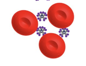
IgM

IgG

Anti-IgG

Describe AHG antisera
Monoclonal or polyclonal
Directed at Ig’s, complement, whatever
Originally from animals
Now commercially made
Depending on manufactuer, can be IgG or IgM
What are some potentiators (ubstance or drug that increases the strength, effectiveness, or duration of action of another substance)
Temperature (at body temp 37C)
Time
Medium
Concentration
pH
Enzymes (ish)

What is Prozone
Occurs when there are far too many antibodies relative to antigens. stopping proper cross-linking
falsely low or false-negative test results

Equivalence
optimum proportions of antigen and antibody

Postzone
Antigen excess. A similar false-negative issue caused by an overwhelming excess of antigen rather than antibody.

Zeta potential
net negative electrical charge at the slipping plane of the cell membrane that causes erythrocytes to repel each other and stay separated in the bloodstream.
What need to be overcome for agglutination
What are ways to enhance the agglutination (based on the lecture)
Standard saline incubation 37C – 30-60 min
LISS - Low Ionic Strength Saline
22% Albumin
PEG - Polyethylene Glycol
Combination of the above
Searching for the perfect combination of specificity and sensitivity
Describe LISS – Low Ionic Strength Saline
0.2% saline
Decreases negative repulsion (Zeta Potential)
Decreases incubation time to 5-15 min
Describe 22% Albumin
Also decreases zeta potential
Incubation time 15-60 min
Describe PEG – Polyethylene Glycol
Decreases water between RBCs
Decreased hydrophobic repulsive force
Brings cells closer together
Incubation time at 37C is 10-30 min
What are the Indirect Antiglobulin Test Principle
Central question for IATs: does this patient have antibodies that could bind to RBCs?
Patient sample (plasma) is the unknown while the screening cells are the known
If antibodies are present, cells will be sensitized
This test is in vitro and does not reflect patient status
Describe the tube method for Indirect Antiglobulin Test
Patient plasma is incubated with screening cells in an enhancement media
Cells are than washed to removed excess IgG
AHG is added and sample is centrifuged
Test is positive if cells agglutinate
If no agglutination is seen, check cells are added and should agglutinate upon centrifugation
Check cells are sensitized RBCs

Was this a good wash?
This picture is a bad wash because it has free floating antibodies lead to a false negative. These are neutralize and are binding to free floating antibodies instead of the RBCs.
What is a another name for check cells
as control cells, Coombs cells
What are check cells
RBCs coated with IgG antibody
Usually Rh pos RBC coated with anti-D
Also available in RBCs coated with C3
They are added to every AHG reaction tube with a negative result
They are a guard against false negatives
Bad washing, no AHG added
What is the appearance for Check cells
Mixed field appearance
Checks will agglutinate, screening cells will not
Two different cell populations
Check cells test are vaild if
they agglutinate
If CC do not agglutinate, then it can be cause by
Bad washing
No AHG added
Test is invalid and must be repeated
What are the pros for tube method?
gold standard, inexpensive, flexible
What are the cons for tube method?
Labor intensive, not stable
Describe the GEL
Patient plasma, LISS and screening cells are incubated in a microtube
Microtube is filled with gel which is laced with AHG
If cells are sensitized, red cells will be held in gel, otherwise will form pellet at bottom
What are the pros of GEL?
Smaller sample size
No cell washing step
Clear, stable endpoint
Sensitivity comparable
Can be automated
What are the cons of GEL?
Expensive
More false positive
What method is this?
Patient plasma, LISS and screening cells added to microtiter well lined with Protein A which has a high affinity for Fc portion of AHG and incubated.
Wash
AHG added
Centrifugation
Graded via computer system.
Positive will show as cells lining entire well
Solidphase
What is solidphase?
Red blood cell membranes or specific antigens/antibodies are bound (immobilized) to the bottom surface of small plastic microplate wells (typically 96-well plates). - all labs are different
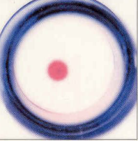
What does this mean in solidphase?
Neg. (solid pellet); tube was 0

What does this mean in solidphase?
tube was 1+

What does this mean in solidphase?
tube was 2+

What does this mean in solidphase?
tube was 3+

What does this mean in solidphase?
Pos. diffuse; tube was 4+
Pros of solidphase?
Reduced volume needed
Increased sensitivity
Less technique needed
cons of solidphase
Learning curve
Expensive
Reagent expiration
Check cells only check for if the
AHG was added or if there was a improper wash
Direct Antiglobulin Test Principle
Central question for DATs : does this patient have antibodies or complement already stuck to their red cells in vivo?
Test Procedure: Washed patient red blood cells + AHG
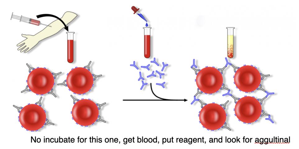
Is this direct or indirect antiglobulin test
Direct Antiglobulin Test
Why use DATS?
Ordered less frequently
Transfusion reactions
Suspected hemolysis
Can tell you if IgG and/or complement is bound to RBC
Neonates:
Do NOT have functional complement
Only test for IgG
Looking for maternal IgG causing hemolysis (HDFN)
Complete redrawing of the thing we did in class (on ipad)
complete