Sect 6 - WBC Disorders
 Call Kai
 Learn
 Practice Test
 Spaced Repetition
 Match
 Flashcards
 Knowt Play
1/67

There's no tags or description
Looks like no tags are added yet.
68 Terms
< 1 week
lifespan of neutrophils
2 weeks
lifespan of eosinophils
12-18 months
lifespan of basophils
years
lifespan of T-cells
few months
lifespan of B-cells
3 days
lifespan of monocytes
Myeloblast
Promyelocyte
Myelocyte
Metamyelocyte
Band
Segmented Neutrophil
6 stages of neutrophil maturation
Myeloblast
cytoplasm: blue
no granules
nucleoli present in nucleus
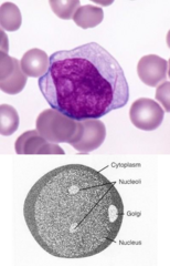
Promyelocyte
Nucleus: round or oval
Nucleoli: present in nucleus
Cytoplasm: pale blue with dark-purple to red-purple
Non-specific primary granules

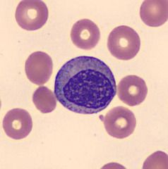
Myelocyte
Nucleus: circular-shaped
No nucleoli present
Granules: specific secondary granules present
last stage of myeloid cell division
Metamyelocyte
Nucleus: kidney-bean shaped
Granules: pink-purple present in cytoplasm

Band
Nucleus: horse-shoe shaped
Granules: pink-purple present in cytoplasm

Segmented neutrophil
Nucleus: segmented
Granules: pink-purple present in cytoplasm

storage pool
marginal pool
circulating pool
WBCs can be found in 3 places:
storage pool
WBCs found in bone marrow
marginal pool
WBCs found in walls of the blood vessels in lungs, liver and spleen
circulating pool
WBCs found in peripheral blood
conditions with Neutrophilia
Leukemoid reactions
Bacterial infections
Chronic myelogenous leukemia (CML)
Myelofibrosis
Pregnancy
conditions with Neutropenia
Viral illnesses
Malaria
Thyroid disorders
Aplastic anemia
Typhoid fever
conditions with Eosinophilia
Parasitic infections
Allergic reactions
Connective tissue disorders
Asthma
CML
Hodgkin's lymphoma
Polycythemia vera
Myelofibrosis
conditions with Eosinopenia
Shock
Trauma
Surgery
Administration of Corticosteroids/Epinephrine
conditions with Basophilia
Allergic reaction
Basophilic leukemia
Polycythemia vera
CML
Myelofibrosis
conditions with Basopenia
Generally not an issue
Granulocytopenia
Granulocytosis
Leukemias
3 Granulocyte Disorders
Granulocytopenia
too few granulocytes/myelocytes
causes of Granulocytopenia
1. Reduced granulopoiesis with myeloid hypoplasia
2. Increased ineffective granulopoiesis
3. Reduced granulocyte survival
combo of all three mechanisms
What is the most common cause of Granulocytopenia?
Leukemoid reaction
Increased WBC count that can mimic leukemia, usually caused by bacterial infection
LAP score >118 units
Granulocytosis
too many granulocytes/myelocytes
Acute and Chronic
2 types of Granylocytosis
Acute Granulocytosis
increase granulocytes in a quick amount of time -- rxn happens in a matter of min
stress, exercise, administration of epinephrine
3 causes of Acute Granulocytosis
mechanisms of Acute Granulocytosis
increased marrow storage pool shift (coming out of bone marrow into circulation)
increased marginal pool shift
Chronic Granulocytosis
increase granulocytes caused by recurrent or sustained physiological stimulus
ex: pregnancy, pinworm
mechanisms of Chronic Granulocytosis
increased proliferation and production from the bone marrow
Increased granulocyte survival
Decreased movement from the circulating pool
Leukemias
abnormal cells
Eosinophilia
NAACP is related to:
Neoplasms
Allergic Reactions
Asthma
Connective Tissue Disorders
Parasitic infections
What does NAACP stand for?
heparin and histamine
The blue/black granules of basophils contain
Basophilia
Allergic reaction
Basophilic leukemia
Polycythemia Vera
Chronic Myelogenous Leukemia (CML)
Myelofibrosis
lab tests to evaluate Granulocytosis
Bacterial, Viral, Ricketsial Parasitic studies
CBC
Bone marrow
LAP
Chromosome studies
Pelger-Huet
May-Hegglin Anomaly
Chediak-Higashi Syndrome
Alder-Reilly Anomaly
Chronic Granulomatous Disease
Name the five inherited WBC disorders
neutrophils
What cell are the 5 WBC disorders commonly found in?
Pelger-Huet
cells called Pence-Nez
no hematological disorders
no physical characteristics
inherited autosomal dominant

hypo-segmentation
characteristics of neutrophil in Pelger-Huet
Pelgeroid
an acquired form of Pelger-Huet that can be induced by drugs or seen in leukemia (not true inherited)
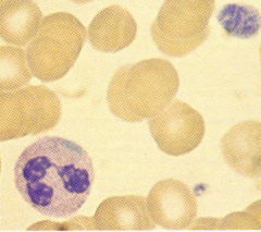
May-Hegglin anomaly
anomaly that presents with low WBC, low and deficient PLTs
inherited autosomal dominant
Dohilod bodies & giant bizarre platelets
Inclusion that can be identified in May-Hegglin anomaly
Mild to severe hemorrhagic disease (bleeding and epistaxis)
No physical characteristics
Clinical features of May-Hegglin anomaly
Chediak-Higashi syndrome
the ineffective killing of bacteria due to impaired release of enzymes by enlarged granules
CHS1 gene
gene mutation associated with Chediak-Higashi syndrome
inherited autosomal recessive
Clinical features of Chediak-Higashi syndrome
Partial ocular cutaneous albinism
Seizures
Immunocompromised
Recurrent infections
WBC and platelet count decreased
Lab Features of Chediak-Higashi Syndrome
death usually occurs before early teens from recurrent pyogenic infections
What is the prognosis of Chediak-Higashi syndrome?
abnormal granulation of both granulocytic and lymphocytic cells
Chediak-Higashi syndrome morphology

primary and secondary granules fuse together
Granulocytic cell characteristics seen in Chediak-Higashi syndrome
azuerophilic granules in NK cells
Lymphocytic cell characteristics seen in Chediak-Higashi syndrome
Alder-Reilley anomaly
caused by a metabolic disorder of mucopolysaccharides
autosomal inheritance -- very rare
Hurlers
other name for Alder-Reilly
characteristics of Alder-Reilly anomaly
Lysosomal storage disorder with giant granules in neutrophils, lymphocytes, and monocytes
Associated with gargoylism
Chronic Granulomatous Disease
Condition where bacteria are ingested but not killed
Cells can't produce hydrogen peroxide in conjunction with other oxygen metabolites which are necessary to kill microorganisms
inherited autosomal recessive or sex-linked recessive
catalase positive organisms
CGD is associated with infections caused by
Catalase positive organisms
Staph
Salmonella
Nocardia
Pseudomonas
Listeria
Aspergillus
Candida
E. coli
Serratia.
micro-abscess and granuloma formation
CGD infections are characterized by
septicemia or chronic pulmonary disease in early childhood
What is the common cause of death in Chronic Granulomatous Disease?
Nitro Blue-Tetrazolium Test
What does NBT stand for?
NBT
Semiquantitative assay that tests phagocytic function of polymorphonuclear leukocytes
Normal: cells reduce colorless NBT into blue-black deposit within the cell

negative/no color change
results in NBT for a patient with CGD