Exam 3: CT Protocol
 Call Kai
 Learn
 Practice Test
 Spaced Repetition
 Match
 Flashcards
 Knowt Play
1/48

There's no tags or description
Looks like no tags are added yet.
49 Terms
A patient comes in a CT of the chest. The reason for the exam on the requisition states: abnormality seen on chest x-ray". What exam protocol is the most appropriate?
CT Chest with contrast
______________ protocol of the chest is used for the assessment of lung parenchyma in patients with diffuse lung disease such as fibrosis and emphysema.
High-Resolution Chest CT
Most pulmonary emboli are caused from thrombi originating in the ____________ known as deep vein thrombosis (DVT).
Lower extremities

What thoracic pathology is shown on the image?
Pulmonary nodule
High resolution CT protocols may include three series of scans: inspiratory supine, expiratory supine, and ______________.
Inspiratory prone

What anatomy is displayed by the letter 'A' on the image?
Right coronary artery
This lab test is considered valuable when diagnosing a patient with PE. It is an inexpensive screening tool. If this lab test is within normal limits, a PE is highly unlikely. However, if the lab test is positive, it doesn't necessarily indicate that the patient has a PE. What lab test is this?
D-dimer

What thoracic pathology is shown on the image?
Pulmonary embolism
It is recommended for protocols to detect PE scan are scanned in a caudal to cranial direction. Why is this recommended?
To reduce respiratory motion in the bases of the lungs
One complete cardiac cycle and is sometimes referred to as the ____________.
R-R interval

What thoracic pathology is shown on the image?
Lung mass

Which method of ECG tracing is depicted in the image?
Prospective ECG gating
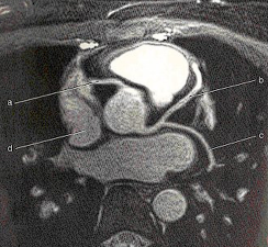
Based on the image, what thoracic exam was performed?
CTA for Coronary Arteries

What is labeled as 'C' on the image?
Left ventricle
Which of the following is incorrect about a High-Resolution Chest CT.
It requires contrast.
Accurate demonstration of _______________ would most likely require the intravenous injection of an iodinated contrast agent during a CT study of the chest.
Pulmonary embolism
CT images of a routine chest should be acquired with the patient:
At full inspiration
Which of the following pathologic processes may be considered an interstitial disease of the lung?
Bronchiectasis
Which of the following is a lung window:
WW 2000
WL -600
The use of a saline flush after the injection of the iodinated contrast is recommended for a CTA pulmonary artery study. Why?
To reduce or eliminate beam hardening artifacts from dense contrast media within the superior vena cava that may obscure small emboli in adjacent vessels
In addition to imaging the pulmonary arterial system for patients with suspected PE, CT venography is sometimes performed to assess for venous thrombosis within the pelvis and lower extremities. How is this accomplished?
A second scan series is obtained in a delayed venous phase approximately 180 seconds after IV contrast injection from the iliac crest to the knees.
Which is not a technical parameter used in CT imaging of the chest?
Thick slices
Which of the following pharmaceuticals may be administered prior to a cardiac CT procedure in an effort to improve visualization of the coronary vessels?
1. beta blockers
2. nitroglycerin
3. metformin
1 and 2 only
What does a CT calcium score of 1-10 indicate?
Minimal plaque
Beta-blockers are used as part of a CT cardiac protocol to lower the heart rate to less than how many beats per minute (bpm)?
65-70 bpm
Cardiac computed tomography applications include all of the following except:
Assessments of aspiration
For high resolution CT of the thorax _____________ images can help to differentiate actual disease from densities owing to the effects of gravity that mimic disease.
Prone
Which cavity of the heart sends blood only to the lungs?
Right ventricle
Which helps to reduce artifacts created by respiratory motion?
Utilizing the shortest scan time possible
Why is it recommended that cardiac CT calcium scoring be restricted to patients with risk factors for coronary artery disease, rather than using it as a screening tool for all patients?
Risks related to the radiation dose are a concern.
For a routine CT of the chest, the range of suggested IV contrast flow rates is:
2.0ml/s to 4.0ml/s
Match the common ECG gating techniques below:
Acquires images only during selected phases of the cardiac cycle
Prospective ECG Triggering
Match the common ECG gating techniques below:
Continuously acquires images throughout the cardiac cycle, allowing reconstruction at desired phases
Retrospective ECG Gating

Which of the following thorax pathologies is indicated on the image below?
Aortic dissection
The start location for a routine chest CT is ______________ and the end location for a routine chest CT is ___________________.
Just above the apices of the lungs, just below costophrenic angles
Choose all of the indications below that would warrant a CT of the chest:
Abnormality on Chest X-ray
Pulmonary Nodules
Lung Cancer/Metastasis
Pulmonary Embolism
Aneurysm/Dissection
Interstitial Bronchiectasis
Cardiac computed tomography applications include all of the following except:
Gated stress imaging
Retrospective ECG gating utilizes which of the following portions of the cardiac cycle for image reconstruction?
1. atrial systole
2. ventricular systole
3. cardiac diastole
3 only
What is cardiac gating?
A technique for cardiac CT acquisition that attempts to use only those images acquired during periods of lowest cardiac motion
What is the leading cause of cancer deaths in the U.S?
Lung cancer
What are common symptoms that appear later in patients with lung cancer? What are the common sites of metastasis?
Symptoms: Cough, hemoptysis, chest pain
Common sites: Brain, liver, and skin
Match description:
A localized dilation of the aorta
Aortic Aneurysm
Match description:
A tear in the aortic wall allows blood to flow between layers, separating them.
Aortic Dissection
What is the center, start location, and end location for a routine chest?
Center: @ level of shoulders
Start: Just above apices of lungs
End: Just below costophrenic angles of the lungs
What is the center, start location, and end location for a CTA for PE?
Center: @ level of shoulders
Start: Just below costophrenic angles of the lungs
End: Just above apices of lungs
How much IV contrast is typically administered for the CTPA?
75-100 mL
What are the reconstruction slice thickness and interval for CTA PE?
1.25 / 0.625 mm
Where is the ROI placed for bolus tracking during CTA for PE?
Main pulmonary artery
What is the center, start location, and end location for cardiac calcium scoring?
Center: @ level of shoulders
Start: Just below carina
End: Just below apex of the heart