A&P - Chapter 8: Movement
 Call Kai
 Learn
 Practice Test
 Spaced Repetition
 Match
 Flashcards
 Knowt Play
1/102

Earn XP
Description and Tags
long cards...
103 Terms
Gliding movement
occurs in plane joints
slight movement
angular movement
angle between bones changes
circular motion
rotation around an axis
flexion
angular movement
decreases angle between two bones
movement of a body part anterior to the coronal plane for the top half of the body
extension
angular movement
straightening movement that increases angle of two bones
movement of a body part posterior to the coronal plane for the top half of the body
hyperextension
angular movement
extension of joint beyond anatomical position
can be normal or caused by injury
plantar flexion
angular
flexion of the foot
movement of the foot toward plantar surface
EX: standing on toes
dorsiflexion
angular
flexion of the foot
movement of the foot toward the shin
EX: walking on the heels
abduction
angular motion
movement away from the midline
abducted by aliens
adduction
angular motion
movement toward the midline
rotation
circular motion
turning of structure around long axis
EX: shaking head no
pronation
circular motion
rotation of the forearm
prone = facedown
palm faces posteriorly in anatomical position
radius and ulna are crossed
supination
circular motion
rotation of the forearm
supine = faceup
palms face anteriorly in relation to anatomical position
radius and ulna are parallel
circumduction
Combination of flexion, extension, and abduction and adduction
occurs at freely movable joints like the shoulder
circular motion
arm moves in an arc as if tracing the base of a cone
special movements
Movements unique to only one of two joints
elevation
special movement
moves the structure superiorly
EX: shrugging shoulders
depression
special movement
move structure inferiorly
EX: opening the mouth
protraction
special movement
Gliding motion that moves the structure in an anterior direction
EX: jutting out the jaw
retraction
special movement
Gliding motion that moves the structure in a posterior direction
EX: pulling the scapulae back toward the vertebral column
lateral excrusion
special movement
moving mandible either right or left of the midline
medial excrusion
special movement
moving the mandible back to the midline position
opposition
special movement
unique to the thumb
thumb + tio of finger on same hand are brought toward each other across the palm
reposition
special movement
returns thumb to neutral anatomical position
inversion
special movement
turns ankle so that the plantar surface of foot faces medially toward the opposite foot
weight on the outside edge of the foot
rolling out
eversion
Turns the ankle so the plantar surface of the foot faces laterally
weight is on the inside edge of the foot
rolling in
range of motion
describes amount of mobility that can be given
active range of motion
amount of movement that can be accomplished by contracting muscles that normally act across a joint
passive range of motion
amount of movement that can be accomplished when structures that meet at the joint are moved by an outside force
Chad doing his PT and forcing a patient to flex their arm
passive for normal joints is greater than active
dislocation
caused by movement of joint beyond normal range of motion
AKA luxation
occurs when articulating surfaces of bone are moved out of proper alignment
results in damage to supporting ligaments + articular cartilage
subluxation
a partial dislocation
sprain
occurs when ligaments are damaged
degree can range from stretched to completely torn
results in inflammation, swelling, and pain
what influences range of motion for a given joint
shape of articular surfaces of the bones forming the joint
amount and shape of cartilage covering those articular surfaces
strength + location of ligaments, tendons, and muscles associated with the joint
bulk of surrounding tissues like muscle and adipose tissue
amount of fluid in and around the joint
amount of pain in and around the joint
amount of use or disuse the joint has received over time
abnormalities that impact range of motion
damage to ligament associated with a joint can increase range of motion
torn piece of cartilage within a joint can limit its range of motion
nerve supply to a muscle is damaged —> muscle is weakened so the active range of motion may decrease but the joint passive range is unchanged
fluid buildup and/or pain in or around the joint can limit both active + passive ranges
Occurs when soft tissue around joint develops edema following an injury
Disuse can decrease both active + passive
joints
AKA articulations
Named according to bones or portions of bones that join together
EX: temporomandibular joint is between temporal + mandible
three types of joints
fibrous
cartilaginous
synovial
*named according to the major CT type that holds bone together + the presence of a fluid-filled joint capsule
functional categories of joints
according to degree of motion
synarthroses
amphiarthrosis
diarthrosis
synarthroses
nonmoveable joint
fibrous + cartilaginous joints
amphiarthrosis
slightly movaeble joints
fibrous + cartilaginous joints
diarthrosis
freely moveable joint
synovial joints
fibrous joints
connection between two bones that are held together by fibrous CT
have no joint cavity + exhibit little or no movement
three subdivisions
sutures
syndesmoses
gomphoses
sutures
seams found only between the bones of the skull
some sutures become completely immovable in older adults
Few are smooth, and the opposing bones often interdigitate (interlocking finger-like processes)
Interdigitation adds stability
margins of bones within sutures are sites of continuous intramembranous bone growth that eventually become ossified
When a suture fully becomes ossified, it becomes a synostosis
results when two bones grow together across a joint to form a single bone
fontanels
membranous area on a suture in newborns
soft part of the skull that is flexible for the birthing process + allows for growth of the head after birth
suture tissue between each bone
dense, regular collagenous CT
periosteum on inner + outer surfaces of adjacent bones continues over the joint
two layers of periosteum + dense fibrous CT in between for a sutural ligament
syndesmosis
a slightly movable type of fibrous joint
Bones are farther apart than in a suture
Two bones are joined by ligaments (sheets of CT - collagen = interosseous membrane)
some movement can occur because ligaments are flexible
occurs in the radioulnar syndesmosis that binds the radius + ulna together
movement can occur in one direction but not other
gomphosis
specialized joints that consists of pegs that fit into sockets + held in place by fine bundles of regular collagenous CT
only located between teeth + sockets of the mandible + maxillae
CT bundles between the teeth + sockets are periodontal ligaments
allows a slight amount of “give” to the teeth during mastication
allows realignment by braces
cartilaginous joints
Holds two bones together by a pad of cartilage
exhibits little to no movement
two subdivisions based on the type of cartilage:
synchondroses
hyaline cartilage
symphyses
fibrocartilage
synchondrosis
consists of two bones joined by hyaline cartilage with little or no movement occuring
most are temporary
EX: epiphyseal plate in growing bones
synchondrosis is converted to a synostosis as bone replaces existing cartilage
synchondrosis can be converted to synovial joints
EX: costochondral joints located between the ribs + costal cartilages
The change occurs because movement occurs between the ribs/costal cartilage and the sternum
EX: breathing
symphysis
consists of two bones joined by a pad of fibrocartilage
EX: intervertebral disks, junction between the manubrium + body of the sternum, pubic symphysis
the fibrocartilage is meant to withstand a lot of force and absorb shock
made out of a lot of organized cartilage
synovial joints
typically unite bones of appendicular skeleton
covered in a thin layer of hyaline cartilage (articular cartilage)
provides a smooth surface where bones meet
The joint capsule that surrounds the joint has two layers
fibrous capsule
synovial membrane
only blood supply and nerve supply to the synovial membrane
how does articular cartilage receive nourishment
BV that form a vascular circle at peripheral margin of cartilage but do not penetrate the cartilage or enter joint cavity
nutrients and O2 must diffuse from surrounding vessels into cartilage due to lack of innervation
may receive nutrients from underlying spongy bone + synovial fluid covering the cartilage
sensory nerves of joints
enters fibrous capsule + synovial membrane (but less)
supplies brain with information about pain + sends information about joint position + degree of movement
does not enter cartilage or joint cavity
joint cavity
the space around the articular surfaces of bones in a synovial joint
filled with synovial fluid
includes hyaluronic acid which makes it slippery which prevents friction
includes proteins + fats
surrounded by joint capsule
helps bones together while still allowing movement
has two layers: fibrous capsule (outer), synovial membrane (inner)
fibrous capsule
the outer layer of the joint capsule
consists of dense irregular CT + continuous with fibrous layer of periosteum that covers the bones united at the joint
portions may thicken + collagen fibers may be regularly arranged to form ligaments
ligaments + tendons can be present outside fibrous capsule which contributes to the strength + stability of joint while limiting movement in some directions
synovial membrane
inner layer of the joint capsule
lines the joint cavity except over the articular cartilage + articular disks
thin delicate membrane
consists of a collection of modified CT, either intermixed with a part of the fibrous capsule or separated from it by a layer of areolar tissue or adipose tissue
fat pads
prominent areas of adipose tissue that lie between fibrous capsule + synovial membrane
helps protect articular cartilage by acting as a cushion around the joint
EX: elbow + knee
synovial fluid
viscous lubricating film that covers the surface of a joint
complex mixture of polysaccharides, proteins, lipids, and cells derived from serum filtrate (serum = blood fluid) and secretions from synovial cells
Hyaluronic acid is the major polysaccharide that provides much of the slippery consistency + lubricating qualities of synovial fluid
bursae
pockets of synovial membrane + fluid that extend from joint
found in areas of friction
tendons rubbing on bones or other tendons
subcutaneous olecranon bursae
provides cushion between skin + underlying bony prominences where friction could damage the tissue
bursitis: inflammation of bursa
ligaments +tendons
used for stabilization in joints
articular discs
flat pad of fibrocartilage that lies in between the articular cartilages of bone
the circumference of the disk is attached to fibrous capsule
helps adjust the connection between articulating bones which improves joint stability + reduces wear of articular cartilage
EX: temporomandibular, sternoclavicular, acromioclavicular
menisci
fibrocartilaginous pads in the knee
only partially spans synovial cavity where there is an opening in the center
tendon sheath
synovial sacs that surround tendons as they pass near or over the bone
type of bursae
type of synovial joints
plane
saddle
hinge
pivot
ball-and-socket
ellipsoid
three types of movement in synovial joints
uniaxial
biaxial
multiaxial
uniaxial movement
occurring around one axis
biaxial
occurring around two axes at right angles to each other
multiaxial
occurring around several axes
plane joints
consist of two flat bone surfaces of about equal size where a slight gliding motion occurs between
facet joints
considered uniaxial
some rotation is possible
limited by ligaments and adjacent bone

examples of plane joints
acromioclavicular joint
carpometacarpal joints
articular processes between vertebrae
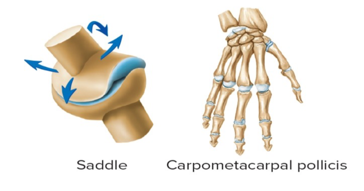
saddle joints
shape of a saddle - convex + concave movement
biaxial
the two saddle-shaped articulating surfaces oriented at right angles to one another so their complementary surface articulates
example of saddle joints
carpometacarpal joint of the THUMB
sternoclavicular joint
hinge joint
rare —> can be found at the joint at the elbow
uniaxial
convex cylinder in one bone is applied to a corresponding concavity in the other bone
the only hinge joint in the entire body
knee somewhat counts but also not.

pivot joints
a relatively cylindrical bony process that rotates within a ring composed partly of bone + ligament
uniaxial
restricts movement to a rotation around a single axis

examples of pivoit joints
proximal radioulnar joint
C1 + C2
ball-and-socket joint
socket that articulates with the ball head
multiaxial joint
wide range of motion in almost any direction
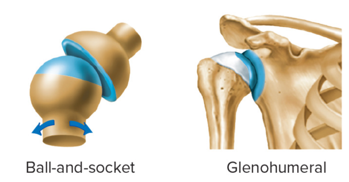
examples of ball-and-socket joints
glenohumeral joint
coxofemoral joint
ellipsoid joint
two slightly rounded joints on top of one another
AKA condyloid joint
modified ball-and-socket joint
articular surface is in an ellipsoid in shape
biaxial
the shape of joint limits the range of movement almost to a hinge motion in two axes + restricts rotation

examples of ellipsoid joints
atlantooccipital joint of neck
joint between C1 and C2
metacarpophalangeal joints
temporomandibular joint
between temporal bone + mandible
mandibular condyle fits into the mandibular fossa of the temporal bone
fibrocartilage articular disk is located between mandible + temporal bone
divides joint into superior + inferior joint cavities
articular disk that may pop
surrounded by fibrous capsule where the articular disk attaches at its margin
strengthened by lateral + accessory ligaments
combination of planar + ellipsoid joint
mostly ellipsoid

process of moving the temporomandibular joint
opening of jaw involves inferior cavity of TMJ below the articular disk
mandibular condyle rotates anteriorly on disk in the hingelike movement of jaw
second portion of jaw opening occurs in superior cavity of TMJ
articular disk + condyle glide together anteriorly
similar to the motion that occurs in protraction of mandible
medial lateral movements of the mandibular condyle allow lateral excursion (side-to-side motion of the jaw)
allows for chewing + grinding in mastication
glenohumeral joint
ball-and-socket
less stability but more mobility than the one in the hip
flexion, extension, abduction, adduction, rotation, and circumduction can occur here
rounded head of humerus articulates with glenoid cavity of the scapula
subacromial bursa and subscapular bursa are present
the stability of this joint in maintained by four sets of ligaments + four muscles
four muscles = rotator cuff
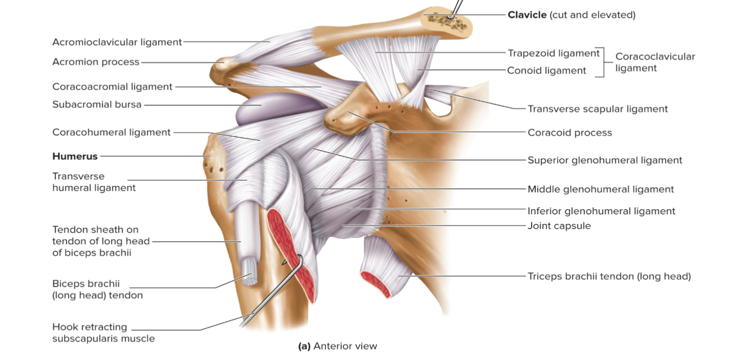
rotator cuff of glenohumeral joint
holds the humeral head tightly within glenoid cavity
head of humerus is also supported with tendon from biceps brachii muscle against the glenoid cavity in the anterior part of the arm
this tendon passes through articular capsule of shoulder joint before crossing the head of the humerus + attaching to scapula at the supraglenoid tubercle
subacromial bursa of glenoid cavity
near the joint cavity but is separated from the cavity by the joint capsule
subscapular bursa of glenoid cavity
opens into joint cavity
glenoid labrum
the rim of the glenoid cavity that has a ring of fibrocartilage
dislocation of glenohumeral joint
most common injury of the shoulder
because major ligaments that cross the superior part of the shoulder joint but no major ligaments or muscles in the inferior side
easy to dislocate inferiorly
can result in damage to nerves and arteries
elbow joint
AKA cubital joint
compound hinge joint
consists of humeroulnar joint and humeroradial joint and proximal radioulnar joint
surrounded by a joint capsule
prevents abduction of the elbow
ulnar collateral ligament (UCL) prevents this on the medial side
radial collateral ligament (RCL) prevents this on the lateral side
has olecranon bursa

humeroulnar + humeroradial joint
flexion + extension due to the shape of trochlear notch + trochlea interaction
proximal radioulnar joint
pronation + supination occur due to the head of radius rotates in the radial nothch of ulna + against capitulum
olecranon bursa
subcutaneous and covers up the proximal + posterior surfaces of the olecranon process
hip joint
coxofemoral joint
femur of head articulates with the relatively deep, concave acetabulum of hip bone
iliofemoral ligament can support the entire weight of the body
hard to disarticulate
has acetabular labrum
has transverse acetabular ligament
fovea capitis holds a ligament from acetabulum to femur
houses BV
capable of flexion, extension, abduction, adduction, rotation, and circumduction
has a strong joint capsule that is reinforced by several ligaments + extends from the rim of the acetabulum to the neck of the femur

acetabular labrum
located on the lunate surface of acetabulum
lip of fibrocartilage
transverse acetabular ligament
crosses the acetabular notch on the inferior edge of acetabulum
strengthens + deepens the acetabulum
ligament of the head of the femur
located inside the hip joint between the femoral head + acetabulum
does not contribute much toward strengthening the hip joint
carries a small nutrient artery to the head of the femur in most people

knee joint
modified hinge joint
forms a complex ellipsoid union that allows flexion, extension, and small leg rotation
distal femur has two large ellipsoid surfaces with a deep fossa between them
femur only articulates with tibia NOT fibula
fibula articulates with the lateral side of tibia
margin of tibia are built up by menisci
four major ligaments that stabilize the knee
joint strength is also provided by popliteal ligaments + tendons of thigh muscles that extend around the knee
has suprapatellar bursa

four ligaments that stabilize the knee
anterior cruciate
posterior cruciate
medial collateral
lateral collateral
suprapatellar bursa
largest bursae of the knee
superior extension of the joint capsule that allows the anterior thigh muscles to move over the distal end of the femur
other bursa in the knee
subcutaneous prepatellar bursa + deep infrapatellar bursa
popliteal bursa
gastrocnemius bursa
subcutaneous infrapatellar bursa
anterior cruciate
extend between intercondylar eminence of tibia + fossa of femur
prevents anterior displacement of tibia relative to the femur
posterior cruciate
extend between intercondylar eminence of tibia + fossa of femur
prevents posterior displacement of the tibia
medial collateral
stabilizes the medial side of knee