Ch 12: The Somatic Sensory System: Touch, and Pain
0.0(0)
Studied by 0 people Call Kai
 Learn
 Practice Test
 Spaced Repetition
 Match
 Flashcards
 Knowt Play
Card Sorting
1/57

Earn XP
Description and Tags
FNTB Exam 3
58 Terms
1
New cards
What are mechanoreceptors, and what types of sensory information do they detect?
Somatic sensory receptors that respond to physical distortion of the skin (indentation, stretch, bending, vibration). Low-threshold: activated by even light mechanical force. Detect touch, pressure, vibration, stretch, and texture. Skin mechanoreceptors use Aβ axons (6-12 µm, 35-75 m/s).
2
New cards
What are the major types of mechanoreceptors in the skin, and where are they located?
Merkel's disks: epidermal-dermal border (hairy and glabrous skin). Meissner's corpuscles: upper dermis of glabrous skin. Pacinian corpuscles: deep dermis/subcutaneous tissue, onion-like layered capsule. Ruffini's corpuscles: deeper dermis. Also hair follicle receptors, free nerve endings, and Krause end bulbs.

3
New cards
What does a Merkel's disk detect?
Small receptive field, slowly adapting. Signals sustained light pressure, fine spatial detail, edges, and texture.
4
New cards
What does a Meissner's corpuscle detect?
Small receptive field, rapidly adapting. Signals light touch, skin movement across the surface, low-frequency vibration (flutter), and slip of a held object.

5
New cards
What does a Pacinian corpuscle detect?
Large receptive field, rapidly adapting. Signals high-frequency vibration and rapid pressure transients, even from deep tissue.

6
New cards
What does a Ruffini's corpuscle detect?
Large receptive field, slowly adapting. Signals sustained pressure and skin stretch (helps sense finger/hand shape and position).
7
New cards
What is a receptive field?
The patch of skin where a stimulus changes the firing of a particular sensory neuron (found by touching the skin with a probe while recording from the nerve).
8
New cards
How does receptive field size influence touch sensitivity?
Small receptive fields (Merkel, Meissner) give high spatial resolution/precise localization (e.g., fingertips). Large receptive fields (Pacinian, Ruffini) give poor localization but broad sensitivity; better for detecting that something happened than where.
9
New cards
What is the difference between rapidly adapting and slowly adapting mechanoreceptors?
Rapidly adapting: fire at stimulus onset and offset, silent during steady pressure; signal change/movement. Slowly adapting: keep firing throughout a sustained stimulus; signal ongoing contact, pressure, or stretch.
10
New cards
How do receptive field size and adaptation rate differ among the four major cutaneous mechanoreceptors (Fig. 12.3)?
Meissner's: small RF, rapid. Pacinian: large RF, rapid. Merkel's: small RF, slow. Ruffini's: large RF, slow.

11
New cards
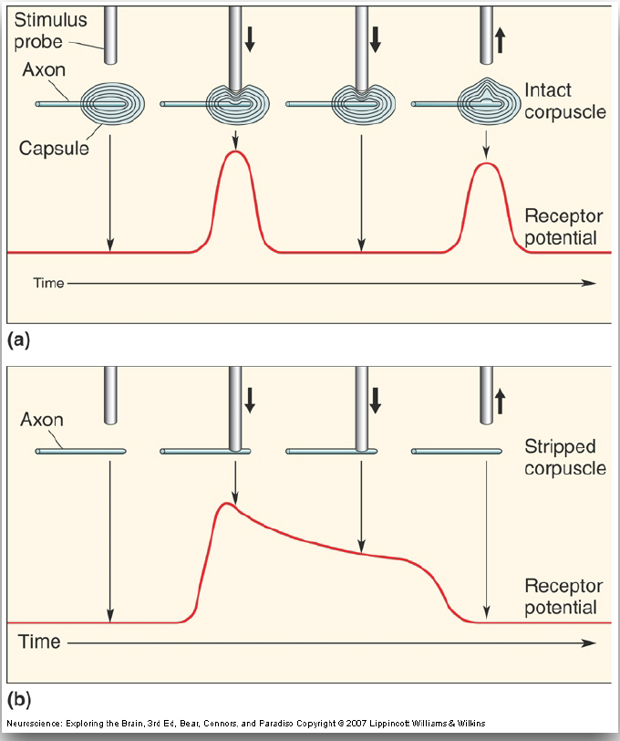
What explains the rapid adaptation of the Pacinian corpuscle?
Largely its layered capsule, which acts as a mechanical filter. Intact corpuscle: brief receptor potential only at probe onset and release. Stripped corpuscle (bare axon): sustained receptor potential for as long as the probe is down.
12
New cards
What is two-point discrimination?
The smallest distance at which two simultaneous touches are perceived as two separate points rather than one; a measure of tactile spatial acuity.

13
New cards
Why does two-point discrimination sensitivity differ across body regions?
(1) Receptive field size and receptor density: fingertips/lips have many receptors with small RFs, so nearby points activate different afferents; wrist, back, calf have large overlapping RFs. (2) Cortical magnification: hand, fingers, and lips have disproportionately large S1 representations. Example: index finger/thumb a few mm; back ~42 mm.

14
New cards
What is mechanotransduction, and why is it important for touch?
Conversion of a mechanical force (pressure, bending, stretch) into an electrical signal in a sensory neuron. It is the first step of touch; without it, skin contact would never become neural activity.
15
New cards
What are mechanosensitive ion channels, and where are they located?
Membrane proteins that open in response to mechanical force. Located in the plasma membrane of mechanoreceptor sensory nerve terminals. Nonselective cation channels (Piezo2 is a key touch channel in mammals).
16
New cards
How can pressure, stretching, or bending of a cell membrane activate mechanosensitive channels (Fig. 12.5)?
Tethered model: protein tethers link the channel to extracellular proteins and intracellular cytoskeleton; force pulls the channel open. Lipid bilayer model: force changes membrane tension/curvature around the channel and opens it directly.

17
New cards
What happens to ion movement when mechanosensitive channels open?
Na+ and Ca2+ flow into the cell down their electrochemical gradients (nonselective cation channels), producing net inward positive charge.
18
New cards
How does opening mechanosensitive channels change the membrane potential?
Inward cation current depolarizes the sensory terminal, producing a graded receptor potential. Size depends on how many channels open (stronger deformation = larger receptor potential).
19
New cards
What is the relationship between a receptor potential and action potential generation?
The receptor potential is graded and local. If it depolarizes the axon's spike-initiating region to threshold, voltage-gated Na+ channels open and action potentials fire. Larger receptor potential = higher firing rate (intensity coded as spike frequency); below threshold = no spikes.
20
New cards
How does the nervous system use mechanoreceptor signals to detect mechanical stimulation?
Which neurons fire = receptor type and location (receptive field, somatotopy). Firing rate = intensity. Timing/adaptation = onset, offset, vibration, duration. Population pattern across skin = shape and texture. Signals travel via the dorsal column-medial lemniscal pathway to the thalamus (VP) and S1.
21
New cards
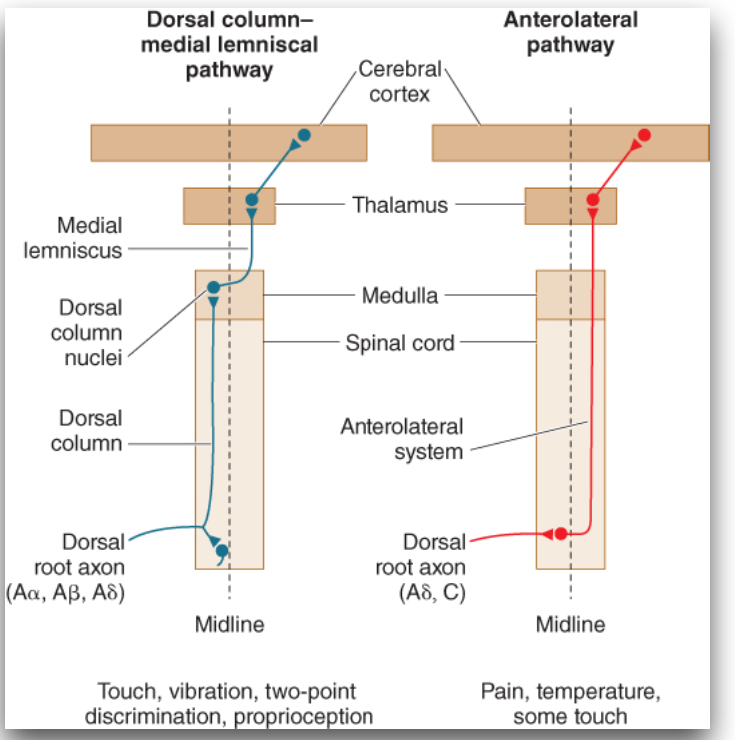
What are the two major ascending somatosensory pathways?
(1) Dorsal column-medial lemniscal (DCML) pathway. (2) Anterolateral pathway (anterolateral system). Both carry information to the thalamus and then the cerebral cortex, preserving distinct types of sensory information.
22
New cards
What sensory information does the DCML pathway transmit?
Fine touch, vibration, two-point discrimination, and proprioception. Uses large fast axons (Aα, Aβ, some Aδ) from skin mechanoreceptors and muscle proprioceptors.
23
New cards
What sensory information does the anterolateral pathway transmit?
Pain, temperature, itch, and some (crude) touch. Uses small slow axons (Aδ and C) from nociceptors and thermoreceptors.
24
New cards
What are the axon types and properties for Aα, Aβ, Aδ, and C fibers (Fig. 12.11)?
Aα (Group I): 13-20 µm, 80-120 m/s, muscle proprioceptors. Aβ (II): 6-12 µm, 35-75 m/s, skin mechanoreceptors. Aδ (III): 1-5 µm, 5-30 m/s, pain and temperature. C (IV): 0.2-1.5 µm, 0.5-2 m/s, temperature, pain, itch.

25
New cards
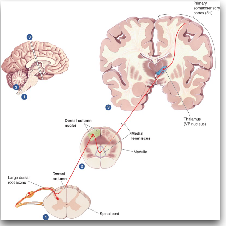
What are the major anatomical structures of the DCML pathway (Fig. 12.16)?
(1) First-order neuron: cell body in dorsal root ganglion; large axon enters dorsal root and ascends ipsilaterally in the dorsal column (no cord synapse). (2) Dorsal column nuclei in the medulla (first synapse). (3) Medial lemniscus (crossed second-order axons). (4) VP nucleus of the thalamus. (5) Primary somatosensory cortex (S1).
26
New cards
What are the major anatomical structures of the anterolateral pathway (Figs. 12.29, 12.31)?
(1) First-order neuron: dorsal root ganglion; small Aδ/C axon enters dorsal root (zone of Lissauer) and synapses in the dorsal horn (substantia gelatinosa). (2) Second-order neuron crosses and ascends in the anterolateral columns/tract through medulla and brainstem. (3) Thalamus: intralaminar and VP nuclei. (4) S1 and other cortex.

27
New cards
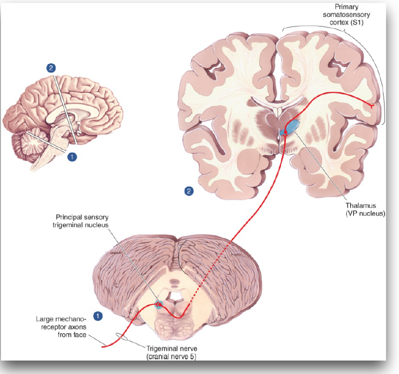
What is the pathway for touch from the face (Fig. 12.17)?
Large mechanoreceptor axons run in the trigeminal nerve (CN V) to the principal sensory trigeminal nucleus in the brainstem; second-order axons cross to the VP nucleus of the thalamus, then S1. The face equivalent of the DCML pathway.
28
New cards
Where does the DCML pathway cross the midline?
In the medulla. The first-order axon stays ipsilateral up the cord; after synapsing in the dorsal column nuclei, second-order axons cross and form the medial lemniscus.
29
New cards
Where does the anterolateral pathway cross the midline?
In the spinal cord. The second-order neuron crosses soon after the first synapse in the dorsal horn and ascends contralaterally.
30
New cards
What roles do the spinal cord, brainstem, thalamus, and somatosensory cortex play in somatic sensory processing?
Spinal cord: entry via dorsal roots; segmental (dermatome) organization; dorsal horn is first relay for pain/temp/itch and site of gate control. Brainstem: first synapse and crossing for DCML (medulla); trigeminal nuclei for face; descending pain control (PAG to raphe nuclei). Thalamus: relay to cortex (VP, intralaminar). Cortex: S1 gives conscious topographic representation; posterior parietal cortex integrates with vision, attention, planning.
31
New cards
How do the DCML and anterolateral pathways differ in organization and function?
DCML: fine touch/vibration/proprioception; large myelinated fast axons; first synapse in medulla; crosses in medulla; ipsilateral dorsal columns then medial lemniscus; thalamic VP. Anterolateral: pain/temp/itch/crude touch; thin slow axons (Aδ, C); first synapse in dorsal horn; crosses in spinal cord; contralateral anterolateral columns; thalamic intralaminar and VP.
32
New cards
What is the clinical consequence of the different crossing points of the two pathways?
A lesion of one half of the spinal cord causes loss of touch/proprioception on the SAME side below the lesion and loss of pain/temperature on the OPPOSITE side.
33
New cards
What is a dermatome?
The area of skin served by a single spinal nerve (segment of the spinal cord): cervical, thoracic, lumbar, sacral. Face is served by the trigeminal nerve.
34
New cards
What is the gate control theory of pain (Fig. 12.34)?
Nociceptive C fibers excite dorsal horn projection neurons (to the anterolateral tract). An inhibitory interneuron suppresses the projection neurons; Aβ touch fibers excite it (closing the gate) and C fibers inhibit it (keeping the gate open). Explains why rubbing an injury relieves pain.

35
New cards
What is the descending pain-control pathway (Fig. 12.35)?
Periaqueductal gray (PAG) in the midbrain projects to the raphe nuclei in the medulla, which project to the dorsal horn to suppress pain transmission.

36
New cards
What is the primary somatosensory cortex (S1), and where is it located?
Primary cortical area for somatic sensation in the parietal lobe, in the postcentral gyrus just behind the central sulcus. Comprises Brodmann areas 3a, 3b, 1, and 2. Posterior parietal cortex (areas 5, 7) is behind it.

37
New cards
How are neurons organized within S1?
In columns: columns of slowly adapting neurons alternate with columns of rapidly adapting neurons, and each column receives input from the same body region (e.g., a particular fingertip).

38
New cards
What is somatotopy, and how is the body represented in S1?
Orderly point-to-point mapping of the body surface onto the cortex; adjacent skin is represented by adjacent cortex. From the midline top to lateral: toes, foot, leg, hip, trunk, head, arm, forearm, hand, fingers, thumb, eye, nose, face, lips, teeth, gums, jaw, tongue, pharynx, intra-abdominal. Genitals near the midline beside the foot.
39
New cards
What is the sensory homunculus, and why do some body regions have larger cortical representations?
A distorted 'little man' drawn over S1 showing cortex allotted to each body part. Hands, fingers, lips, tongue are huge; trunk, back, legs small. Area reflects innervation density and sensory importance (many receptors, small RFs), not body surface area; matches two-point discrimination.

40
New cards
What is cortical plasticity?
The brain's ability to change its organization with experience or injury; the somatotopic map is not fixed but is continually shaped by sensory input.

41
New cards
How can repeated stimulation or learning change the somatosensory cortex (Fig. 12.24D)?
In owl monkeys, selectively and repeatedly stimulating digits 2 and 3 (tactile discrimination training) expands their cortical representations. Practice and use enlarge the cortical territory for the stimulated skin.
42
New cards
How can peripheral nerve damage or loss of a body part affect the somatotopic map (Fig. 12.24C)?
Deprived cortex is taken over by neighbors. After digit 3 is removed, the digit 3 territory is gradually invaded and the representations of digits 2 and 4 expand.
43
New cards
What is phantom limb sensation, and how is it related to sensory reorganization?
Perception that an amputated limb is still present (touch, position, pain). In S1 the face map sits next to the hand map; after amputation the deprived hand area is taken over by adjacent face and residual arm input. Touching the face or arm can evoke sensations in specific parts of the phantom hand, because the cortex still interprets that area as 'hand'.

44
New cards
What functions does the posterior parietal cortex serve, and what happens with damage?
Integrates somatic sensation with vision, attention, and planning. Damage causes agnosia (can't recognize objects despite normal basic senses), astereognosia (can't recognize objects by feel, often contralateral), and neglect syndrome (hemispatial neglect: ignoring the side opposite the injury).
45
New cards
What are nociceptors, and how do they contribute to pain?
High-threshold free nerve endings of small-diameter axons that signal tissue damage or risk of damage. Strong mechanical stimuli, temperature extremes, oxygen deprivation, or chemicals open ion channels, producing receptor and action potentials that travel via the anterolateral pathway to thalamus and cortex. Activation can lead to the conscious experience of pain.
46
New cards
What is the difference between nociception and pain?
Nociception: the sensory process that detects and transmits potentially damaging stimuli (provides the signals that trigger pain). Pain: the conscious feeling of sore, aching, throbbing sensations, constructed by the brain. One can occur without the other.
47
New cards
What are the major types of nociceptors and what activates them?
Mechanical: damaging pressure or deformation. Thermal: harmful heat (burning) or extreme cold. Chemical: substances released by injury or inflammation (e.g., histamine). Polymodal: more than one stimulus type.
48
New cards
What are polymodal nociceptors, and how do they differ from other nociceptors?
Nociceptors that respond to more than one type of noxious stimulus (mechanical, thermal, and chemical), whereas other types are tuned to a single kind. They signal that something harmful is happening regardless of cause.
49
New cards
What chemical substances released during injury or inflammation activate or sensitize nociceptors (Fig. 12.27)?
Bradykinin (formed by proteases), ATP, K+, prostaglandins, serotonin, histamine (from mast cells), and substance P and CGRP (released from nociceptor terminals; promote swelling and amplify the response). CGRP is also implicated in migraine. This 'inflammatory soup' opens channels or sensitizes nociceptors (hyperalgesia).

50
New cards
How do Aδ fibers and C fibers differ in transmitting pain (Fig. 12.28)?
Aδ: 1-5 µm, thinly myelinated, 5-30 m/s; first pain: fast, sharp, well localized. C: 0.2-1.5 µm, unmyelinated, 0.5-2 m/s; second pain: delayed, dull, aching/burning, poorly localized; also carry temperature and itch.

51
New cards
What are hyperalgesia and allodynia, and how do they differ?
Hyperalgesia: increased pain from a normally painful stimulus (primary = within damaged tissue; secondary = supersensitivity in surrounding tissue). Allodynia: pain caused by a normally nonpainful stimulus (e.g., light touch on sunburned skin). Difference is whether the stimulus was originally painful.
52
New cards
What are thermoreceptors, and what do they detect?
Free nerve endings (Aδ and C fibers) that contribute directly to the perception of temperature, detecting warming and cooling. Sensitivity varies across the body surface, and hot and cold have distinct receptive fields. Signals travel in the anterolateral pathway.
53
New cards
What is the difference between warm-sensitive and cold-sensitive thermoreceptors?
Warm receptors increase firing when skin temperature rises; cold (cool) receptors increase firing when it falls. They occupy distinct spots (receptive fields) on the skin.
54
New cards
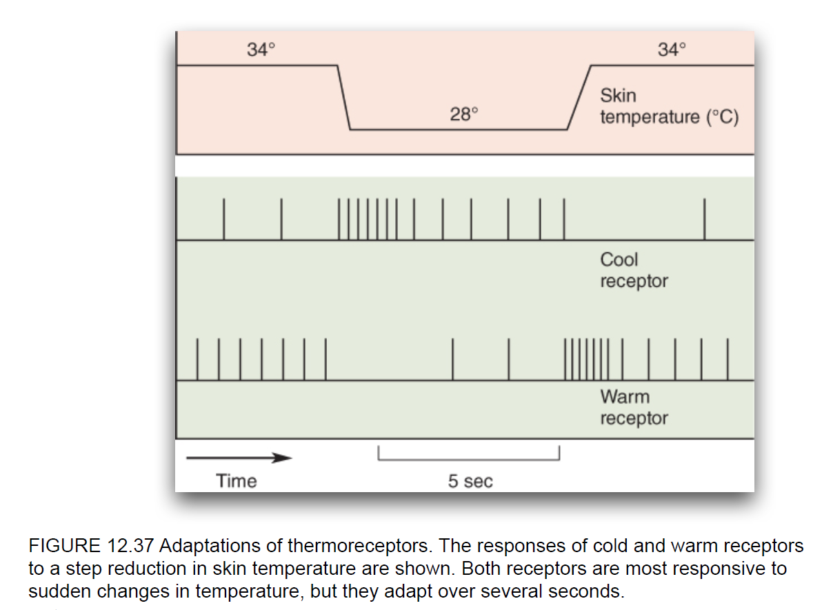
How do thermoreceptors respond to changes in skin temperature (Fig. 12.37)?
Both are most responsive to sudden changes. A cool receptor bursts when skin cools (34 to 28 °C) then settles; a warm receptor falls nearly silent during the drop and bursts when warming returns to 34 °C. Both fire at a modest background rate at steady temperature and adapt over several seconds.
55
New cards
What temperature ranges are associated with mild warmth, mild cool, and potentially harmful temperatures?
Above ~42 °C: painful, potentially harmful. 31-42 °C: mild warmth. 16-30 °C: mild cool. ~15 °C and below: noxious, potentially harmful.
56
New cards
Why are some thermoreceptors classified as nociceptors?
Receptors responding to extreme temperatures (burning heat or extreme cold) signal potential tissue damage, so they act as thermal nociceptors. Those responding to non-damaging temperatures only contribute to temperature perception.
57
New cards
How do thermoreceptors for harmless temperatures differ from those for damaging temperatures?
Innocuous thermoreceptors respond across mild warm (31-42 °C) and mild cool (16-30 °C) ranges and support temperature perception, not pain. Thermal nociceptors have high thresholds, respond only above ~42 °C or at ~15 °C and below, and their activation can produce pain.
58
New cards
What is sensory adaptation, and how does it affect warm and cold receptors?
Decline in a receptor's response during a maintained, unchanging stimulus. Warm and cold receptors respond most to sudden temperature changes and adapt over several seconds if the new temperature persists (why a pool feels cold at first, then comfortable).