med surge theory
 Call Kai
 Learn
 Practice Test
 Spaced Repetition
 Match
 Flashcards
 Knowt Play
1/179

There's no tags or description
Looks like no tags are added yet.
180 Terms
what is COPD
t
Chronic obstructivbe pulmonary disease. respiratory disease that has persistant airflow obstruction
mainly involves two processes
Chronic bronchitis
Chronic bronchitis
inflammation and excessive mucus production in teh bronchi
ariwasys become sweollend and filled with mucus, narrowing the passage available for airflow
Emphysema
destruction of alveolar walls and loss of lung elasticiyt
lungs lose ability to recoil dyring expiration, resulting in air trapping
Why does air trapping occur
lungs are elastice
during expiration recoil helps push air out
with emphysema
aveolar walls destroyed
recoil decreases
airways collpase more easily
air remains trapped
pt works harder to breahte
This explains several classic COPD findings:
Finding | Why it happens |
|---|---|
Barrel chest | Chronic hyperinflation |
Prolonged expiration | Difficulty moving air out |
Pursed-lip breathing | Helps prevent premature airway collapse |
Accessory muscle use | Increased work of breathing |
Dyspnea | Air trapping and increased respiratory effort |
Diminished breath sounds | Reduced airflow |
why is there low oxgyen and high co2
Hypoxemia means decreaseed oxygen in arterial blood
Hypercapnia means increased carbon dioxide in arterial blood
Nursing priorities during an exacerbation
imagine pt with COPD develops sudden shortness of breath, a respiraroty rate of 34 and an spo2 of 85%
Priorities
assess airways, breathing oxygenation and mental status
position the pt upright often in high fowlers
adminsiter and titrate oxygen according to orders and clinical condtion
administer prescribed short acting bronchodilators
reassess work of breathing lung sounds, cital signs, and response
escelate immediately if pt has sever distress, delcniing consciousness, or signs of respiratory failure
Falling respiratory rate is not always improvement
going from 36 to 8 is not alwasy great. might be fatigue
Inhalers and how they work
primarily work by
relaxing smooth muslce, reducing inflammation, or both
major medication classes (NOT needed for exam)
Class | Examples from your lecture | Mechanism and purpose |
|---|---|---|
SABA — Short-acting beta₂ agonist | Albuterol | Stimulates beta₂ receptors → relaxes bronchial smooth muscle → rapid bronchodilation |
LABA — Long-acting beta₂ agonist | Salmeterol | Same receptor mechanism, but provides prolonged bronchodilation for maintenance |
SAMA — Short-acting muscarinic antagonist | Ipratropium | Blocks muscarinic receptors → reduces cholinergic bronchoconstriction |
ICS — Inhaled corticosteroid | Fluticasone | Reduces airway inflammation; used in selected patients, generally in combination therapy |
Combination inhalers | Advair, Combivent | Combine different mechanisms to improve symptom control |
dry powder inhaler vs metered dose inhalers
Metered does inhaler
release a measured aerosol dose using a propellant
dry powder inhaler
uses pt inhalation to draw powdered medication into lungs
Feature | MDI | DPI |
|---|---|---|
Medication form | Aerosol | Dry powder |
Inhalation | Slow and deep | Fast, forceful, and deep |
Coordination | Requires timing unless using spacer | Breath-actuated |
Spacer | Often beneficial | Not used |
Main limitation | Poor hand-breath coordination | Insufficient inspiratory effort |
proper inhaltion tech for meteered dose inhaler
Metered-dose inhaler (MDI)
Remove the cap and shake the inhaler if required by the product.
Attach a spacer when appropriate.
Exhale fully, away from the inhaler.
Seal lips around the mouthpiece.
Begin inhaling slowly and press the canister once.
Continue inhaling slowly and deeply.
Hold breath for approximately 10 seconds if tolerated.
Exhale slowly.
Wait as directed before another puff.
Rinse mouth and spit after an inhaled corticosteroid.
proper tech for dry powder inhaler
Dry powder inhaler (DPI)
Open the device and load the dose according to manufacturer instructions.
Exhale fully away from the device.
Seal lips around the mouthpiece.
Inhale quickly, forcefully, and deeply.
Hold breath for approximately 10 seconds if tolerated.
Exhale away from the device.
Close the device.
Rinse mouth and spit if using an inhaled corticosteroid.
Nutritional needs of pt with COPD
Pt need more energy breathing becasue their respiratory msucles work hard to breathj
leads tto
increased caloric expenditure
weght loss and. uscle wasting
fatigue during meals
decreased appetite
malnutrition and respiratory muscle weakness
inadequate diet leads to expeniture and fatigue
administration of oxygen to patients with copd
Target o2 range is 88-92
indicators that inhalres adminsted were effectivbe
Assessment | Before treatment | After effective treatment |
|---|---|---|
Respiratory rate | Elevated | Moves toward patient's baseline |
Wheezing | Present | May decrease |
Dyspnea | Significant | Patient reports easier breathing |
Accessory muscle use | Increased | Decreases |
Breath sounds | Reduced airflow | Improved air movement |
SpO₂ | May be decreased | May improve |
Heart rate | Often elevated from distress | May decrease as distress improves |
changes in vitals over time and concerns related rto changes
Vital sign | Value | Interpretation |
|---|---|---|
Blood pressure | 160/96 | Hypertension |
Heart rate | 130 bpm | Significant tachycardia |
Respiratory rate | 36/min | Severe tachypnea |
Temperature | 37.6°C | Slightly elevated |
SpO₂ | 86% on room air | Hypoxemia |
how vital signs change as respiratory failure develops
Early deterioration
Tachypnea, tachycardia, anxiety, restlessness, and increased accessory muscle use. The patient is working harder to compensate for impaired gas exchange.
Worsening respiratory failure
Increasing hypoxemia, respiratory fatigue, worsening CO₂ retention, confusion, and decreasing effectiveness of breathing.
Impending respiratory arrest
Decreased respiratory effort or rate despite ongoing distress, altered consciousness, weak respiratory effort, and possible hemodynamic instability. Requires immediate emergency response.
Interventions for shortness of breath
Priority | Intervention | Rationale |
|---|---|---|
1 | Assess airway, respiratory effort, RR, SpO₂, and mental status | Determines severity and whether emergency support is needed |
2 | Position in high Fowler's or tripod position | Improves diaphragmatic movement and lung expansion |
3 | Administer or titrate prescribed oxygen | Corrects hypoxemia |
4 | Administer prescribed short-acting bronchodilators | Relieves bronchoconstriction |
5 | Encourage pursed-lip breathing when appropriate | Prolongs expiration and reduces airway collapse |
6 | Reassess respiratory status and response to treatment | Determines effectiveness and need for escalation |
why does pursed lip breathing help
pt with emphsema lose elastic recoul. making airways more likely to collapse during expiration
pursed lips create mild positve expiratory pressure, helping keep airwasys open long
this allows more air to escape, reducing air trapping and potentially decreasing dyspnea

what happens in pneumonia
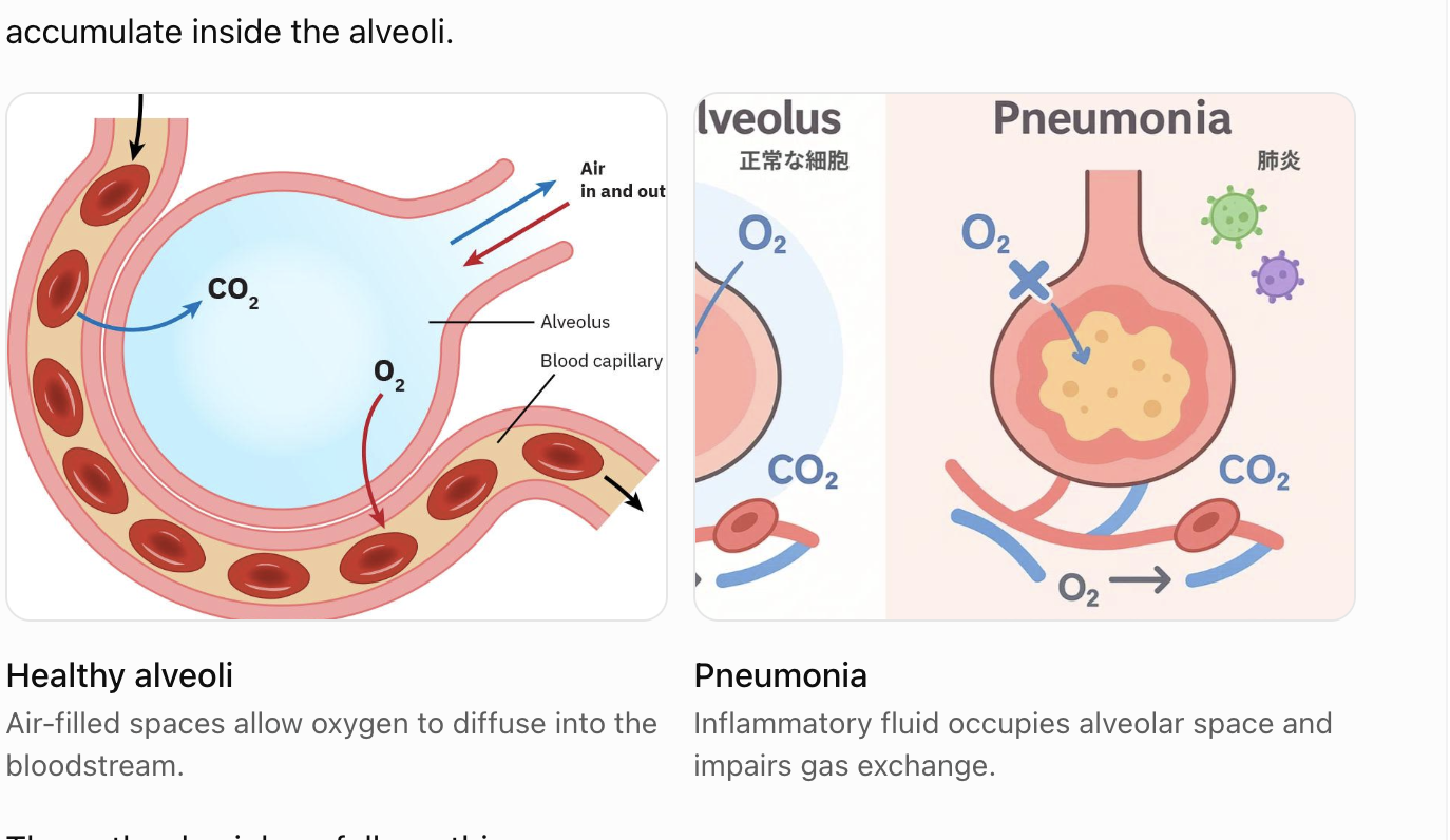
infection of lung tissue that causes inflammation in alveoli and surroudnign structures
normally alveoli are filled with air allowing oxygen to diffuse into pulmonary capillaries
inflammation causes fluid, inflammatory cells and someties pus to accumulate inside teh alveoli
priority action when a pneumonia pt complains of increasing shortness of breath despite oxygen
assess pt’s airway and breathing, including respiratory rate, work of breathing, spO2, mental status, and whetehr the o2 delivery system is functioning
position pt upright if you can and escelate in pt has signs of severe respiratory distress
signs that exacerbation is improving
decreased respiratory
from 33 to 20
decreased work o fbreathing
less accessory muslce use, tripof positioning, retractions
improved oxygenation
spo2 moes toward pt target, often 88-92
improved lung sounds
decreased dyspnea
breathing is easier
improved mental status
less confusion drowziness
improved activityu tolerance
improving ABGs
Wheezing occurs when air moves through narrowed airways. If airflow becomes severely reduced, wheezing can disappear even though the patient is deteriorating.
A patient with very diminished breath sounds, worsening hypoxemia, exhaustion, or altered mental status requires immediate assessment.
vesicular breath sounds
what you want to hear
Location: Most peripheral lung fields.
Sound: Soft, low-pitched, gentle rustling or blowing.
Inspiration vs. expiration: Inspiration is longer and louder than expiration.
Normal finding: Yes, over most lung tissue.

bronchial breath sounds
Location: Normally heard over the trachea.
Sound: Loud, harsh, high-pitched, tubular.
Inspiration vs. expiration: Expiration is longer and often louder than inspiration.
Normal finding: Yes, over the trachea; abnormal over peripheral lung fields.
caused by lung consolidatoin

what causes lung consolidation
1. Fluid or pus (pneumonia): The alveoli fill with inflammatory fluid and cellular debris, creating consolidation. This is a classic cause of bronchial breath sounds in peripheral lung fields.
2. Blood (pulmonary hemorrhage): Blood fills the alveoli, increasing tissue density and improving sound transmission.
3. Other causes of consolidation: Cells or other material replace air within the alveoli, producing similar findings.
what is the relationship between hemoglobin and oxygen saturation
oxygen saturation and oxygen carrying capacity are not the same thing
Hgb binds oxygen and transports throughout body
SpO2 measures estimated percentage of availbable hemoglobing binding sites occupied by oxygen
hemoglobin concentration measures how much hgb is available to carry oxygen
high hgb can partially compensate for reduced satruation by increasing amt of oxygen carrying protein, but does not correct hypoxemia, high hgb can increase blood viscosity as well
Signs and symptoms of pneomonia
Finding | Why it occurs |
|---|---|
Fever and chills | Inflammatory response to infection |
Productive cough | Secretions and inflammatory exudate in the airways |
Crackles | Air moving through or reopening fluid-affected small airways |
Bronchial breath sounds over affected lung tissue | Consolidated lung transmits airway sounds more effectively |
Tachypnea | Compensation for impaired oxygenation |
Tachycardia | Response to fever, hypoxemia, and physiological stress |
Dyspnea | Impaired gas exchange and increased work of breathing |
Pleuritic chest pain | Inflammation involving the pleura |
Fatigue and weakness | Systemic illness and increased metabolic demand |
Confusion | Can reflect infection, hypoxemia, or delirium, particularly in older adults |
assessment findings that indicate pneomonia is resolving
Assessment | Improvement looks like |
|---|---|
Temperature | Fever resolves |
Respiratory rate | Returns toward normal |
SpO₂ | Improves or requires less supplemental oxygen |
Work of breathing | Decreased accessory muscle use |
Lung sounds | Improving air movement; crackles may diminish |
Cough | Less frequent or less productive |
Sputum | Decreasing amount and purulence |
Heart rate | Returns toward baseline |
Mental status | Improved alertness and orientation |
WBC | Trends toward normal if initially elevated |
Activity tolerance | Less fatigue and dyspnea with activity |
which findings are most important for pneomonia
improvements in oxygenation and work of breathing are especially important
A patient initially requires 4 L/min of oxygen to maintain SpO₂ of 93%. Two days later, they maintain 95% on 1 L/min.
This suggests improving oxygenation.
interventions to decrease shortness of breath: initial priority and easiest action
high fowlers is easiest
Intervention | Rationale |
|---|---|
High Fowler's positioning | Improves lung expansion |
Supplemental oxygen as prescribed | Corrects hypoxemia |
Encourage coughing and deep breathing | Mobilizes secretions |
Incentive spirometry when appropriate | Encourages sustained inspiration and lung expansion |
Encourage hydration when appropriate | Helps maintain secretion clearance |
Administer prescribed antibiotics | Treats bacterial infection |
Bronchodilators when indicated | Relieve associated bronchospasm |
Mobilization as tolerated | Improves ventilation and secretion clearance |
Monitor SpO₂, RR, and lung sounds | Evaluates respiratory status |
position with good lung down!
Gravity directs blood flow to dependent lung, improving ventialtion, also helps pull away bad stuff from bad lung
key things for pneomonia
Study-guide topic | Key takeaway |
|---|---|
Worsening dyspnea despite oxygen | Immediately assess airway, breathing, SpO₂, and respiratory effort; escalate if unstable |
Hemoglobin vs. SpO₂ | Hemoglobin = carrying capacity; SpO₂ = percentage saturated |
Pneumonia symptoms | Fever, cough, crackles, tachypnea, dyspnea, pleuritic pain |
Resolving pneumonia | Improving oxygenation, decreased respiratory effort, resolving fever |
Easiest initial intervention | Position upright in high Fowler's while assessing and supporting breathing |
ekg/ecg
records electrical activity of the heart not the mechanical pumping itself
normal ekg wave

Component | Normal value | Electrical activity | What's happening physiologically? |
|---|---|---|---|
P wave | <0.12 sec | Atrial depolarization | The SA node has initiated an impulse that spreads through the atria. Atrial muscle cells become electrically activated, triggering atrial contraction shortly afterward and pushing blood into the ventricles. |
PR interval | 0.12–0.20 sec | Electrical conduction from atria toward ventricles | The impulse travels through the atria and AV node, where it is briefly delayed, then through the His-Purkinje system. This delay allows the ventricles more time to fill before contracting. |
QRS complex | <0.12 sec | Ventricular depolarization | The electrical impulse spreads through the ventricular myocardium, triggering ventricular contraction shortly afterward. Pressure rises, eventually opening the aortic and pulmonary valves and ejecting blood. |
ST segment | Normally near baseline | Ventricles remain largely depolarized | Ventricular muscle cells are in the plateau phase of their action potentials. The ventricles are mechanically contracting and ejecting blood during much of this period. |
T wave | No single fixed duration | Ventricular repolarization | Ventricular muscle cells restore their electrical resting state. Ventricular relaxation follows, pressure falls, and the ventricles enter diastole. |
QT interval | Heart-rate dependent | Ventricular depolarization through repolarization | Represents the total electrical activation and recovery period of the ventricles, encompassing much of ventricular systole. |
what is ventricular tachycardia (v-tach)
occures when rapid electricla impusles orginate in teh ventricles rather than following normal condiction through SA nose
Recognition
usually rapid rate, often aroudn 150-250 bpm
wide QRS complexes, trypically .12 seconds or longer
often regular
p waves mauy be absent, difficult to see, or dissociated from QRS complexs

why is V-tach dangerous
when ventricles beat extremely quickly they have less time to fill with blood
can cause
decreased stroke volume adn cardiac output
hypotension
reduced cerebral perfusion
loss of consciousness
progression to cardiac arrest
what to do if there is vtach on the monitor
assess responsivenss, breathing, and pulse immediately
No pulse
begin CPR, activate code response adn defib as soon as possible
if pulse
assess BP, mental status, chest pain, shock, and signs of heart failure
Unstable
urgen synchronized cardioversion for regular monomorphic VT, with emergency team support
stable
monitoring, IV access, 12-lead ECG when feasible and ordered antiarrhythmic treatment.
sinus tachycardia
SA node is firing faster than normal
How to recognize
heart rate
greater than 100 bpm
rhthm
regular
P waves
present, one before each QRS
PR interval
0.12-0.20
QRS
usually narrow, under 0.12
looks like nomral sinus rhthym except is faster
why does sinus tachycardia occur
often response to another problem rather than primary primary problme itsel
Common causes
fever or infection
dehydration or hypovolemia
pain or anxjety
hypoxemia
anemia
hemorrhage
pulmonary embolism
myocardial ischemia
what is priority concern for sinus tachycardia
Treating underlying cause
at higher heart rate, ventricles have less time to fill, stroke volume may decrease, myocardial oxygen deman increases, coronary perfusion time decreases
Nursing interventions for sinus tachycardia
Asses vitals, BP, SpO2, temp, pain, perfusion, and possible causes
what is atrial fibrillation (A-fib)
Atrial fibrillation occurs when the atria experience chaotic electrical activity rather than coordinated depolarization; instead of one organized impulse originating from the SA node, multiple disorganized impulses activate the atria
What happens in the heart
normally, atria contract in a coordinated manner
during a-fib
atria fibrillate rather than contract effectively
organized P waves disappear
the AV node conducts impulses irregularly
the ventricular rhythm becomes irregularly irregular

how to recognize A-fib
Feature | Atrial fibrillation |
|---|---|
Heart rate | Variable; can be rapid or controlled |
Rhythm | Irregularly irregular |
P waves | No distinct, consistent P waves |
PR interval | Cannot be measured reliably |
QRS | Usually narrow unless another conduction abnormality exists |
why is A-fib dangerous
decreased cardiac output
atria loses coordinated contraction, sometimes called atrial kick
can reduced ventricular filling and cardica output, especially in patients iwth impaired ventricular relazation
stroke risk
becasue the atria are not contracting effectively, blood can stagnate, particularly in the left atrial appendage
this can increase risk of thrombus formation
a clot can dislodge, travel through the arterial circulation, and cause and ischemic stroke
Treatment for A-fib
Treatment goal | Examples |
|---|---|
Control ventricular rate | Metoprolol, diltiazem, sometimes digoxin |
Restore or maintain sinus rhythm | Cardioversion, selected antiarrhythmics, catheter ablation |
Prevent thromboembolic stroke | Warfarin or a direct oral anticoagulant when indicated |
Warferin teaching for patients with A-fib
wafarin is an anticoagulant used to reduce risk of thrombboembolic stroke in selected patients wiht a-fib
it works by inhibiting vitamin k-dependant clotting factor syntehsis
does not directly dissolve existing lcots
teaching
Teaching | Why? |
|---|---|
Monitor INR regularly | Determines anticoagulation intensity |
Typical INR goal 2.0–3.0 for A-fib | Balances clot prevention with bleeding risk |
Maintain consistent vitamin K intake | Sudden dietary changes can affect INR |
Report unusual bleeding or bruising | May indicate excessive anticoagulation |
Avoid starting NSAIDs without medical advice | Increased bleeding risk |
Discuss new medications and supplements | Many interactions affect warfarin |
Use a soft toothbrush and electric razor | Helps reduce bleeding injuries |
Do not stop warfarin independently | Stopping can increase thromboembolic risk |
Avoid warfarin during pregnancy | Can cause fetal harm |
ischemia
The myocardium is receiving inadequate oxygen rich blood
injury
acute myocardial damage is occuring
infarction
Myocardial tissue has undergone irreversible cell death
EKG changes indicateing ischemia
ST sesfment depression can indicate myocardial ischemia, it may be seein with NSTEMI, unstable angina or demand ischemia, among other causes

EKG changes indicating acute injury
ST segmetn elevatoin in an apporpriate pattern can indicate acute myocardial injry, including a STEMI
this requires urgent evaluation for cornary arter occlusion

EKG changes indicating Infarction
Pathologial Q waves can indicate prior mysocardial infarction adn scar tissue
they may also develop during an evloling MI, althoubh not every infarction produces Q waves

what about T wave inversions
Can indicate myocardial ischemia, although it is not specific to ischemia
acute coranory suynromes may produce evolving changes over tiem, so serial ECGs are important
Condition | EKG finding |
|---|---|
Ischemia | ST depression |
Injury | ST elevation |
Infarction | Pathological Q waves, potentially with ST/T-wave changes |
Nursing priority: New ischemic EKG changes accompanied by chest pain, dyspnea, diaphoresis, or instability require immediate assessment, urgent provider notification, and a 12-lead ECG/chest pain protocol as appropriate.
what is sinus bradycardia
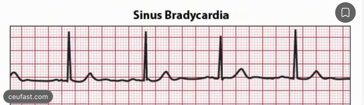
occurs when SA node fires fewer than 60 times per minute
Feature | Sinus bradycardia |
|---|---|
Heart rate | Under 60 beats/min |
Rhythm | Regular |
P waves | Present before every QRS |
PR interval | 0.12–0.20 sec |
QRS | Usually narrow |
is bradycardia always dangerous
No. bradycardia can be normal in healthy, well conditions indiciduals or during sleep
BUT
does the pt have adequate perfusion
Nursing interventions for symptomatic bradycardia
Assess airway, breathing, pulse, BP, mental status, and perfusion.
Call for immediate assistance if unstable.
Apply cardiac monitoring, obtain IV access, and support oxygenation when needed.
Prepare to administer atropine, according to the ACLS protocol or provider orders.
Prepare for transcutaneous pacing or other advanced treatments if atropine is ineffective.
administration of diabetic miedications the morning of surgery
Biggest concern is hypoglycemia while the pt is NPO (nothing by mouth)
normally, pt eat carbohydrates that riase their blood glucose, it pt takes insulin or certain oral diabetes medications but doesnt eat, their blood glucose can fall dangerously low. bnecaue your body ztill needs insulin, and srugery increases glucose beacsue of stress.
however, wihtholding all diabetes medications can also cause hyperglycemia, so teh nurse follows the perioperative medication plan rather than making blanket decisions.
Medication | Typical morning-of-surgery approach | Why? |
|---|---|---|
Rapid-acting insulin (lispro, aspart) | Usually hold the routine mealtime dose while NPO; correction insulin may still be ordered | No meal is being consumed |
Short-acting regular insulin | Hold scheduled nutritional doses while NPO; follow correction orders | Prevent hypoglycemia |
Long-acting basal insulin (glargine, detemir, degludec) | Usually continue at a reduced or individualized dose | Prevent excessive hyperglycemia and ketosis |
NPH insulin | Often give a reduced morning dose | Has an intermediate peak that can cause hypoglycemia |
Sulfonylureas (glipizide, glyburide) | Usually hold on surgery day | Stimulate insulin secretion even without food |
Metformin | Often held on surgery day; follow the perioperative plan | Discussed below |
SGLT2 inhibitors (empagliflozin, dapagliflozin) | Typically stop 3 days before surgery; ertugliflozin 4 days | Risk of euglycemic diabetic ketoacidosis |
GLP-1 medications (semaglutide, dulaglutide) | Individualized plan based on aspiration risk and current guidance | May delay gastric emptying |
nursing priorities for diabetes pre-op
Nursing priorities
Check the patient's blood glucose before surgery.
Confirm NPO status and the last time they ate.
Review which diabetes medications were taken.
Follow the anesthesia or perioperative medication orders.
Report significant hypoglycemia, hyperglycemia, or discrepancies in the medication plan.
Metformin before surgery
pt is NPO so does not need glucose
what should nurse do with metformin before surgery
For an exam question in which the patient is NPO and scheduled for surgery that morning, the expected response is commonly to hold metformin according to perioperative orders.
If the patient has already taken it, notify the appropriate perioperative team rather than independently canceling the procedure.
In current practice, some protocols allow metformin to continue for selected low-risk procedures, so the actual order and clinical situation matter.
Preoperative labs: which ones should be reported
Lab | Typical reference range | Why it matters before surgery |
|---|---|---|
Hemoglobin | Approximately 12–17.5 g/dL, varies by sex/lab | Low Hgb means reduced oxygen-carrying capacity and possible anemia or blood loss |
Hematocrit | Approximately 36–53% | Helps evaluate anemia, blood loss, or hemoconcentration |
Platelets | 150,000–400,000/µL | Low levels increase bleeding risk |
INR (not on anticoagulants) | Approximately 0.8–1.2 | Elevated INR may indicate increased bleeding risk |
aPTT | Approximately 25–35 seconds | Prolongation may indicate anticoagulant effect or coagulation abnormalities |
Potassium | 3.5–5.0 mEq/L | Low or high potassium can cause dangerous dysrhythmias |
Sodium | 135–145 mEq/L | Significant abnormalities can affect neurological function |
Creatinine | Approximately 0.6–1.3 mg/dL | Helps evaluate renal function and medication clearance |
Glucose | Depends on clinical context | Severe hypo- or hyperglycemia increases perioperative risk |
WBC | Approximately 4,000–11,000/µL | Elevation may indicate infection or inflammation |
labs to be reported Hgb
Lab | Typical reference range | Why it matters before surgery |
|---|---|---|
Hemoglobin | Approximately 12–17.5 g/dL, varies by sex/lab | Low Hgb means reduced oxygen-carrying capacity and possible anemia or blood loss |
Hematocrit labs to be reported
Lab | Typical reference range | Why it matters before surgery |
|---|---|---|
Hematocrit | Approximately 36–53% | Helps evaluate anemia, blood loss, or hemoconcentration |
platelet labs to be reported
Lab | Typical reference range | Why it matters before surgery |
|---|---|---|
Platelets | 150,000–400,000/µL | Low levels increase bleeding risk |
INR labs to be reported
Lab | Typical reference range | Why it matters before surgery |
|---|---|---|
INR (not on anticoagulants) | Approximately 0.8–1.2 | Elevated INR may indicate increased bleeding risk |
aPTT labs to be reported
Lab | Typical reference range | Why it matters before surgery |
|---|---|---|
aPTT | Approximately 25–35 seconds | Prolongation may indicate anticoagulant effect or coagulation abnormalities |
Potassium labs to be reported
Lab | Typical reference range | Why it matters before surgery |
|---|---|---|
Potassium | 3.5–5.0 mEq/L | Low or high potassium can cause dangerous dysrhythmias |
sodium labs to be reported
Lab | Typical reference range | Why it matters before surgery |
|---|---|---|
Sodium | 135–145 mEq/L | Significant abnormalities can affect neurological function |
creatinine labs to be reported
Lab | Typical reference range | Why it matters before surgery |
|---|---|---|
Creatinine | Approximately 0.6–1.3 mg/dL | Helps evaluate renal function and medication clearance |
gliucose labs to be reported
Lab | Typical reference range | Why it matters before surgery |
|---|---|---|
Glucose | Depends on clinical context | Severe hypo- or hyperglycemia increases perioperative risk |
WBC labs to be reported
Lab | Typical reference range | Why it matters before surgery |
|---|---|---|
WBC | Approximately 4,000–11,000/µL | Elevation may indicate infection or inflammation |
Priority lab to report
Potassium
PCA for postoperative pain management
PCA stands for patient controlled anelgesia
allows pt to administer a prescribed dose of IV pain medication, usually an opiod such as morphine or hydromorphone, by pressing a button
Setting | Meaning |
|---|---|
Basal rate | Continuous medication infusion, whether or not the patient presses the button |
Bolus or demand dose | Medication administered when the patient presses the button |
Lockout interval | Minimum time before another demand dose can be delivered |
mL/hour | Volume of medication solution infused each hour |
Maximum dose limit | Maximum medication allowed during a specified period, if programmed |
nursing considerations for PCS button
ONLY THE PT presses the button, family members and staff should not activate demand doses for them
assess pain and whether the PCA is providing relife
monitor respiratory rate, sedation, SpO2 and BP
verify medication concetration adn pump settings against the order
monitor for nasuea, itching, constipation, urinary retentoin and respiratroy depression
Exam takeaway: The lockout interval prevents repeated doses too close together, but it does not eliminate the risk of opioid overdose.
pre/post op care interpretation of o2 saturation
spo2 measuers estimated perfentage of hemoglibin binding sites occupied by o2
does not measure amt of hemoglobin
95–100% | Usually normal for a healthy adult |
92–94% | May indicate reduced oxygenation; assess clinical context |
90–91% | Concerning in many patients |
Below 90% | Significant hypoxemia requiring prompt assessment and intervention |
Priority actions when receving a pt from teh PACU
PACU stands for post anesthesia care unit
when pt returns from PACU your immediate priopiryt is identifying complications from anesthesia or srugery
1. Airway and breathing
Check airway patency, respiratory rate and depth, SpO₂, lung sounds, and work of breathing. Assess for residual anesthesia or opioid-related respiratory depression.
2. Circulation
Check BP, HR, skin perfusion, peripheral pulses, and signs of hemorrhage or shock.
3. Level of consciousness
Determine whether the patient is awake, oriented, and responding appropriately.
4. Surgical site, drains, and lines
Assess dressings, bleeding, drain output, IV fluids, Foley output, and device function.
5. Pain, nausea, and ongoing recovery
Assess pain, review PCA settings, provide prescribed symptom management, and implement complication-prevention measures.
when can pt resume eating after surgery
depensd on pt condition adn postoperative orders
Assessment | Why it matters |
|---|---|
Awake and able to swallow | Reduces aspiration risk |
Nausea and vomiting | May indicate intolerance |
Abdominal distention | Could suggest ileus or obstruction |
Abdominal pain | Worsening or severe pain may indicate complications |
Flatus or bowel movements | Useful signs of returning bowel function, but not always required before feeding |
Surgical orders | Determine permitted diet and progression |
General surgery
once pt is sufficiently awake, can protext theri airway and has no contraindications, oral intake may often resume realtively early
you should asses for nasuea, comiting and swallowing safety
Bowel surgery
However, modern enhanced recovery protocols frequently recommend early oral intake after uncomplicated bowel surgery, sometimes within 24 hours, as tolerated.
what is postoperative ileus
temporary reduction or cessation of intestinal motility after surgery
possible findings include ditention, nuasea, vomiting, and inabiliyt to tolerate food
TAKEAWAY: do not automatically feed a pt who has sings of ileu or bowl obstruction ubt also remember that waiting for bowel sounds or flatus is not universally required after uncomplicated bowel surgery
orthostatic vital sings and orthostatic hypotension
hypotension occurs when blod pressure drops significantly after a person moves from lying down to standing
when a person stands up gravity casues blood to pool in lwoer extremities,
temporarily decerases venous return to heart
Normally, baroreceptors detect the decrease in blood pressure and trigger a sympathetic response:
Heart rate increases.
Blood vessels constrict.
Blood pressure is maintained.
If the body cannot compensate adequately, blood pressure drops and the patient may become dizzy or faint.
technique for orthostatic hypotension
What indicates orthostatic hypotension?
Within 3 minutes of standing:
Systolic BP decreases by 20 mmHg or more, or
Diastolic BP decreases by 10 mmHg or more.
heart rate up by twenty

lab findings indicating orthostatic hypotension
Lab finding | Possible explanation |
|---|---|
Elevated BUN-to-creatinine ratio | May suggest dehydration or reduced renal perfusion |
Elevated sodium | May accompany water deficit |
Elevated Hgb/Hct | Can suggest hemoconcentration from dehydration |
Low Hgb/Hct | May suggest anemia or blood loss |
Low potassium | Can accompany vomiting or diuretic use |
Nursing interventions
Assist the patient to sit or lie down.
Implement fall precautions.
Assess HR, BP, symptoms, hydration status, and possible bleeding.
Review fluid balance and medications such as diuretics and antihypertensives.
Notify the provider about significant findings and administer ordered fluids or other treatments as appropriate.
what is a DVT
blood clot that forms in a deep vein, most commonly in legs

why does a DVT form
thre major factors that promote clot formation
Venous stasis
blood moves too slowly, such as during prolonged bed rest or after surgery
endothelial injury
damage to inner lining of a blood vessel
hypercoagulability
blood has an increased tendency to clot, such as with cancer, pregnancy or certain inherity disorders
Signs and sympotms of a DVT
unilateral leg swelling
pain or tenderness, often in calf
warmth and redness
sometiems no symptoms at all
The biggest concern is that part of the clot can break off, travel through the venous circulation, and lodge in a pulmonary artery, causing a pulmonary embolism (PE).
Sudden dyspnea, chest pain, tachycardia, hypoxemia, or collapse in a patient with suspected DVT requires immediate evaluation for PE.
what is heperain therapy
heparin is an anitcoagulant, prevents blood clots from getting larger
does not dissolve existing clot
Mechanism
enhances activity of antithrombin, a naturally occuring portein that inhibnits clotting factors
why give heperain to someone who already has a DVT
prevents blood clot from growin while body gradually breaks down the clot
nursing considerations for heparin therapy
monitor for bleeding: brusing hematuria, black stools, bleeding gums, and unexpecrted hypotension
monitor hemoglobin, and hematocrit for possibel blood loss
monitor platelet counts for heparin induced thrombocytopenia (HIT)
assess for symptoms of pulmonary embolism
verify infusion rate and ordered laboratory monitoring
what is heparin induced thrombocytopenia
immune mediated complcation in which antibiodies activate platelets
causes platelet consumption adn can paradoxically lead to new dangerous blood clots despit a falling platelet count
a platelt decrease of 50% or more is concerning even if platelet count remians within normal range
warferin therapy how it owrks and is monitored
oral anticoagulant
interferes with bodys ability to produce certain clottin gfactors
inhibits vitamin K reducing activation of vitamin K dependent clotting factors
II,VII,IX. and X
because existing clotting factors must be cleared from circulation, warferin takes several days to produce its full anticoagulatn effect
why might heparin and warferin be given together
for acute DVT treatment with warferin, pt genearlly receive an immediately acting anticoagulant such as heparin while warferin becoems therapeutic
called bridging
how is warferin monitored
using PT/INR
PT
prothrombin time
INR
international normalized ratio
NR | Meaning |
|---|---|
Below 2.0 | Often insufficient anticoagulation for DVT treatment |
2.0–3.0 | Typical therapeutic range |
Above 3.0 | Increased bleeding risk as INR rises |
nursing considerations for warferin
monitor INR as ordered
assess for bleeding
monitor for medicatoin interactions
teach consisten vitamin K intake
ensrue pt understatns the importance of taking warferin consistently
avoidd pregnany while taking warferin becasue it can cause fetal harm
labs to monitor for wafarin and heparin
Medication | Primary monitoring | Important additional labs |
|---|---|---|
Unfractionated heparin (IV) | aPTT or anti-Xa | CBC, platelets, Hgb/Hct |
Warfarin | PT/INR | CBC, Hgb/Hct |
Low-molecular-weight heparin (enoxaparin) | Usually no routine aPTT monitoring | CBC, platelets, creatinine; anti-Xa in selected patients |
what is aPTT
activated partial thromboplastin time
A typical normal aPTT is approximately 25–35 seconds, depending on the laboratory.
Why does heparin increase aPTT?
Unfractionated heparin works differently from warfarin.
Instead of reducing the liver's production of clotting factors, heparin enhances the activity of antithrombin.
measures how long it takes blood to clot through the intrinsic and common coagulatoin pathways
heparin prolongs this time
40-70 seconds is therapeutic rang
what is INR
warferin prolongs PT (prothrombin time), whihc is standerdized ad INR (international normalized ratio)
PT measures the time, in seconds, for plasma to form a fibrin clot after tissue factor and calcium are added in the laboratory.

low molecular weight heprarin versus unfractionated heparin
eature | Unfractionated heparin | LMWH (enoxaparin) |
|---|---|---|
Mechanism | Enhances antithrombin; inhibits IIa and Xa | Enhances antithrombin; predominantly inhibits Xa |
Administration | IV infusion or subcutaneous injection | Usually subcutaneous |
Duration | Shorter acting | Longer acting |
Monitoring | aPTT or anti-Xa for therapeutic IV use | Usually no routine aPTT |
Dose response | Less predictable | More predictable |
Kidney impairment | Often preferred in severe renal impairment | Requires renal assessment and possible dose adjustment |
Reversal | Protamine | Protamine only partially reverses its activity |
Home use | Less convenient | Commonly used at home |
patient teaching enoxaparin ijections
Wash hands and check the prescribed dose.
Select an abdominal injection site at least 2 inches from the umbilicus, avoiding bruises, scars, and irritated areas.
Rotate injection sites.
Clean the skin and allow it to dry.
Pinch a fold of skin and insert the needle into the subcutaneous tissue, typically at 90 degrees for a standard prefilled syringe.
Inject the medication as instructed, remove the needle, and dispose of the syringe in a sharps container.
do not massage or rub injection site afterwar it can cause bruising
Take injections at the prescribed times.
Do not double doses if one is missed; contact the care team for instructions.
Watch for bleeding, bruising, black stools, or blood in urine.
Avoid aspirin and NSAIDs unless approved by the prescriber.
Keep follow-up appointments for CBC and renal function monitoring.
Seek emergency care for sudden chest pain, shortness of breath, coughing blood, or fainting.

nursing care for patient with dvt
Assess swelling, warmth, tenderness, and color of the affected leg.
Measure leg circumference consistently to monitor changes.
Elevate the affected leg for comfort when appropriate.
Administer prescribed anticoagulation.
Monitor for bleeding.
Encourage early ambulation once anticoagulation is effective and the patient is clinically stable, as appropriate.
Do not massage the affected leg.
Do not apply sequential compression devices directly over an affected limb with known acute DVT unless specifically directed by the treating team.
what is acute coranary syndrome
occurs when blood flow through a coronary artery suddenly decreases, depriving part of the heart muscle of oxygen
heart requires constant supply of oxygen, because it is constantly contracting
what is stable angina
stable angina is predictbale chest discomfort casued by a temporary imabalnace between myoscardial oxygen suppply and deman
for ex
pt with coronary artery disease might experience chest pain while climbing stairs
during excersie
hr increases
myocardial contractility increases
heart requires more oxygen
narrowed coronary artery cannot increase blood flow sufficiently
myocardium becomes ischemic, producing chest discomcfort
nursing priority when pt with stable angina complains of chest pain with activity
stop activity and have pt rest
asses pain location, severity, quality, duration adn radiation
assess vital signs, BP, HR, RR and SpO2
administer prescribed sublingual nitroglycerin if appropriate and BP is adequate
reassess the pt sympotms and vital sings
if pain persists, unusual or occurs at rest, activate the appropriate chest-pain or emergency response protocol becaseu ACS may be developing
what labs are indicative of myocardial infarction
when myocardial cells are damaged, intracellular proteins are released into the bloodstream
Lab | What it indicates | Importance |
|---|---|---|
Cardiac troponin I or T | Myocardial cell injury | Most important and cardiac-specific biomarker used for diagnosing MI |
CK-MB | Muscle injury, particularly cardiac muscle | Less specific than troponin; occasionally useful in selected circumstances |
Myoglobin | Muscle injury | Rises early but is not cardiac-specific; not routinely used to diagnose MI |
troponin
porotein involved in muscle contraction
cardiac tropomnin I and T are found in cardiac muscle cells when those cells are injured, troponin enters blood
elevated troponin does not necessarily mean pt is having an MI.
physicians look for rise or fall in troponin with evidence of ischemia such as symptoms. ECG changes or imaging findingd
serial troponins
troponin may not be elevated immediately after symptoms begin
blood samples are collected at specified intervals to identify whether levesl are rising or fallin
nitroglycerin
nitrate vasodilator used to relieve angina
how it works
converted to nitric oxide, —> relaxes smooth muslce
venodilation
veins dilate
more blood to remain in venous circulation
decreased preload
reduced ventricular wall tension
reduced myocardial oxygen demand
nitroglycerin teaching
at onset of angina stp activity and lie down
place one tablet under tongue and allow it to dissolve or use prescribend sublingual spray
not not chew or swallow
if pain not relieved in 5 minutes, call 911
additional doess may be taken every 5 minutes up to three totoal according to prescription instructions
do not drive to hospital