Facial bones 2- NASAL BONES, ORBITS, AND ZYGOMAS
 Call Kai
 Learn
 Practice Test
 Spaced Repetition
 Match
 Flashcards
 Knowt Play
1/26

There's no tags or description
Looks like no tags are added yet.
27 Terms
MAXILLAE articulations
Skull bones?
Facial bones?
Frontal & Ethmoid
7 facial bones - opposite maxillae, same side zygoma, lacrimal, nasal, palatine, inferior nasal conchae, vomer
ZYGOMA articulations
Skull bones?
Facial bones?
Frontal and Temporal
Maxillae
LACRIMAL articulations
Skull bones?
Facial bones?
Frontal and Ethmoid
Maxillae, inferior nasal conchae
NASAL articulations
Skull bones?
Facial bones?
Frontal and Ethmoid
Maxillae, opposite nasal bone
INFERIOR NASAL CONCHAE articulations
Skull bones?
Facial bones?
Ethmoid
Maxillae, lacrimal and palatine
PALATINE articulations
Skull bones?
Facial bones?
Sphenoid and Ethmoid
Maxillae, inferior nasal conchae, vomer, opposite palatine
VOMER articulations
Skull bones?
Facial bones?
Sphenoid and Ethmoid
Right and left Maxillae & right and left palatine

Frontal process of maxilla
Nasion
Lacrimal bone
Nasal bone
*in order→ nasal bone→ frontal process of maxilla→lacrimal
Nasal Bones radiographs
Waters
Bilateral nasal bones
What makes up the bony nasal septum?
Perpendicular plate of ethmoid
Vomer

Septal cartilage
Perpendicular plate of ethmoid
Vomer
Parietoacanthial (Waters) for Nasal Bones
MML & MSP perpendicular
Raise OML to form 37 degree perpendicular to IR
Petrous Pyramids below maxillary sinuses
Rotation- outer edge of orbit to lateral border of skull, looking for symmetry

View?
Label
Parietoacanthial (Waters) for Nasal Bones
Orbit
Zygomatic arch
Maxillary sinus
Maxilla
Petrous ridge
Mandibular angle
Lateral Nasal Bones
2 laterals
No grid, small focal spot, close collimation
-Interpupillary line perpendicular to IR
-MSP parallel to IR
-IOML parallel to top edge or perpendicular to side edge
-CR perpendicular, centered to ½ inch inferior to nasion
Include anterior nasal spine
What are the orbit orientations?
30° superior angle
37° medial angle
Important for the optic foramina positioning

Base of orbit
Label the 3 bones
Frontal (orbital plate)
Zygoma
Maxilla
Bony Composition of Orbits
7 bones
SPELMFZ- from apex to base
• Sphenoid
• Palatine
• Ethmoid
• Lacrimal
• Maxilla
• Frontal
• Zygoma

A. Frontal
B. Lacrimal
C. Ethmoid
D. Sphenoid (most posterior)
E. Palatine - vertical portion
F. Zygoma
G. Maxilla
H. Maxilla
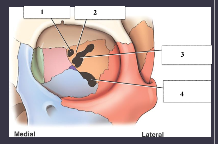
Openings in the orbit
Optic foramen
Sphenoid strut - little line
Superior orbital fissure
Inferior orbital fissure
BLOW OUT Orbital Fracture
teardrop sign
blunt trauma to the eye, pressure builds up and floor collapse = bones break up
Orbit/Optic Foramina views
Parietorbital oblique (Rhese)
Parietoacanthial (Waters)
PA (Caldwell)
*Can replace waters with modified waters*
Parietoorbital Oblique Projection (Rhese method)
Orbits
Rotate head 37 degree toward affected side (3 point landing -chin cheek and nose)
-MSP will form a 53° to IR
-AML perpendicular to IR
-CR perpendicular to downside orbit
-Use grid 75-85 kVp
-Bilateral projections taken for comparison
Optic foramen in the lower outer quadrant
right orbit= right chin, cheek, nose
Modified Parietoacanthial (Modified Waters)
-kissing board (hint)
Orbits
-Raise OML to form a 55° angle to IR
-LML & MSP perpendicular to IR
-CR perpendicular, exits at acanthion
Petrous ridges projected in lower 1/2 of maxillary sinuses
Orbital floors not distorted
Zygomatic Arch Routine
Submentovertex (SMV) or Oblique inferosuperior (tangential)
AP axial (modified Towne)
Special (commonly part of facial bone study)
• Parietoacanthial (Waters)
• Lateral
SMV
Zygomatic arches
CR perpendicular to IOML , Center right below mentum
-IOML is parallel to IR
-Bilateral zygomatic arches
-if patient cant go all the way back= angle to match
Zygomatic arches well demonstrated in profile and symmetric
Oblique Inferosuperior (tangential)
Zygomatic arch
-Start in SMV -IOML parallel to IR
-Rotate and tilt skull 15° toward affected side
-CR perpendicular to IR and IOML
-CR to arch of interes
AP Axial (modified Townes)
Zygomatic arch
-Have patient tuck chin down
-OML perpendicular = CR 30° caudal
-Settle for IOML perpendicular = 37° caudal
-CR 1 inch superior to Nasion (to pass through mid arches)