Anatomy & Physiology I - EXAM 4
 Call Kai
 Learn
 Practice Test
 Spaced Repetition
 Match
 Flashcards
 Knowt Play
1/91

There's no tags or description
Looks like no tags are added yet.
92 Terms
What is the cerebrum and what are its lobes?
Largest portion of the brain
Frontal, Parietal, Temporal, Occipital, Insula
Compare gray matter vs. white matter
Gray matter
UNmyelinated neurons making NERVES
makes cortex, nuclei, horns for INFO PROCESSING
nucleus is functional group of soma deep in brain
White matter
myelinated axons making TRACTS for TRANSMISSION
Name three major white matter tracts
Projection - connects brain and spinal cord
Commissural - connects L & R (Ex. corpus callosum)
Association - connects regions in the hemisphere
What are the functions of the cerebrum?
sensory awareness & sensation
voluntary motor function
association integrates information
Majority of cerebral cortex
Receive/send information from/to multiple regions
Features of Frontal Lobe
Primary motor cortex
Premotor cortex
Prefrontal cortex
Broca’s area
Describe the pre-frontal cortex
Part of the Frontal Lobe
Largest association cortex
planning, decision making, behavioral regulation, self-awareness
Develops until 25+
What is the precentral gyrus?
Part of the Frontal Lobe
Primary motor cortex for voluntary movement
communicates with muscles
action potential generated by its neurons \
Communicates with premotor cortex (planning & coordination)
What is the premotor cortex?
Part of the Frontal Lobe
Motor planning & coordination
Receives visual, auditory information used to decide movement
Communicates with precentral gyrus (primary motor cortex)
What is Broca’s area?
Part of the (left) Frontal Lobe
Speech production by motor coordination and grammatical word linking
What is Broca’s Aphasia?
Inability to produce speech, NOT impairing comprehension
Speech w/ effort in short phrases or words
Can impact writing and sign language (may not be understandable)
*Caused by damage from TBI, stroke, tumor, infection, or neurodegenerative conditions
Describe the Motor Homunculus.
Represents the proportions of cortex devoted to each VOLUNTARY function

What cortexes are present in the parietal lobe?
Visual association cortex
spacial awareness and visual attention
Postcentral gyrus
primary somatosensory cortex (general sensation)
Somatosensory Association cortex
integration of stimuli
tactile memory
Describe the sensory homunculus
Represents the proportions of cortex devoted to each sensory reception

What cortexes are part of the temporal lobe?
Primary auditory cortex & olfactory cortex
Auditory association cortex
classification of sounds
Visual association cortex
visual recognition, patterns
What is Wernicke’s area?
Part of the (left) temporal Lobe
Language comprehension by linking words to common meaning
What is Wernicke’s Aphasia?
Fluent, nonsensical speech
Reading and writing severely impaired
May not be recognized by pt but not understandable language to others
*Caused by damage from TBI, stroke, tumor, infection, or neurodegenerative conditions
What cortexes are found in the occipital lobe?
Primary visual cortex & visual association cortex
color, orientation, shape
What cortexes are found in the Insula?
Vestibular cortex - conscious balance
Gustatory cortex - taste perception
Visceral sensory cortex - hunger, nausea, organ pain
What forms the basal nucleus? What do they do?
Caudate, putamen, and globus pallidus
Induce & suppress motor neuron function (DO NOT communicate with muscles directly)
control voluntary movement by initiating and executing
inhibit antagonistic & unnecessary movement
Integrate info from many brain regions
Procedural learning/muscle memory, habitual behaviors, decision making

What can Tourette’s Syndrome or addiction be linked to?
Dysfunction of the basal nucleus that induces or inhibits motor neuron function
What makes up the limbic system?
“Emotional visceral brain”
Cingulate gyrus - emotional expression
Amygdala - fear responses
Hippocampus - long term memory consolidation
What does the limbic system connect to?
Olfactory bulb - emotional response to odor (triggers memory formation and retrieval)

Types of memory
Memory - encoding, storage, retrieval of stimuli & info
Declarative - conscious recollection (Ex. numbers, facts) consolidated in STAGE 3
Nondeclarative - unconscious performance (Ex. skills, procedures) consolidated in REM
What is memory consolidation and how does it work?
Consolidation - transfer to long term memory due to association, rehearsal or emotion
Where?
Hippocampus of limbic system- organizes stimuli into unified memory
induces long-term potentiation (LTP) to increase synaptic activity between neurons
increases dendritic growth
greater neurotransmitter release
memory stored in respective sensory regions (candle smoke, birthday song, visuals)
Describe spatial navigation
Hippocampus processes spatial memories to forms cognitive map – mental representation of environment
What structures make up the diencephalon?
Thalamus, Hypothalamus, Epithalamus (PINEAL GLAND), Subthalamus
Describe the thalamus
“Gatekeeper” that edits, sorts, routes signals going to the cortex
Location where 2nd order neurons synapse w/ 3rd
All BUT olfaction
Describe the hypothalamus
“Thermostat” for homeostatic control
thermoregulation and fever
hunger & fullness via blood chemistry
thirst & water retention via blood osmolarity
endocrine regulation/pituitary via antidiuretic hormone and oxytocin
circadian clock & regulates autonomic nervous system
How does the hypothalamus regulate emotional responses?
Integrates limbic system w/ cerebrum for perception and ANS for visceral response
Describe the epithalamus
Circadian release of melatonin from PINEAL GLAND regulates circadian timing
What is an emotion?
Sensation generated by limbic system and hypothalamus along with:
Visceral motor response - heart racing, blushing, crying
Somatic motor response - smiling, frowning
Cognitive response - recognition or regulation
How does the circadian rhythm regulate?
Eyes send photic signal to hypothalamus to induce wakefulness
Lower evening light induces “sleep pressure”
What happens during sleep onset?
Hypothalamus decreases brainstem communication with thalamus
“Disconnects” cerebral cortex
Brain stem functions and environmental monitoring continue
What happens upon waking up?
Hypothalamus stimulates brainstem to promote wakefulness
In response to circadian pattern, hormones, stimuli
Describe sleep stages (non REM)
LIGHT Stage 1: 1-5 min of transition into sleep
LIGHT Stage 2: 10-60 min
Heart/respiratory rate, blood pressure, and body temp lower
Eye movements stop
DEEP Stage 3: 20-40 min
Muscles fully relax & reduced brain activity
Tissue growth and repair processes
Consolidation of declarative memories
Describe sleep stages (REM)
10 – 60 min where eyes oscillate back and forth
Heart and respiratory rate increase
Increased brain activity
Limbic system very active, prefrontal cortex is not → strange/intense dreams
Sleep paralysis - inhibition of voluntary muscular activity
Consolidation of nondeclarative memories
Describe the cerebellum
Three lobes in each hemisphere w/ HALF neurons in brain
Afferent fibers from cerebrum (prefrontal cortex) trunk, inner ear, eyes, limbs
Efferent fibers to cerebrum & brainstem
What does the cerebellum do?
Coordinates planned actions and produces smooth, coordinated movement/balance
Motor learning, cognitive/language processing (working memory, executive function)
Emotional regulation
Describe motor processing (cerebellum)
Cerebral premotor cortex send info
Cerebellum evaluates current body position
Determines necessary adjustments
Plan sent to motor cortex & brainstem
What are the meninges?
3 layers of connective tissue to cover & protect CNS
Encloses blood vessels & contains CSF
What are the layers of the meninges?
Dura mater
Arachnoid mater
Pia mater
Describe the dura mater
HIGHLY vascular
Periosteal layer adhered to inner surface of cranial bones
Meningeal layer continuous with dura of spinal cord
Describe the arachnoid mater
Thin, web like layer
Subarachnoid space transmits blood vessels
Describe the pia mater
Deepest layer that directly follows contour of brain
Describe meningitis
Infection of the meninges diagnosed via CSF
Fever, headache, neck stiffness, light sensitivity
Viral - mild, heals in 2 weeks
Bacterial - body rash, rapid progression, extensive abx tx
Why does the brain have such a high blood demand?
15% of cardiac output to brain
High oxygen & glucose demand
Interruptions cause LOC or neural impairment
What vessels supply the brain?
Vertebral arteries (form basilar artery)
branch from subclavian
travel through transverse foramena
supply cerebellum & posterior cerebrum
Internal carotid arteries (form anterior/middle cerebral arteries)
branch from common carotid
supply anterior cerebrum

What is the cerebral arterial circle?
“Circle of Willis”
Anastomosis - redundant circulation
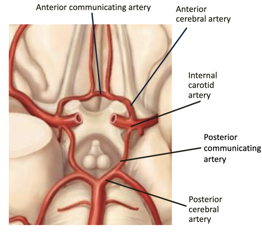
Describe what arteries supply what parts of the brain
Anterior cerebral artery - frontmost and medial superior ridge
Middle cerebral artery - deep middle & lateral side
Posterior cerebral artery - posterior

What is the blood brain barrier?
Regulates transport out of capillaries permeable to water, glucose, some ions, hydrophobic lipid-solubles
Astrocytes induce capillary endothelial cells to form tight junctions
Can impede delivery of medication & trauma or inflammation can impair function
Components of the Brain Stem
Midbrain
Pons
Medulla Oblongata
Components of the Midbrain
Superior Colliculi - coordinates head and eye movement (tracking moving object)
Inferior Colliculi - relays auditory info from inner ear
Pons
“Bridge”
Tracts connecting cerebrum to spinal cord & cerebellum
Medulla Oblongata
All ascending & descending fibers pass through
Pyramids - voluntary motor output from cerebrum (efferent)
Decussation of Pyramids - descending axons/fibers cross sides, so R/L cortices control opposite sides
Reticular Formation
Functional system in brain stem
Widespread connection ot cerebrum, diencephalon, cerebellum
Reticular Activating System (RAS)
Continuous stream of stimuli to cerebrum
Inhibited by hypothalamic sleep center
Severe injury leads to coma
HABITUATION - repetitive stimuli filtered out
Spinal Cord
Connects brain & body, continuous w/ brainstem
(Ends at L2)
Describe the gray matter horns of the spinal cord.
DEEP
dorsal horn - sensory neurons
dorsal nerve root - only sensory fibers
ventral horn - motor neurons
ventral nerve root - only motor fibers
lateral horn (T2 -L1) - sympathetic neurons
Describe the ascending white matter tracts of the spinal cord.
Sensory to brain
Three neuron relay
1st order synapses at cord or brainstem
2nd order synapse at thalamus
3rd order synapse at sensory cortex
Name two ascending spinal tracts.
Dorsal column-medial lemniscal (DCML)
vibration, visceral pain, touch, proprioception
Spinothalamic
somatic pain, pressure, temp, itch
NOTE: these are contralateral
Describe the descending white matter tracts of the spinal cord.
Motor from brain
Two neuron relay
Upper motor neurons - originate in cortex/stem and synapse in cord
Lower motor neurons - synapse in muscle fiber
Name two descending spinal tracts.
Corticospinal Tract
voluntary motor
decussate at pyramidal decussation
Tectospinal Tract
originate in midbrain
coordinate head movement in response to stimuli
Name the Cranial Nerves
olfactory, optic, oculomotor
trochlear, trigeminal, abducens
facial, vestibulocochlear, glossopharyngeal
vagus, accessory, hypoglossal
Describe CN - II and its relay pathway.
Optic nerve
Receptors in retina
1st order neurons
2nd order neurons - soma in retina, axon behind eye
3rd order neurons - soma in thalamus & project to primary visual cortex in occipital lobe
Describe CN - V and its divisions
Trigeminal Nerve (LARGEST)
v1 - opthalmic sensation (upper)
v2 - maxillary sensation (middle)
v3 - mandibular motor innervation and mastication (lower)
NOTE: General tongue sensation
Describe CN - VII
Facial Nerve
*Special sensory - taste sensation of anterior 2/3 tongue*
Somatic motor - facial expression
Visceral motor - lacrimal, nasal, salivary glands
Describe CN - X
Vagus Nerve (LONGEST)
Somatic motor - swallowing, phonation
Visceral motor - HR, breathing, digestion, urination
General sensation - outer ear, pharynx, larynx
Visceral sensation - Aortic ANS w/ chemoreceptors & baroreceptors
Describe CN - XI
Accessory Nerve
Soma in C1-C5 ventral horn
Somatic motor fibers to traps & sternocleidomastoid

Describe CN - XII
Hypoglossal Nerve
Somatic motor - chewing, swallowing, speech
How many spinal nerves? How are they formed?
31 pairs of nerves - (8C, 12T, 5L, 5sciatic, 1coccygeal)
Formed by both dorsal and ventral roots and divided into rami
Compare dorsal rami and ventral rami of spinal nerves.
Dorsal rami - muscles & skin adjacent to spine
Ventral rami - trunk & limbs
NOTE: Networks form plexuses for overlapping innervation in event one is damaged.
Compare the dorsal & ventral roots of spinal nerves.
Dorsal root - afferent only for sensory to spinal cord
dorsal root ganglion - soma of pseudounipolar 1st order neurons
Ventral root - efferent only for motor output

Describe Shingles
Caused by varicella dormant in dorsal root ganglion (suppressed until 50+)
Rash along path of sensory neurons w/ pain, itching, blister in torso band
Tx w/ antibiotics, OTCs, vaccine!
Phrenic Nerve
Fibers C3-C5 - “C3,4,5 keeps diaphragm alive”
Primary innervation of diaphragm
What are the spinal nerves of the arm?
Axillary Nerve - deltoid & teres minor for shoulder stabilization/abduction
Musculocutaneous - anterior arm
Radial - posterior arm/forearm, tricep
Ulnar - wrist, finger flexors (funny bone - through middle of finger 4 to 5)
Median - pronator, wrist/finger flexors, thumb (sensory all other fingers)
What is carpal tunnel syndrome?
Carpal tunnel - carpal bones spanned by flexor retinaculum, where median nerve and flexor tendons enter
Inflammation compresses structure in tunnel
Patterns of weakness, numbness, pain
Long Thoracic Nerve
Serratus anterior & retraction of scapula
Superficial to easy injury results in “winged scapula”
What are the spinal nerves of the leg?
Femoral - anterior thigh
Superior Gluteal - medius/minimus for stability
Inferior Gluteal - maximus for extension
Tibial - posterior leg/thigh
Common Fibular - anterior/lateral leg
Sciatic nerve
Combo of tibial & common fibular nerve in common sheath
Diverge proximal to knee

What is sciatica?
Irritation or compression of nerve caused by herniated disc
Pain radiates down posterior thigh/leg
Dermatomes
Area of skin supplied by single sensory spinal nerve
Patterns of numbness can localize injury
Referred Pain
Visceral pain perceived as originating from a dermatome
Show cross-communication between sensory neurons
Steps of spinal reflex arc.
Reflex - involuntary motor response w/out brain involvement
muscle/tendon receptor
afferent neuron to spinal cord
interneuron connects in gray matter
efferent neuron for motor impulse
muscle effector
Two types of somatic reflex arcs
Flexor reflex
rapid contraction of flexor muscle to pull limb away (IPSILATERAL)
relaxation of extensors
Crossed extension reflex
extension of CONTRALATERAL limb to maintain balance
relaxation of flexors
How does visceral reflex arc differ from somatic?
Same steps, but instead of spinal cord, neurons relay to CNS
Somatic - sensory from skin, motor to muscle via one neuron
Visceral - sensory from organs, motor to muscle via two neurons (preganglionic, postganglionic)
Describe the sympathetic division.
“Fight or flight” & homeostasis
Fibers from spinal levels T1 - L2
SHORT Preganglionic soma in lateral horn
LONG Postganglionic soma in sympathetic trunk
Describe the sympathetic trunk.
Series of sympathetic ganglia next to vertebral column
Connects to spinal roots/nerves via rami
Preganglionic fibers can ascend or descend if not synapsing at T1-L2

Describe the parasympathetic division.
“Rest and digest”
SHORT Preganglionic soma in brainstem, spinal levels S2-S4
LONG Postganglionic soma near target organ
What nerves are parasympathetic?
Oculomotor - ciliary muscles, constrictor pupillae
Facial - all glands
Glossopharyngeal - parotid gland
Vagus - thoracoabdominal viscera to proximal 2/3 colon
Pelvic splanchnic - pelvic viscera
Types of dual innervation
Antagonistic effects
same effector cell - HR
different effector cell - pupil dilation
Cooperative effects
both induce salivation
What hormones do each division of ANS produce?
Sympathetic - norepinephrine (can diffuse into blood)
Parasympathetic - acetylcholine (degraded at synapse)
What is autonomic tone?
Sympathetic - constant baseline input
increase causes vasoconstriction, decrease causes vasodilation
Parasympathetic - maintains smooth intestine muscle, resting HR