Signal Transduction
 Call Kai
 Learn
 Practice Test
 Spaced Repetition
 Match
 Flashcards
 Knowt Play
1/61

There's no tags or description
Looks like no tags are added yet.
62 Terms
What is an example of how hormones signal responses in different tissues?
Hormones are generated in different cells —> the brain senses a need for fight or flight. As a result, epinephrine is secreted by the adrenal medulla. These adrenal glands will secrete this hormone, which will then bind to a receptor on cells that are present on certain tissue types to allow a fight or flight response.
What does insulin cause
glucose uptake by cells
What does glucagon cause?
glycogen breakdown
Basic steps of signal transduction:
Cells respond to external stimuli —> signals are transmitted through the membrane without transporting a molecule through the membrane —> cellular response
What is the first messenger?
This is a small molecule (like the hormone insulin or a larger molecule of protein) binds to a receptor.
The receptor (membrane protein) is integral in the membrane and will bind to this first messenger and cause a conformational change —> this activates a transducer (another integral membrane protein or membrane associated protein) —> this protein will interact with an effector enzyme (also an integral membrane protein) will then be activated to produce a second messenger which interacts with other cytoplasmic or nuclear effectors in the cell to cause a cellular response —> for example turn a gene on or off, turn off certain proteins to be functional, secrete components outside the cell in a defense response (vesicles will be activated and transported to the membrane and the molecules will be released).
See signal transduction pathway in many cell functions, but the details of the receptors, transducers, effector enzymes, second messegner, and cytoplasmic effectors are different and the responses will be different.
Singals are often _____ in a cell
amplified
The signals that are produced when the first messenger binds to its receptor start the __________
signal transduction event! You just need a few molecules to bind to the receptor to start the process!
in the steps that are involved to initiate a cellular response, it gets amplified —> it becomes a cascade event!
Cascade event = where one event causes multiple events to occur and each one of those events causes multiple events to occur.
These activation steps can activate many different proteins once the 2nd messenger is produced in the cell.
__________ is a key regulator of enzyme activitiy
phosphorylation
How are kinases activated?
through second messengers: essentially, proteins that are not active will become active if phosphorylated. OR, proteins that are active will become inactivated if phosphorylated
T/F: every signalling pathway must have a strategy to turn the signal off!
TRUE! Failure to turn the signal off may lead to cancer, eg mutations in the G-protein Ras that prevent GTP hydrolysis —> the cell thinks it must keep growing!
Things to know about signals and signal transduction:
A single receptor can interact with multiple tranducer molecules
2) A tranducer can interact with multiple effector enzymes
3) Second messengers can activate many kinases
4) Kinases can phosphorylate many target molecules
Each step in the pathway also provides an opportunity to regulate the signal

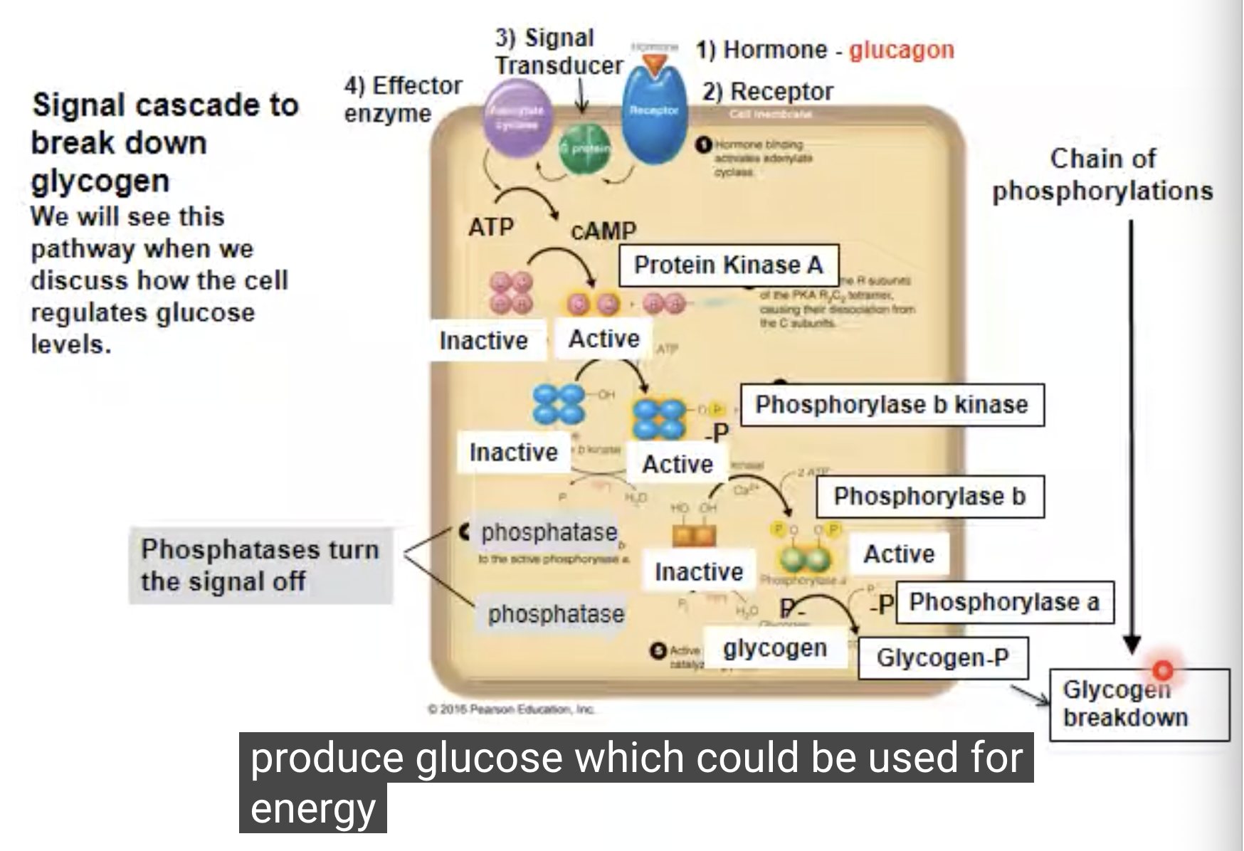
Explain this image:
the transducer is the G protein —> Translates that binding event into a cellularly understandable format, often through a cascade of biochemical changes.
Hormone is the primary messenger: hormone signals to breakdown glycogen
Receptor binds hormone —> signal is transmitted into the cell.
Adenylate cyclase is the effector enzyme that converts ATP to cAMP (second messenger) —> cAMP activates a phosphorylation cascade by binding to other proteins !
T/F: cAMP can activate a series of different kinases?
true!!!!! It can also activate a bunch of phosphorylases!
What do kinases do?
phosphorylate proteins
What do phosphorylases do?
hydrolyze/remove the phosphate from proteins
The ultimate effect of the cascade of phosphorylation is
to break down glycogen! The result is glucose that can be used for energy!
This process is initiated by glucagon: if you need energy, glucagon will initiate this cascade to break down glycogen to create glucose!
How did G-protein couple receptors get their name?
their transducers are g proteins!
GCPRs have 7 transmembrane alpha helices (it is an integral membrane protein)
It has various loop regions involved and a long N and C terminus also!
The N-terminus has some oligosaccharides on it and the C terminus has a lot of recognition areas —> particularly in regards to the phosphorylation of some of these residues!
T/F: Humans have around 800 GCPRs
true
About half of all prescription, non-antibiotic drugs target GCPRs

What is this
Schematic of a G-protein coupled receptor (GPCRs)
Antagonists do what
block a response
Agonists do what?
stimulate a response
Hormones can act as _______ or ________
Hormones can act as agonists or antagonists

What is happening in this picture?
This is describing the structure of beta2 Adrenergic Receptor
The hormones are binding in green
Cholesterol present in the lipid bilayer
Binding of the hormone causes a conformational change and the receptors may dimermerize as a result —→ this transduces the signal to the G-protein
There is structural information available to these proteins
Signal is transduced across the membrane to the G proteins.
G proteins
G proteins have 3 subunits (alpha, beta, and gamma); alpha and gamma are lipid anchored membrane proteins (fatty acid and prenyl).
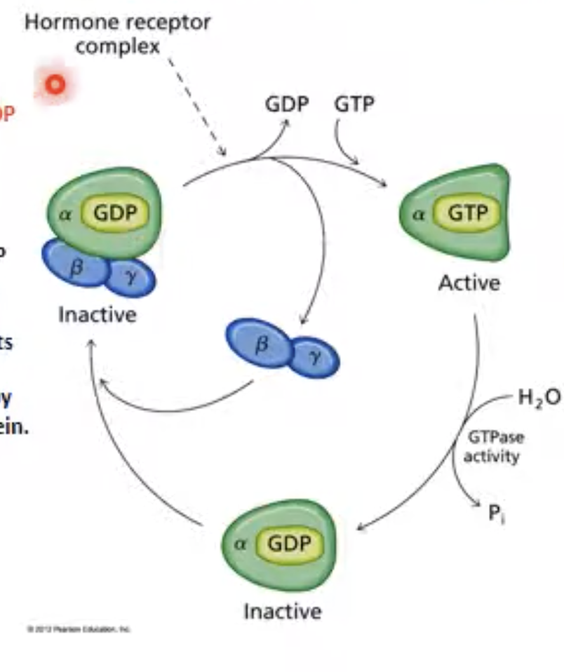
G proteins are inactive when bound to GDP, and active when bound to GTP
G proteins are ______ when bound to GDP, and _______when bound to GTP
G proteins are inactive when bound to GDP, and active when bound to GTP
How do you turn the signal on when working with G-protein?
Turning the signal on:
Activation occurs when the G-GDP interacts with a
hormone receptor complex, activating the G protein by:
1) replacement of GDP with GTP
2) conformational change in a subunit
3) dissociation of b and g subunits: G-GTP now propagates a signal by
interacting with an effector protein.
How do you turn the signal off when working with G-protein?
The a subunit slowly hydrolyzes GTP to GDP, dissociating from the effector protein. G-GDP associates with b and g subunits and is ready for another cycle of activation
What are the three subunits that make up G protein?
alpha, beta, and gamma
T/F: When the hormone binds the G protein coupled receptor and causes a conformational change, the G protein is changing its conformation in a way that it now releases GDP and is able to bind GTP
true! When this happens, the beta and gamma subunits of the trimeric G protein dissociate away as a pair! Now you are left with an active alpha subunit. \
This GTP alpha subunit will be able to interact with the effector enzymes and activate those effector enzymes
After a certain period of time, the GTP is hydrolyzed by the intrinsic hydrolysis activity of the alpha subunit itself (after a certain period of time, GTP would start to be hydrolyzed and you see accumulation of the GDP or inactive form!) —> intrinsic upon the particular G-protein involved!
There are also other proteins like effector proteins that can help to increase the hydrolysis rate to inactivate the G-protein sooner!
Once the effector molecule has been activated, the signal is shut down by shutting down the active G-protein form.
then, the inactived form of G-protein that is bound to GDP will bind to the beta and gamma subunits again! It is now in the inactive state, ready for the next signal, transduction event!
G-GTP =
ON
G-GDP =
OFF

What does the active form do
active form will interact with its target to activate the effector enzyme or become inactivated through GTP hydrolysis
What assists activation of G protein?
GEF (Guanine nucletotide exchange factor, exchanging GDP for GTP
G protein interacts with what to form cAMP?
Adenylate cyclase
Adenylyl Cyclase signalling pathway?
Adenylyl cyclase is the effector enzyme and it is activated or inactivated by G-proteins based on the hormone that is secreted!
Activation pathway:
Hormone binds to and activates a stimulatory receptor (Rs).
Rs activates a stimulatory G protein (Gas)-GTP.
Gas-GTP activates adenylyl cyclase (it is an enzyme integral membrane protein that takes ATP and forms cyclic AMP) which synthesizes cAMP (5’,3’ AMP).
cAMP binds protein kinase A (PKA) and activates it; this releases C subunits from R subunits and PKA is activated.
PKA phosphorylates target proteins (transcription factors) at Ser Thr, activating them.
Phosphorylation of the proteins either activates or inactivates them, thus providing a cellular response based on the hormone!
Inhibitory pathway:
An inhibitory hormone binds a receptor Ri, which activates a (Gia) → this is a G-protein that is inhibitory, by stimulating exchange of GTP in place of GDP.
Gia -GTP inhibits adenylyl cyclase, and the subsequent activation steps are reversed —> this stops the production of cAMP
The other way to stop cAMP is to have a phosphodiesterase hydrolyze cAMP to form 5’-AMP
What are the two ways to shut the signal off in the adenylyl cylase signalling pathway?
Gia -GTP inhibits adenylyl cyclase, and the subsequent activation steps are reversed —> this stops the production of cAMP
The inhibitory hormone binds to its receptor to activate a G-protein that is inhibitory (Gi) —> Gi-alpha will interact with adenylate cyclase to inhibit it and stop the production of cAMP
The other way to stop cAMP is to have a phosphodiesterase hydrolyze cAMP to form 5’-AMP
what is adenylyl cyclase?
it is an enzyme integral membrane protein that takes ATP and forms cyclic AMP
What is CAMP?
You have the 3’ hydroxyl group that will form a phosphodiester linkage with the alpha phosphate to generate cAMP and the leaving group is pyrophosphate!
To turn off cAMP: The hydrolysis reaction that was initiated through the enzyme, phosphodieserase or to have water come in will generate a 3’ hydroxyl group and the 5’ phosphate group —> hydrolyze that phosphoester linkage at the 3’ carbon and forms AMP!!!
How does cAMP activated PKA (protein kinase A)?
2 regulatory subunits that are dimerized and two catalytic subunits
when cAMP is present, it binds to the regulatory subunits and that binding causes a conformational change to allow dissociation of the catalytic subunits that become active and can use ATP to phosphorylate other protein substrates.
To shut down the activity of pka, we need to get rid of cAMP (cAMP is bound in free forms —> when you degrade the free form of cAMP, there is dissociation of cAMP from these regulatory subunits. This makes them inactive and they there associate with the catalytic subunits to turn off pka activation.
How is PKA turned off
Get rid of cAMP: Enzymes called phosphodiesterases (PDEs) destroy cAMP by breaking it down into inactive 5'-AMP.
cAMP leaves the regulatory subunits: As free cAMP is degraded/removed from the cell's fluid, the cAMP molecules attached to the regulatory subunits fall off.
Reassociation and shutdown: Without cAMP holding them in an altered state, the regulatory subunits change back to their original shape. They eagerly re-associate (glue themselves back) to the catalytic subunits.
Back to inactive: Once the regulatory and catalytic subunits snap back together, the regulatory subunits block the catalytic activity once more, turning PKA off. [1, 2, 3]
NIH: https://pmc.ncbi.nlm.nih.gov/articles/PMC9692727/
PKA when turned on looks like
Inactive PKA: Normally, PKA is made of two catalytic subunits (the parts that do the work) bound to two regulatory subunits (the brakes). While attached, the regulatory subunits block the catalytic subunits, keeping PKA off.
Turning PKA On: When cAMP builds up in the cell, it binds to the regulatory subunits. This changes their shape, causing them to dissociate (release/fall off) from the catalytic subunits. The free catalytic subunits are now active and go on to phosphorylate other proteins.
NIH: https://pmc.ncbi.nlm.nih.gov/articles/PMC1765484/
What type of pathway is Inositol-phospholipid signaling pathway?
G-protein coupled receptor mediated pathway.
What happens in the Inositol-phospholipid signalling pathway?
When the ligand binds to its cognitive receptor, a G-protein is activated, this G-protein is G_q and it interacts with phospholipase C to cause it to be active (phospholipase C is an enzyme that cleaves glycerolphospholipids to remove the acyl component of the glycerolphospholipids to release the phospho-head group (hydrolyze the phosphoester linkage to generate inositol 1,4,5-trisphosphate or IP3 and diacylglycerol (DAG).
So PIP2 (phosphotidylinositol 4-5 bisphosphate in the lipid) is hydrolyzed by phospholipase C to form DAG and IP3 —> IP3 goes into the cytosol and gets degraded by phosphatases that take off one phosphate at a time and eventually generates your sugar alcohol inositol.
IP3 itself can bind to a calcium channel that aligns in the lumen. The ligand gated activation of the channel allows calcium to leave the ER and enter the cytosol because cytosolic Ca²+ levels are low, but high in the ER (endoplasmic reticulum) —> Calcium can then go into the cytosol and bind to kinases and activate them.
Calcium binds to PKC and PKC also needs DAG to bind to it in order to be active!
PKC is named this way because it is a kinase that is activated by phospholipase C activity!
PKC is then activated and is membrane associated and phosphorylates certain proteins on their hydroxyl groups to cause them to be activated or inactivated to promote a cellular response!
What are the two secondary messengers of the Inositol-phospholipid signalling pathway?
he two secondary messengers of the inositol-phospholipid signalling pathway are diacylglycerol (DAG) and inositol 1,4,5-trisphosphate (IP3)
Receptor Tyrosine kinase signalling (_____________)
insulin receptor
Receptor Kinase Signaling:
these pathways are activated by these receptors that are kinases that are integral membrane proteins that are activated by ligand binding!
The insulin receptor starts out in an inactive state, but when it binds insulin, it becomes active! It phosphorylates itself and tyrosine residues on the receptor become phosphorylated through transfer of the gamma phosphate from ATP to the tyrosine residue —> autophosphorylation!
Autophosphorylation only occurs when the hormone is binding to the receptor
Once the receptor has autophosphorylated itself, its now able to interact with other insulin receptor substrates and phosphorylate those —> in insulin signalling, it activates phosphitidyl 4-5 diphosphate kinase (PI kinase) to phosphorylate PIP2 to become PIP3!
PIP3 is the second messenger that activates PIP3 dependent protein kinases to propogate the signal
When does autophosphorylation occur?
Autophosphorylation only occurs when the hormone is binding to the receptor
Is there a g protein involved with insulin
NOPE
Summary of Receptor Try Kinase Signalling:
Ligands bind to the receptors and they dimerize to become activated and autophosphorylate their kinase domains to allow interaction with other protein substrates in which they will promote the signal transduction
What is this describing:
Many growth factors use tyrosine kinase pathways for regulation.
The receptor, transducer and effector functions of tyrosine kinases residue in a single membrane protein
The tyrosine kinase domains of the receptor in the inactive state are separated.
ligand binding causes a conformational change, bringing 2 proteins together to form a dimer
When the kinases are close together inside the cell, they phosphorylate each other - called autophosphorylation, activating the kinase
The kinase phosphorylates other cytosolic proteins, transmitting the signal
What is the insulin signal transduction
Dimerization = Requires lateral movement through the membrane.
What is insulin’s effector enzyme:
Autophosphorylation
What is the structure of a tyrosine kinase: actually dont really need to know this
Single, spanning alpha helical transmembrane spanning region. There is an extracellular domain that binds the ligand. On the cytoplasmic side, adjust the membrane segment: you have a kinase domain that has an activation loop and a C terminal tail —> lots of regualtory aspects of the cytoplasmic domain
For Try ligand signalling, when the ligand binds, what happens?
You have a dimerization of the two monomeric units that are ligand bound and these kinase domains will start to interact and autophosphorylate one another and promote transphosphorylation!!!
Insulin, epidermal growth factor, and fibroblast growth factor are examples of what
tyrosine kinases
T/F: if we look at the different types of tyrosine kinases, the extracellular domain is much more conservative compared to the intracellular/kinase domain
true
What is unique about the insulin receptor?
there is a disulfide between the alpha subunits that form upon the dimer → this gives you a covalent linkage between the monomeric units that do not exist for the other singalling mechanisms in the family!
What is diabetes?
result of the the loss of control of insulin siganling
Type 1 diabetes:
insulin is no longer produced. Also called insulin dependent diabetes. Insulin not made by the pancreas
Type 2 diabetes:
often results from insulin resistance due to reduced binding or signaling from the insulin receptors —> person is still making insulin tho
T/F: the disulfide bond is formed between the two tyrosine kinase receptors upon insulin binding
true! The autophosphorylation occurs in the kinase domains. Then the phosphorylation of downstream substrates to propogate signal
T/F: does the concept of receptor dimerization and autophosphorylation occurs in plants
true! The plants have all 3 residues that involve phosphorylation. BUT, in mammals it is just tyrosine phosphorylation.
There are a lot more genes in plants, do there is a lot more receptor kinases that exist as well.