Eukaryotic Module 13
 Call Kai
 Learn
 Practice Test
 Spaced Repetition
 Match
 Flashcards
 Knowt Play
1/40

Earn XP
Description and Tags
chapter 13 and 14
41 Terms
DNA replication model
proposed by Watson and Crick
replication occurred by gradual separation of the strands of the double helix
each strand can act as a template to direct the synthesis of the complementary strand and restore the double-stranded state
as a result, each of the daughter duplexes consist of one complete strand inherited from the parental duplex and one complete strand that has been newly synthesized
this is called semiconservative replication

conservative vs. dispersive replication
other two DNA replication models
have been disproved

meselson and stahl experiment
supported the semiconservative model of replication in bacterial cells
used heavy (15N) and light (14N) isotopes of nitrogen to distinguish between parental and newly synthesized DNA strands
grew bacteria in a medium with 15N as the sole nitrogen source, making the nitrogen-containing bases of the DNA only contain the heavy nitrogen isotope
cultures of “heavy” bacteria were washed free of the old medium and incubated in fresh medium with light 14N-containing compounds
samples were removed at increasing intervals over a period of several generations
DNA was extracted from the samples of bacteria and subjected to equilibrium density-gradient centrifugation (DNA is mixed with cesium chloride)
the double-stranded DNA will reach equilibrium according to their density
in this experiment, the density of the DNA molecule was directly proportional to the percentage of 15N or 14N atoms it contained
for semiconservative replication, after one generation DNA molecules would be hybrids of 15N and 14N, so they would be in the middle
as replication continues, the newly synthesized strands would contain only the light isotopes, and the two types of duplexes would appear in the gradients: those containing hybrids and those containing only light

experiment for DNA replication in eukaryotes
cultured mammalian cells underwent replication in bromodeoxyuridine (BrdU), incorporated into DNA in place of thymidine
after one round of replication in BrdU, both chromatids of each chromosome pair contained BrdU
after two rounds of replication, one chromatid of each chromosomes was composed of two BrdU-containing strands, the other chromatid was a hybrid of a BrdU-containing strand and a thymidine-containing strand
demonstrated semiconservative replication

replication in bacterial cells
origin: a specific site (oriC in E. coli) where a number of proteins bind to initiate the process of replication
replication proceeds bidirectionally, meaning the once initiated, replication proceeds outward from the origin in both directions
replication forks: the sites where the pair of replicated segments come together and join the non-replicated segments

the unwinding problem
the separation of the DNA strands causes two topological problems:
unwinding strands causes torsional stress, in which the unseparated portion becomes more tightly wound
this overwound DNA then becomes positively supercoiled
overall, movement of the replication fork generates positive supercoils in the unreplicated portion of the DNA ahead of the replication fork

DNA gyrase
DNA gyrase is a type II topoisomerase
relieves the mechanical strain that builds up during replication
works by cleaving both strands of the DNA duplex, then it passes a segment of DNA through the double-stranded break to the other side, and seals the cuts
the process is driven by the energy released by ATP hydrolysis
DNA polymerase
synthesizes new DNA strands
DNA pol requires a template DNA strand to copy, and a primer to add nucleotides on to
DNA pol cannot initiate the formation of a DNA strand
primer is a DNA strand that provides the necessary 3’ OH terminus
can only synthesize DNA in a 5’→ 3’ direction
synthesis of new strands
both newly synthesized strands are assembled in a 5’→3’ direction
one grows toward the replication fork and the other grows away
semi-discontinuous replication: one strand is synthesized continuously and the other discontinuously
leading strand: the strand that is synthesized continuously
lagging strand: the strand that is synthesized discontinuously

okazaki fragments
Reigi Okazaki incubated bacteria in [3H]thymidine and extracted DNA at different time points
okazaki fragments: the small segments synthesized during DNA replication in the lagging strand
1000-2000 nt in bacteria, about 150 nt in eukaryotes
formation of okazaki fragments
the initiation of okazaki fragments is accomplished by a distinct type of RNA pol (primase) that constructs a short RNA primer
primer is at the 5’ end of each fragment
a short RNA primer is also synthesized at the 5’ end of the leading strand by primase
the RNA primers are subsequently removed, and the resulting gaps are filled with DNA and then sealed by DNA ligase

machinery of replication fork
helicase: an enzyme that uses energy from ATP hydrolysis to unwind a DNA duplex and expose the single-stranded DNA templates; breaks hydrogen bonds
helicase (ex. DnaB) has 6 subunits forming a ring-shaped complex that encircles the lagging-strand template and moves in a 5’→ 3’ direction
DnaB is the major unwinding machine for replication in E.coli
single-stranded DNA-binding (SSB) proteins: proteins that selectively bind to single-stranded DNA, keeping it in an extended state and preventing it from being rewound or damaged
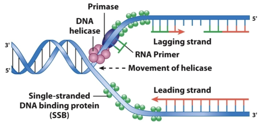
initiation of replication in E. coli
begins when multiple copies of the DnaA protein bind to oriC and separate the DNA strands at that site
DnaB helicase is then loaded onto the single-stranded DNA of the lagging strand of oriC, with the help of DnaC
DnaB helicase then translocates in a 5’→3’ direction along the lagging strand and unwinds the helix as it goes
attachment of SSB proteins help unwind the strands
three DNA polymerases in bacteria
DNA pol III: has polymerizing activity and 3’→5’ exonuclease activity
part of a large protein complex called DNA pol III holoenzyme (replisome) that synthesizes DNA strands during DNA replication
DNA pol II: has polymerizing activity and 3’→5’ exonuclease activity; has unknown functions
DNA pol I: polymerizing activity, 5’→3’ exonuclease activity, and 3’→5’ exonuclease activity
a single polypeptide that has three activities in different domains of the polypeptide, functioning in DNA repair and removing the RNA primers of okazaki fragments and replacing them with DNA
the holoenzyme (DNA pol III) components
β clamp: a non-catalytic component that keeps the pol associated to the DNA template
clamp loader: a multisubunit that aids in the assembly of the β clamp around the DNA
in the ATP bound state, the clamp loader bings to a primer-template junction while holding the β clamp in an open conformation
once DNA has gone through the opening, the ATP bound to the clamp loader is hydrolyzed, causing the release of the clamp, which closes around the DNA and the β clamp can bind pol III

β clamp of the holoenzyme
polymerases must remain associated with the template over long stretches to synthesize a continuous strand, but they also have to be loosely attached enough so they can move from one nucleotide to the next; these properties are provided by the β clamp
the pol on the leading-strand template remains tethered to a single β clamp during replication
the pol on the lagging-strand template completes the synthesis of an okazaki fragment, and then disengages from the β clamp and is cycled to a new β clamp that has been assembled at an RNA primer-DNA template junction closer to the replication fork
replication of leading and lagging strands
the pol synthesizing the lagging strand “hitches a ride” on the pol synthesizing the leading strand in order to move to another site on the lagging-strand template
the two tethered pols can replicate both strands by looping the DNA of the lagging-strand template back on itself, causing the template to have the same orientation as the leading-strand template
once the pol assembling the lagging strand reaches the 5’ end of the okazaki fragment synthesized during the previous round, the lagging-strand template is released and the pol begins work at the 3’ end of the next RNA primer toward the fork (trombone model)
recent studies have shown that there are actually 3 DNA pol IIIs acting in the replisome; one on the leading strand and two on the lagging strand (picture only shows 2)

replication in eukaryotic cells
eukaryotic cells replicate their genome in small portions called replicons
each replicon has its own origin from which replication forks proceed outward in both directions
the timing of replication of a chromosome region is roughly correlated with the activity of the genes in the region and/or its state of compaction
autonomous replicating sequences (ARSs): sequences that promote replication of the DNA in which they are contained
origin recognition complex (ORC): a multi-subunit complex specifically binds to the core element of an ARS
replication in eukaryotic chromosomes begins at many sites along the DNA
mechanism restricting replication to once per cell
the origin of replication is bound by an ORC complex
“licensing factors” bind to the ORC to assemble a prereplication complex
activation of key protein kinases leads to the initiation of replication
the activity of Cdk (a cyclin-dependent kinase) remains high from S phase through mitosis, which suppresses the formation of a new prereplication complex
the “licensing factors” move with the replication fork to help complete replication of a replicon
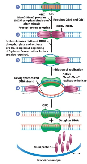
the eukaryotic replication fork
replication activities are similar in eukaryotes and prokaryotes
all replication systems require helicases, single-stranded DNA-binding proteins, topoisomerases, primase, DNA polymerase, sliding clamp and clamp loader, and DNA ligase
DNA is synthesized in a semi-discontinuous manner, and the leading and lagging strands are synthesized in a coordinate manner by a single replicative complex, or replisome
left picture shows proteins required for eukaryotic replication and right shows the replisome protein interactions

5 eukaryotic DNA polymerases
pol α: tightly associated with primase, and helps initiate the synthesis for each okazaki fragment
pol β: functions in DNA repair
pol γ: replicates mitochondrial DNA; has 3’→ 5’ exonuclease activity
pol δ: the primary DNA-synthesizing enzyme during replication of the lagging strand; has 3’→ 5’ exonuclease activity
pol ε: the primary DNA-synthesizing enzyme during replication of the leading strand; has 3’→ 5’ exonuclease activity
other DNA pols: η, κ, and ι have a specialized function that allows cells to replicate damaged DNA
all of the eukaryotic pols elongate DNA strands in the 5’→ 3’ direction
replication foci
in eukaryotes, replication forks that are active at a given time are not distributed randomly throughout the cell nucleus, but instead are localized within 50-250 sites, called replication foci
meaning that replication activities are confined to distinct sites in the nucleus
replication of chromatin structure
the assembly of DNA onto nucleosomes is a very rapid event
the nucleosomes on both daughter duplexes are very close to the replication fork
the (H3H4)2 tetramers present prior to the replication remain intact and are distributed randomly between the two daughter duplexes
the two H2A/H2B dimers of a nucleosome separate from one another and bind randomly to the new and old (H3H4)2 tetramers present on the daughter duplexes
3 broad categories of cells
cells that are highly specialized and lack ability to divide
cells that normally do not divide but induced to divide
cells that normally have relatively high dividing activity
cell cycle
the cycle of cell growth, replication of genetic material, and nuclear and cytoplasmic division
two major phases:
M phase: mitosis (duplicated chromosomes are separated into two nuclei) and cytokinesis (the entire cell divides into two daughter cells)
lasts about an hour in mammalian cells
interphase: the period between cell divisions when the cell grows and engages in diverse metabolic activities
can last days, weeks, or longer depending on cell type and conditions

interphase periods
G1: the first gap; the period following mitosis and preceding DNA synthesis
cell grows and carries out normal metabolism; organelles duplicate
S phase: the period in which DNA is replicated and additional histones are also synthesized; DNA replication and chromosome duplication
G2: the second gap; the period between the end of DNA synthesis and beginning of M phase where the cell grows and prepares for mitosis
cell fusion experiments
to understand if the transitions from G1 → S and G2 → M were controlled by an stimulatory factors, cell fusion experiments were performed
when G1 and S phase cells were fused, the G1 nuclei initiated DNA synthesis
when G2 and S phase cells were fused, the G2 phase nuclei did not initiate another round of DNA synthesis
when G1 and M-phase cells were fused, the G1 phase chromatin underwent premature chromosomal compaction (PCC) to form a set of elongated compacted chromosomes
when G2 and M phase cells were fused the G2 phase chromosomes underwent PCC to form doubled compacted chromosomes
when S phase and M phase cells were fused, the S phase chromosomes underwent PCC for form pulverized chromosome fragments
conclusions of fusion experiments
fusion experiments of G and S phases showed that the cytoplasm of a replicating cell contains diffusible factors that stimulate initiation of DNA synthesis in G1 phase nuclei and the G2 phase nuclei can no longer respond to initiation factors present in the S phase cell cytoplasm
suggests that the transition from G1 to S was induced by cytoplasmic factors
G/S and M phase fusion experiments found that the cytoplasm of an M phase cell contains diffusible factors that stimulate premature chromosomal compact of G1, G2, and S phase chromosomes, suggesting that the transition from G2 to M was induced by the cytoplasmic factors
maturation-promoting factor (MPF)
a cytoplasmic factor that initiates the entry of a cell into M phase
includes two subunits: one with kinase activity that phosphorylates specific protein substrates, and another regulatory subunit called cyclin
kinase transfers a phosphate group from ATP onto Ser or Thr residues of specific protein substrates
called cyclin bc the concentration of this protein rises/falls in a predictable pattern with each cell cycle
when cyclin conc. is low, the kinase lacks the cyclin subunit and is inactive
when cyclin conc. rises, the kinase is activated, causing the cell to enter M phase
suggests that the progression of cells into mitosis depends on a kinase and the activity of the kinase is controlled by cyclin whose concentration varies from one stage of the cell cycle to another
cyclin-dependent kinases (Cdks)
not only involved in M phase but also key regulators that coordinate activities throughout the cell cycle
each phosphorylation event occurs at a appropriate point in the cell cycle, which stimulates or inhibits a particular cellular process in cell division
*MPF is not a Cdk but itself, but rather it is a complex that contains a Cdk combined with a cyclin
two distinct stages of regulation in the eukaryotic cell cycle
first transition point: called START, which is near the end of G1, where a call becomes committed to initiating DNA replication and completing the cell cycle
the levels of one or more G1/S cyclins rise during late G1
these cyclins bind to cdc2 (a Cdk) and activate it
second transition point: near the end of G2, where the cell becomes committed to entering mitosis
Cdks containing a mitotic cyclin (ex. MPF) bind and activate cdc2, which phosphorylate substrates that are required for the cell to enter mitosis
these substrates include proteins needed for dynamic changes in chromosomes and the cytoskeleton

regulation of the activities of Cdks
cycling binding: when cyclin reaches a sufficient conc. in the cell, it binds to the catalytic subunit of the Cdk, causing a major change in the conformation of the enzymes active site
the phosphorylation state of Cdks: more on next slide
cyclin inhibitors: Cdk activity can be blocked by a variety of inhibitors; degradation of the inhibitors allows Cdks to be activated and initiate DNA replication
controlled proteolysis: cells regulate the conc. of cyclins by adjusting synthesis and destruction rates; key regulators of cell cycle are degraded by the ubiquitin-proteasome pathway
subcellular localization: cell cycle regulators are moved into different compartments at different stages to either be united with or separated from the proteins they interact with
Cdk phosphorylation and dephosphorylation
one kinase, CAK (Cdk-activating kinase) phosphorylates a critical Thr residue, which is necessary for the Cdk to be active (but not fully sufficient)
a second kinase, Wee1, phosphorylates a key Tyr residue in the ATP-binding pocket of the enzyme, making the Cdk inactive
the effect of Wee1 overrides the effect of CAK
keeps the Cdk inactive until the end of G2
at the end of G2, the inhibitory phosphate is removed by a phosphatase, Cdc25, switching the stored cyclin-Cdk molecules into the active state, allowing them to phosphorylate key substrates and drive the cell into mitosis

Cdks in yeast vs. mammalian cells
in yeast, there is only one Cdk, cdc2
mammalian cells have several different Cdks, and each binds to a specific cyclin
picture shows combinations between various cyclins and Cks at different stages in the mammalian cell cycle

cell cycle checkpoints
checkpoints: mechanisms that halt the progress of a cell cycle
cell cycle can be halted if any of the chromosomal DNA is damaged, or if certain critical processes (like DNA replication during S phase or chromosome alignment during M phase) have not been properly completed
checkpoints are activated throughout the cell cycle by a system of sensors that recognize DNA damage ro cellular abnormalities
if a sensor detects the presence of a defect, it triggers a response that temporarily arrests further cell cycle progress
two processes in M phase
mitosis: a process of nuclear division in which the replicated molecules of each chromosome are faithfully partitioned into two nuclei
cytokinesis: a process by which a dividing cell splits in two, partitioning the cytoplasm into two cellular packages
five stages of mitosis in animal and plant cells
prophase
prometaphase
metaphase
anaphase
telophase

prophase
during this stage, the duplicated chromosomes are prepared for segregation and the mitotic machinery is assembled
formation of the mitotic chromosome
chromosome compaction (condensation)
chromosome scaffold
condensin
cohensin
formation of the mitotic spindle
the dissolution of the nuclear envelope
chromosome compaction (condensation)
before segregating its chromosomes, a cell converts them into much shorter, thicker structures through compaction/condensation
compaction does not alter the nature of the chromatin fiber, just changes its packaging
scaffold proteins: a structural framework of nonhistone proteins that maintain the basic shape of the intact chromosome
during interphase, the scaffold proteins are dispered within the nucleus, forming part of the cellular matrix