PSIO 201 lab practical #1
 Call Kai
 Learn
 Practice Test
 Spaced Repetition
 Match
 Flashcards
 Knowt Play
1/92

There's no tags or description
Looks like no tags are added yet.
93 Terms
Simple Squamous Epithelial tissue
Location: alveoli, mesothelium, glomerular capsule, capillaries
Function: diffusion, filters blood of waste, seccretion
"allows for gas exchange of oxygen and co2 into alveoli"
Key Features: surrounds lumen, one layer, squished/fried egg shaped, center of cell located nuclei, flat

Simple Cuboidal Epithelium
Location: glands, kidney tubules
Function: secretion, absorption
"filters blood and reabsorbs nutrients in the kidney tubules"
key features: one layer, cubed shaped cells, center of cell located nuclei, lines lumen

Simple Columnar Epithelium
location: lining of stomach, small intestine, large intestine, uterine tubes, spinal cord
function: absorption, secretion, move CSF
"secretes mucus to protect stomach lining from stomach acid acidity"
key features: nuclei located at equal layer at bottom of cell, goblet cells present, cilia present, column shaped cells, one layer, lines lumen

Pseudostratified Ciliated Columnar Epithelium
location: lining of nasal cavity, trachea, bronchi
function: secretion of mucus, cilia move mucus
"moves mucus towards throat where it can be expelled or swelled to be killed by stomach acid"
key features: one layer, squished shaped cells, cilia present, goblet cells present, location of nuclei at different levels, illusion of several layers of cells, all cells touch basement membrane, different shaped cells

Stratified Squamous Epithelium
location: lining of mouth/vagina, skin surface, esophagus, rectum
function: protective barrier from abrasion
"in the oral cavity this tissue provides a protective barrier from microorganisms"
key features: many layers of cells, flat/squished shapes of cells, location of nuclei in center of cell, lines a lumen

Transitional Epithelium
location: lining of urinary bladder, uterus and urethra
function: protective barrier permitting distension
"allows bladder to expand and contract to hold urine"
key features: many layers, cuboidal and squamous cell shapes, nuclei located at center of cell, lines lumen, "distended or non-distended version"
- non distended = makes round shapes (takes more space)
- distended = being pushed into flat

myocytes
elongated muscle cells that shorten to cause movement
Skeletal Muscle Tissue
location: attached to bones and skin throughout the body
Function: heat generation, movement of bones and skin
Key features: 3-5 nuclei per cell, striations, cylindrical-shaped muscle fibers, muscle type --> under voluntary control

Cardiac Muscle tissue
location: walls of the heart
function: movement of blood through cardiovascular system
key features: striations, muscle fibers branch, intercalated disks present, muscle type--> involuntary control, 1-2 nuclei per muscle fiber
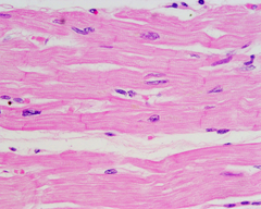
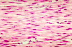
Smooth Muscle Tissue
location: intrinsic eye muscles, walls of arteries, veins, walls of bladder, urethra,
function: in eye muscles--> control pupil size, in arteries and veins--> controls blood flow, blood pressure, in urintray tract--> controls urine movement
key features: muscle type -->under involuntary control, spindle shaped fibers, 1 nuclei per muscle
4 types of connective tissue
1. connective tissue proper (dense and loose)
2. cartilage
3. bone
4. blood
ground substance
allows for movement of nutrients and waste
mast cells
creates immune response by releasing histamine
Loose areolar connective tissue
location: beneath all epithelial tissue, between muscles, lines digestive sytem, respiratory tract, urine tract
function: strength, elasticity, support and diffusion
key features: fibroblasts, mast cells, abundant and viscous ground substance
fiber types: collagen, elastic, reticular

loose adipose connective tissue
location: subcutaneous layer of skin, behind eye, butt, around joints
function: cushions joints and organs, padding, thermal insulation
"absorbs shock in joints"
key features: has adipocytes (fat cells), small nucleus around cells periphery, lipid droplets (adipose)
fiber types: collagen, elastic (little bit)

dense regular connective tissue
location: ligaments, tendons, covering skeletal muscle
function: resist force in one direction
Features: fibroblasts arranged between collagen fibers, ECM is filled with parallel bundles of collagen fibers, little ground substance, little blood vessel
fiber types: collagen fibers, some elastic

dense irregular connective tissue
location: heart valves, reticular region of dermis, capsules around organs
function: resists pulling forces from many directions
features: fibroblasts (very few), around other tissues, little ground substance, avascular
fiber types: collagen

Hyaline cartilage connective tissue
location: nose tip, end of joint, supports larynx, forms septum
function: strength, support, reduces friction
"provides smooth surface for joints to move"
features: chondrocytes, chondroblasts, cells reside in lacunae where they will stay in, avascular, clear/glassy appearence
fiber types: fine collagen fibers (not visible)

fibrocartilage connective tissue
location: menisci of the knee, pubic symphysis, intervertebral discs, parts of tendons
function: provides strength and rigidity, support and joins structures together
"prevents damaging bone to bone contact"
Features: few chondrocytes, cotton candy like appearence, has few lacunae, tough, avascular
fiber types: thick collagen

elastic cartilage connective tissue
location: auditory tubes, auricle of external ear, epiglottis of larynx
function: provides strength and elasticity
"maintains the shape of flexible body parts, example ear"
Key features: chondrocytes in a thread-like network of elastic fibers in the extracellular matrix, "stretchy black fibers" highly visible "spooky"
fiber types: elastic

blood connective tissue
location: in blood vessels, chambers of heart
function: blood clotting, transport, regulation, protection
"prevents blood loss in body by clotting blood in blood vessels"
Key features: has erythrocytes(RBC) , leukocytes(WBC) , thrombocytes(platelets) , extracellular matrix is liquid plasma abd contains diff cell types
Fiber types: none

compact bone connective tissue
location: surface of bones
function: supports body structure, protection, storage, serves as a lever acting with muscle tissue for movement
"acts with muscle tissue as a lever to move body"
Key features: osteocytes, hardest connective tissue, has lacunae, lamellae, canaliculi, central canals, blood vessels
fiber types: collagen, some elastic

key features of epithelial tissue
highly cellular, avascular, can be layered, always lining a lumen
lumen
space lining an organ
Epidermis
made up of stratified squamous epithelium

nervous tissue
location: brain, spinal cord, nerves, white matter on inside of brain
function: neurons recieve and send info while neurogila support the neurons and help them function
key features: neuron is main cell type, has dendritie branches which recieve info, send signal along axon portion, soma is the cell body, white nervous tissue transmits signals faster than gray

dermis
made up of areolar CT in papillary region and dense irregular CT in the reticular region

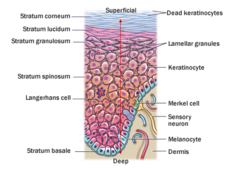
epidermal layers top--> down
1. stratum corneum
2. stratum lucidum
3. stratum granulosum
4. stratum spinosum
5. stratum basale
hair root plexus
free nerve endings wrapped around hair follicle; detects hair movement
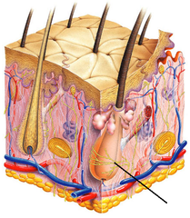
arrector pili
a smooth muscle attached to hair follicles that causes "goose bumps" to appear on the skin when contracted, "errects the hair"
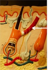
Eccrine gland
on most skin regions, goes from deep dermis all the way up to the epidermis

Apocrine gland
in armpit, pubic area, and around nipples, in deep dermis and opens up to hair follicle

Sebaceous gland
connected to hair follicle, secrete sebum

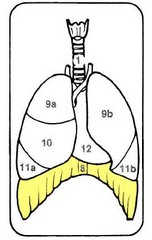
what separates the thoracic cavity from the abnominopelvic cavity
diaphragm
supine
body horizontal lying on the back, face up
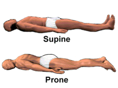
prone
body horizontal lying of stomach, face down

organ system level
Organ systems consist of different organs that work together closely
superior (cranial)
closer to the head

inferior (caudal)
closer to the feet

anterior (ventral)
closer to the front
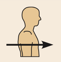
posterior (dorsal)
closer to the back

medial
closer to the midline

lateral
farther from the midline
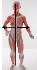
intermediate
between a more medial and more lateral structure
proximal
closer to the trunk (or point of limb attachment)

distal
farther from the trunk (or point of limb attachment)

superficial (external)
closer to or on the surface
deep (internal)
farther from the surface
parietal
pertaining to the wall of a body cavity

visceral
pertaining to the covering of an organ

ipsilateral
on the same side of the body

contralateral
on the opposite side of the body

What is a sagittal plane?
A sagittal plane divides the body or organ vertically into left and right parts.

What is the difference between a midsagittal and a parasagittal plane?
A midsagittal plane divides the body into equal right and left parts, while a parasagittal plane divides it into unequal right and left parts.
What does a frontal plane divide the body into?
A frontal plane divides the body or organ into anterior (front) and posterior (back) parts.

What is another name for the frontal plane?
The frontal plane is sometimes referred to as a coronal plane.
What does a transverse plane divide the body into?
A transverse plane divides the body or organ horizontally into superior (upper) and inferior (lower) portions.

What type of images do transverse planes produce?
Transverse planes produce images referred to as cross-sections.

longitudinal section
a plane along the long axis of an organ

oblique section
cuts made diagonally between the horizontal and the vertical planes

cross section
a diagram that shows what the inside of something looks like

anatomical regions of the body
The body is divided into:
- Cephalic (head)
[Frontal (forehead), Orbital or ocular (eye), Buccal (cheek), Nasal (nose), Oral (mouth), Mental (chin), Cranial (surrounding the brain), Occipital (base of skull), Otic (ear), Temporal (temple)]
- Cervical (neck)
- Thoracic
[Axillary (armpit), Mammary (breast), Pectoral (chest), Sternal (sternum)]
- Upper Extremity
[Scapular (shoulder blade), Brachial (arm), Antecubital (front of elbow), Olecranal (back of elbow), Antebrachial (forearm), Carpal (wrist), Dorsum (back of the hand), Manual (hand), Palmar (palm), Digital (finger)]
- Abdominal (abdomen)
- Pelvic
[coxal (hip), Inguinal (groin)]
- Lower Extremity
[Femoral (thigh), Patellar (kneecap), Popliteal (hollow behind knee), Crural (leg), Sural (calf), Calcaneal (heel), Plantar (sole), Tarsal (ankle), Dorsum (top of foot), Digital (toe), hallux (great toe)

2 major body cavities
dorsal(posterior) and ventral(anterior)
Function of dorsal cavity
protects the nervous system organs
2 sub categories of cavities in dorsal cavity
cranial cavity and spinal cavity
2 sub categories of cavities in ventral cavity
thoracic cavity(enclosed by rib cage) and abdominal pelvic
2 sub categories of cavities in Thoracic cavity
pleural cavity LEFT and RIGHT (left lung and right lung) and pericardial cavity (heart)
2 sub categories of cavities in abdominal pelvic cavity
abdominal cavity and pelvic cavity
what is the protective membrane around the dorsal cavity organs
meninges
what is the fluid around the dorsal cavity organs
cerebral spinal fluid
what is the protective (serous) membrane around the pericardial cavity organs
pericardium
what is the protective (serous) membrane around the pleural (left or right) cavity organs
pleura (left or right)
what is the protective (serous) membrane around the abdominal cavity
peritoneum
what is the fluid around the thoracic and abdominal cavity organs
serous
what is a body cavity?
separation of internal organs, separates to perform their functions without interffering
the abdominal pelvic quadrant has how many separations
4 quadrants
the abdominal pelvic regions has how many separations
9 grid
what are the 4 quadrants of the abdominal pelvic region
right upper quadrant, left upper quadrant, right lower quadrant, left lower quadrant

what are the 9 abdominal pelvic regions
Umbilical
Right lumbar
Left lumbar
Epigastric
Right hypochondriac
Left hypochondriac
hypogastric
Right iliac
Left iliac

what are the 4 major tissue types in the body
connective, muscular, nervous, epithelial
integumentary system includes what
skin, hair, nails, sweat glands, oil glands
skin has what
all 4 tissue types
hypo
below
chondro
cartilage
epi
above
gallbladder function
stores and concentrates bile

bile function
digest food

serous fluid function
Fluid that lubricates the 2 layers and allows organs to move freely within the cavity, protects against friction
how do you name specific tissue type
specific tissue name + primary tissue name
(ex. cardiac muscle)
basic structures of the neuron tissue
dendrites - receive signal
soma = cell body
axon = where signal sent off
**neuroglia = suppor the neuron and help them function
**neuron = receives and send info

white nervous tissue
transmits signals faster than gray, most of brain
myocytes
elongated muscle cells that shorten to cause movement
functions of muscle
body