Interference
 Note
Note Call Kai
 Learn
Learn Practice Test
 Spaced Repetition
 Match
Match Flashcards
 Knowt Play
1/19

There's no tags or description
Looks like no tags are added yet.
Name | Mastery | Learn | Test | Matching | Spaced | Call with Kai | Chat |
|---|
No analytics yet
Send a link to your students to track their progress
20 Terms
What is a lesion?
Damage to brain tissue often studied to understand behavior.
Broca's Area
Area in the left inferior frontal gyrus associated with speech production, named after Paul Broca who studied a patient with a lesion there.
Phineas Gage
A railroad worker whose personality changed dramatically after a brain injury caused by an iron rod passing through his frontal lobe.
Anterograde Amnesia
Inability to form new memories, often associated with damage to the hippocampus, as seen in the case of Henry Molaison.
Group Studies
Method in neuroscience that groups individuals based on behavioral symptoms or lesion locations to study brain function.
Electrical Stimulation
Technique involving the insertion of electrodes in the brain to cause or stop neuronal firing, often used during neurosurgery.
Optogenetics
A method that uses light to control neurons that have been genetically modified to be light-sensitive; mostly used in mice
Transcranial Magnetic Stimulation (TMS)
A non-invasive technique that uses magnetic fields to stimulate nerve cells in the brain, helping to map functions.
Motor Homunculus
A representation of the body in the brain where different body parts are mapped to specific areas that control their movements.
Neural Activity
The electrical and chemical processes that occur in the brain due to neuron firing, which can be measured to study brain functions.
What causes lesions?
caused by injury, surgery, stroke, hemorrhage, tumors, or degenerative diseases
Who is “tan”?
A patient of Paul Broca who could only say “tan” because of a lesion in his IFG (broca’s area)
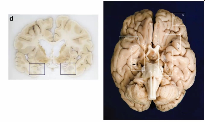
Who is H.M?
Henry Molaison was an epileptic patient that developed anterograde amnesia after his hippocampus was removed to prevent the seizures.
What was different about H.M’s brain?
The hippocampus is missing (notice the holes in his brain)
Orbital damage in part c due to lack of connection to hippocampus
Ventricles are large (white stuff in middle)
Corpus Callosum is thinned out (layer above ventricles)
Cortex is thinner than normal
Caudate looks different
how would you figure out what a brain region does by studying patients with lesions to that area?
observe specific cognitive, sensory, or motor deficits they experience. By comparing their performance on targeted tasks to control groups (patients with lesions in different areas and healthy individuals), you can identify impairments uniquely associated with damage to the target region.
Pros and Cons of Electrical Stimulation?
Pros: allows researchers to map brain regions in real-time which is useful in surgery or research on brain function; can increase or decrease neuronal activity
Cons: invasive, not super precise in targeting specific neurons
What is the process of using optogenetics?
create a genetic construct of light sensitive ion channels for neurons with certain protein
insert it in a virus
put the virus in the mouse brain
fiber optic cable implanted in brain
lasers of specific wavelengths are imputed in the animal
pros/cons of optogenetics?
pros: you can target a specific kind of neuron with precision, you can inc or dec neuronal activity
cons: invasive, can’t be done in humans
In detail, how does a TMS work?
A figure 8 shaped coil creates a magnetic field which disturbs the electrical flow of neurons. Positive charges down towards the cell body can depolarize neurons and lead to excitation
pros/cons of tms?
pros: non invasive, can inc or dec neuronal activity, temporary, focal, and can be randomly assigned
cons: some areas can’t be stimulated (ie. inner parts of brain like the amygdala, hippocampus, basal ganglia, etc.) Lack of precision in terms of how much cortex is being stimulated