Biochem Exam 3 (Ch 10-11): Carbohydrates & Lipids

0.0(0)
Studied by 0 people
call kaiCall Kai
learnLearn
examPractice Test
spaced repetitionSpaced Repetition
heart puzzleMatch
flashcardsFlashcards
GameKnowt Play
Card Sorting

1/319

encourage image

There's no tags or description

Looks like no tags are added yet.

320 Terms

1
New cards

Aldose vs ketose

Aldose: carbonyl at C1 (an aldehyde). Ketose: carbonyl at C2 (a ketone). Both have OH groups on the remaining carbons.

2
New cards

Classify glucose, galactose, ribose, fructose, ribulose as aldose or ketose

Aldoses: glucose, galactose, ribose. Ketoses: fructose, ribulose.

3
New cards

Triose (3 C): examples + role

Glyceraldehyde, DHAP (dihydroxyacetone phosphate). Smallest sugars; metabolic intermediates.

4
New cards

Tetrose (4 C): example + role

Erythrose. Pentose phosphate pathway.

5
New cards

Pentose (5 C): examples + role

Ribose, deoxyribose. Backbone of RNA and DNA (build nucleic acids).

6
New cards

Hexose (6 C): examples + role

Glucose, fructose, galactose. Energy metabolism ("the big one").

7
New cards

Heptose (7 C): example

Sedoheptulose. Less common.

8
New cards

Which two size families do most of the biological work?

Pentoses build nucleic acids; hexoses run energy metabolism.

9
New cards

Name a sugar by carbonyl type + carbon count (e.g., Fischer projection with C=O at C2 and 5 carbons)

Combine the two: ketone at C2 + 5 carbons = ketopentose (e.g., ribulose). Aldehyde at C1 + 6 carbons = aldohexose (e.g., glucose).

10
New cards

Fischer projection: what do horizontal vs vertical bonds mean in 3D?

Horizontal bonds point OUT of the page, toward you. Vertical bonds point BEHIND the page.

11
New cards

How is D vs L assigned?

By the configuration at the chiral carbon FARTHEST from the carbonyl. OH on the right = D; OH on the left = L. (Glucose: C5. Aldopentose: C4. Glyceraldehyde: C2.)

<p>By the configuration at the chiral carbon FARTHEST from the carbonyl. OH on the right = D; OH on the left = L. (Glucose: C5. Aldopentose: C4. Glyceraldehyde: C2.)</p>
12
New cards

Enantiomers

Mirror-image stereoisomers (e.g., D- and L-glyceraldehyde; D- and L-glucose). Every chiral center is inverted.

13
New cards

Which form (D or L) dominates human biology?

D. Almost every sugar in human biology is the D isomer.

14
New cards

L-glucose tastes sweet but human cells can't metabolize it. Why?

It is the mirror image (enantiomer) of D-glucose; enzymes built for D sugars don't fit the mirror-image shape.

15
New cards

Epimers

Stereoisomers that differ at EXACTLY ONE chiral carbon.

<p>Stereoisomers that differ at EXACTLY ONE chiral carbon.</p>
16
New cards

D-glucose vs D-mannose

Epimers at C2

17
New cards

D-glucose vs D-galactose

Epimers at C4

18
New cards

Are D-mannose and D-galactose epimers?

No. They differ at TWO carbons (C2 and C4), so they are not epimers (each is an epimer of glucose, not of each other).

19
New cards

Epimer vs enantiomer: key difference

Epimer = one chiral carbon different. Enantiomer = ALL chiral carbons inverted (mirror image)

20
New cards

How does an aldohexose like glucose close into a ring?

Intramolecular reaction: the C5 hydroxyl oxygen attacks the C1 carbonyl carbon. The ring oxygen comes from the C5 OH

21
New cards

Aldehyde + alcohol → ? / Ketone + alcohol → ?

Aldehyde + alcohol → hemiacetal (glucose). Ketone + alcohol → hemiketal (fructose). New covalent bonds form

22
New cards

Rule for counting ring atoms when a sugar cyclizes

Ring = the carbonyl carbon through the attacking carbon, plus 1 oxygen. Carbons before the carbonyl (e.g., fructose C1) hang OFF the ring as substituents

23
New cards

Pyranose vs furanose

Pyranose = 6-membered ring (5 C + 1 O). Furanose = 5-membered ring (4 C + 1 O)

24
New cards

Fructose furanose ring: which OH attacks, and which atoms are in the ring?

C5-OH attacks C2 (ketone). Ring = C2, C3, C4, C5 + O (5-membered). C1 and C6 are CH2OH groups outside the ring. This is the form in sucrose

<p>C5-OH attacks C2 (ketone). Ring = C2, C3, C4, C5 + O (5-membered). C1 and C6 are CH2OH groups outside the ring. This is the form in sucrose</p>
25
New cards

Fructose pyranose ring: which OH attacks, and which atoms are in the ring?

C6-OH attacks C2. Ring = C2, C3, C4, C5, C6 + O (6-membered). C1 stays outside as CH2OH

26
New cards

Major vs minor ring form (instructor correction!)

PYRANOSE is the major ring form; furanose is minor. (Slide 9 text saying pyranose is minor is WRONG per the instructor.) Fructose is a furanose in sucrose

27
New cards

Aldopentose (e.g., ribose): C4-OH attacks C1. Ring size and anomeric carbon?

Ring = C1, C2, C3, C4 + O = 5-membered (furanose). C5 is outside (CH2OH). Anomeric carbon = C1

28
New cards

Anomeric carbon

The new stereocenter created at the former carbonyl carbon when a sugar cyclizes: C1 in glucose (aldoses), C2 in fructose

29
New cards

Anomers

Ring forms that differ ONLY at the anomeric carbon (α vs β)

30
New cards

α vs β anomer (Haworth)

α: anomeric OH points DOWN (below the ring, opposite the CH2OH). β: anomeric OH points UP (above the ring, same side as the CH2OH)
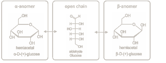
<p>α: anomeric OH points DOWN (below the ring, opposite the CH2OH). β: anomeric OH points UP (above the ring, same side as the CH2OH)</p>
31
New cards

Mutarotation

In water, the ring opens to the open chain and recloses, attacking either face of the carbonyl, so a pure α or β anomer becomes an α/β equilibrium mixture (glucose ≈ 1/3 α, 2/3 β)

32
New cards

Trap: "the anomeric OH swings between faces while the ring stays closed"

FALSE. α ⇌ β interconversion requires the ring to OPEN to the straight chain first, then reclose

33
New cards
[M2] Reducing sugar (definition)
A sugar whose anomeric carbon is FREE, so the ring can pop open into the open-chain aldehyde form. That aldehyde is what does the reducing (donates electrons to oxidizing agents like Cu²⁺ or Ag⁺).
34
New cards
[M2] What actually reduces Cu²⁺ in a reducing-sugar test?
The OPEN-CHAIN ALDEHYDE (not the closed hemiacetal ring, and not the C6 OH). Free anomeric C → ring opens → aldehyde donates electrons → Cu²⁺ reduced.
35
New cards
[M2] Non-reducing sugar
A sugar whose anomeric carbon(s) are LOCKED in a glycosidic bond, so the ring cannot open to an aldehyde (e.g., sucrose).
36
New cards
[M2] D-glucose equilibrium in water: % of each form
β-anomer ≈ 67% (MAJOR), α-anomer ≈ 33%, open-chain aldehyde < 1%.
37
New cards
[M2] Which glucose form is most abundant, and which acts as the reducing agent?
Most abundant = β-anomer (~67%). Reducing agent = the open-chain aldehyde (<1%). Trap: picking α as the major form, or the majority ring as the reducing agent.
38
New cards
[M2] Spotting α vs β in a Haworth ring
Compare the anomeric OH with the CH₂OH on C5: SAME side (both up) = β; OPPOSITE side = α.
39
New cards
[M2] Only <1% of glucose is open-chain, yet excess Cu²⁺ oxidizes nearly all of it. Why?
Le Chatelier: as Cu²⁺ oxidizes the open-chain molecules, more rings open to restore the equilibrium, so aldehyde keeps being supplied until nearly all glucose is oxidized. (Cu²⁺ does NOT push most glucose open at once.)
40
New cards
[M2] Is free galactose (or any free aldose monosaccharide) reducing?
Yes. A free monosaccharide's anomeric carbon is free, so it can open to the aldehyde. Glucose, galactose, maltose, lactose = reducing; sucrose = not.
41
New cards
[M2] How is a glycosidic bond formed?
Condensation (dehydration): the anomeric OH of one sugar (donor) + an OH of another molecule (acceptor) → C–O–C bridge + H₂O.
42
New cards
[M2] How is a glycosidic bond broken?
Hydrolysis: water is added back across the C–O–C bridge (done by glycoside hydrolases in digestion).
43
New cards
[M2] Glycosidic bond: which atoms leave, and whose O becomes the bridge?
The DONOR's anomeric C loses its whole OH; the ACCEPTOR's OH loses only its H. The acceptor's oxygen becomes the bridge.
44
New cards
[M2] Enzymes that BUILD glycosidic bonds in cells
Glycosyltransferases. They use activated sugar donors, typically nucleotide sugars like UDP-glucose.
45
New cards
[M2] Enzymes that BREAK glycosidic bonds in digestion
Glycoside hydrolases (e.g., sucrase, lactase, maltase, amylase). Glycosyltransferases build bonds, they do NOT digest.
46
New cards
[M2] Sucrose: units, linkage, enzyme, source, reducing?
Glucose + fructose; α1→β2; sucrase; plants, fruits, sugar cane (table sugar); NON-reducing.
47
New cards
[M2] Lactose: units, linkage, enzyme, source, reducing?
Galactose + glucose; β1→4; lactase; mammalian milk; reducing (glucose anomeric C1 is free).
48
New cards
[M2] Maltose: units, linkage, enzyme, source, reducing?
Glucose + glucose; α1→4; maltase; starch-digestion intermediate; reducing (right glucose's anomeric C1 is free).
49
New cards
[M2] Match bond → enzyme: α1→β2 (disaccharide)
Sucrase (sucrose).
50
New cards
[M2] Match bond → enzyme: β1→4 (disaccharide)
Lactase (lactose).
51
New cards
[M2] Match bond → enzyme: α1→4 (disaccharide)
Maltase (maltose).
52
New cards
[M2] Match bond → enzyme: α1→4 bonds WITHIN starch chains
Amylase. It acts on the polymer (starch) and produces maltose.
53
New cards
[M2] Naming rule for disaccharidases
Each is named for its substrate: sucrase → sucrose, lactase → lactose, maltase → maltose. Amylase → amylose/starch (polymer).
54
New cards
[M2] Why is sucrose non-reducing?
Both anomeric carbons (glucose C1 AND fructose C2) are locked in the α1→β2 bond, so neither ring can open to expose an aldehyde/ketone.
55
New cards
[M2] Trap: "Sucrose is reducing because each unit has an anomeric carbon"
Wrong. Having anomeric carbons isn't enough; they must be FREE. In sucrose they are joined to each other.
56
New cards
[M2] Glucose α1→α1 glucose (trehalose-type): reducing?
No. C1 of one is bonded to C1 of the other, so BOTH anomeric carbons are in the bond; neither can open.
57
New cards
[M2] C1 of glucose A bonds to C4-OH of glucose B. Which anomeric C is free?
Glucose A's anomeric C1 is locked in the bond; glucose B's anomeric C1 stays free → reducing end on B.
58
New cards
[M2] Reducing end of a disaccharide
The residue whose anomeric carbon is free (not in the glycosidic bond). In maltose/lactose it's the right-hand glucose.
59
New cards
[M2] Lactase deficiency: what happens to milk lactose?
It mostly stays unhydrolyzed in the small intestine, so galactose and glucose are not released for absorption.
60
New cards
[M2] HbA1c: how does it form?
Glucose's OPEN-CHAIN ALDEHYDE reacts non-enzymatically with an amine on hemoglobin → Schiff base → rearranges into a STABLE adduct (glycated hemoglobin).
61
New cards
[M2] HbA1c: what time window does it reflect, and why?
Average blood glucose over the past ~2–3 months, because the adduct is stable for the ~120-day lifespan of the red blood cell.
62
New cards
[M2] Normal fasting glucose today but HbA1c is elevated. Interpretation?
Glucose was often elevated over the past 2–3 months; the stable adduct accumulated over the RBC lifespan. Trap: "the adduct forms and dissociates within hours."
63
New cards
[M2] Which can form HbA1c-type adducts: glucose, maltose, sucrose, methyl glucoside?
Glucose and maltose only (each has a free anomeric C that can open to an aldehyde). Sucrose and the methyl glucoside have their anomeric C locked.
64
New cards
[M2] Link between glycosidic bonds and HbA1c
Same chemistry at the anomeric carbon. In glycosidic bonds the partner is another sugar's OH; in HbA1c the partner is a protein amine.
65
New cards
[M3] Glycogen: organism/tissue, bonds, branching
Animal energy storage (liver, muscle). α-D-glucose; α1→4 chains + α1→6 branches about every ~12 residues. Compact, highly branched.
66
New cards
[M3] Starch: organism/tissue and two components
Plant energy storage (grains, seeds, tubers like potatoes). Amylose + amylopectin.
67
New cards
[M3] Amylose
Starch component: α1→4 glucose chains ONLY, unbranched. Coils into a helix.
68
New cards
[M3] Amylopectin
Starch component: α1→4 chains + α1→6 branches about every ~30 residues. Less branched than glycogen.
69
New cards
[M3] Glycogen vs amylopectin: what differs?
Branching DENSITY (glycogen ~every 12 residues, amylopectin ~every 30), not linkage chemistry. Both are α1→4 + α1→6.
70
New cards
[M3] Which bonds form chains vs branch points in glycogen/amylopectin?
Chains: α1→4. Branch points: α1→6 (C1 of the branch glucose bonds to C6 of a chain glucose).
71
New cards
[M3] Glucose polymer from potatoes, branched (cutting only α1→6 bonds leaves short unbranched chains). Identity?
Amylopectin: plant source = starch, not glycogen; α1→6 branches present. Trap: glycogen is ANIMAL only.
72
New cards
[M3] Reducing end of a polysaccharide
Exactly ONE per molecule: the residue with a free C1 (its C4 is in a bond).
73
New cards
[M3] Non-reducing ends
C1 locked in a bond, C4-OH free. Linear chain = 1 non-reducing end; branched = 1 + number of branches. Enzymes act here.
74
New cards
[M3] Branched polymer with N branches: how many reducing / non-reducing ends?
1 reducing end; N + 1 non-reducing ends (each branch adds one non-reducing end, never a reducing end).
75
New cards
[M3] Why does glycogen branching allow fast glucose release?
Glucose-mobilizing enzymes (e.g., glycogen phosphorylase) work from NON-reducing ends. More branches = more non-reducing ends = more enzymes working at once. Branching also compacts storage.
76
New cards
[M3] Same # glucose, one polymer has more branches. Which releases glucose-1-phosphate faster?
The more-branched one, because extra branches give more non-reducing ends where enzymes act at once.
77
New cards
[M3] Glycogen phosphorylase product
Glucose-1-phosphate (removed from non-reducing ends); liver converts it to free glucose when blood glucose falls.
78
New cards
[M3] Cellulose: monomer, bond, location, function
β-D-glucose; β1→4; plant cell walls (cotton, wood, paper); tensile strength / rigidity. Linear, unbranched.
79
New cards
[M3] Chitin: monomer, bond, location, function
N-acetylglucosamine (GlcNAc); β1→4; insect exoskeletons, crustacean shells, fungal cell walls; rigid armor. Linear.
80
New cards
[M3] α1→4 vs β1→4 chain shape (the big idea)
α CURLS: α1→4 chains coil into hollow helices (storage, compact, digestible). β STRAIGHTENS: β1→4 chains (alternate residues flipped) form straight ribbons that H-bond into rigid sheets (structure).
81
New cards
[M3] Haworth: two glucoses joined C1→C4 with the C1 bond on the same side as its CH2OH (up), every residue alike. Linkage and shape?
β1→4; straight ribbon (cellulose-type). Trap: β1→4 + "coiled helix."
82
New cards
[M3] Why are cotton fibers (cellulose) so strong?
Straight β1→4 chains pack side by side and form MANY hydrogen bonds BETWEEN chains → rigid sheets/fibers. Not coiling, not covalent cross-links, not α1→6 branches.
83
New cards
[M3] Glycogen coils but cellulose is straight. Why, if both are glucose?
Anomeric configuration of the 1→4 bond: α in glycogen/starch, β in cellulose. Same monomer, same carbons joined.
84
New cards
[M3] Why can humans digest amylose but not cellulose?
Human α-amylase (saliva, pancreas) cleaves α1→4; maltase finishes maltose → glucose. Humans make no cellulase; β1→4 is the wrong geometry for our hydrolases, so cellulose passes through as fiber.
85
New cards
[M3] Cow vs human eating cellulose
Human: passes through largely undigested (fiber). Cow (and termites): gut MICROBES make cellulase and hydrolyze it (not the cow's own enzyme).
86
New cards
[M3] Starch digestion path
Starch → α-amylase (cuts internal α1→4 in the polymer) → maltose → maltase → glucose → absorbed.
87
New cards
[M3] Glycolipid: what bonds to what?
The sugar's ANOMERIC C1 bonds to a hydroxyl O of the lipid HEAD GROUP (O-glycosidic, C–O–C). Not to a carbon, not to the fatty-acid tail, not via C6.
88
New cards
[M3] Same glycosidic chemistry, different acceptors
Sugar–sugar: another sugar's OH (di/polysaccharides). Sugar–protein: Asn N or Ser/Thr O (glycoproteins, M4). Sugar–lipid: head-group OH (glycolipids). Always the anomeric C of the sugar.
89
New cards
[M3] Four polysaccharides at a glance
Glycogen: α-Glc, α1→4 + α1→6, branched/compact, animal storage. Starch: α-Glc, α1→4 (+α1→6), linear or branched, plant storage. Cellulose: β-Glc, β1→4, straight H-bonded sheets, plant structure. Chitin: β-GlcNAc, β1→4, straight H-bonded, insect/fungal structure.
90
New cards
[M3] Reading α vs β at a linkage in a Haworth drawing
1) Find the donor ring's C1 (next to the ring O). 2) Does its bond to the bridging O point the SAME way as that ring's CH2OH (both up)? Same side = β; opposite = α. 3) β → straight ribbon; α → coiled helix. Clue: the next residue drawn flipped/zig-zag = β1→4.
91
New cards
[M3] Trap: the sugar's C6 (CH2OH) forms the glycosidic bond
False. The sugar always bonds through its ANOMERIC C1. C6 is an ordinary alcohol that can only be the ACCEPTOR. The partner atom is always an O (or N), never a C.
92
New cards
[M4] Four reasons to glycosylate proteins
1) Solubility & stability (sugar coat prevents aggregation, blocks proteases). 2) Trafficking (sugar "ZIP codes," e.g., M6P → lysosome). 3) Cell-surface recognition (e.g., ABO blood groups). 4) Protection/barrier (mucins → mucus).
93
New cards
[M4] ABO blood types illustrate which glycosylation function?
Cell-surface recognition: displayed glycans act as identity tags the immune system reads.
94
New cards
[M4] N-linked glycosylation: attachment atom + starter sugar
Sugar's anomeric C1 → side-chain AMIDE NITROGEN of ASPARAGINE (Asn). Starter sugar: GlcNAc.
95
New cards
[M4] O-linked glycosylation: attachment atom + starter sugar
Sugar's anomeric C1 → side-chain HYDROXYL OXYGEN of SERINE or THREONINE. Starter sugar (mucin-type): GalNAc.
96
New cards
[M4] Trap atoms for N- and O-linked glycosylation
NOT a backbone amide N, NOT lysine's amino N, NOT Asp/Glu carboxylate O. N-linked = Asn side-chain amide N; O-linked = Ser/Thr side-chain OH.
97
New cards
[M4] N-linked pentasaccharide core
2 GlcNAc + 3 mannose ("2 GlcNAc to anchor, 3 mannose to branch"). GlcNAcs sit at the base on the Asn; mannoses branch on top. Trap: 2 Man + 3 GlcNAc.
98
New cards
[M4] Which enzymes attach sugars to proteins?
Glycosyltransferases (same family as Module 2). Same glycosidic chemistry, different acceptor (Asn N or Ser/Thr O).
99
New cards
[M4] N-glycosylation sequon
Asn-X-Ser/Thr (N-X-S/T), where X is any amino acid EXCEPT proline. Example valid: -Asn-Leu-Thr-, -Asn-Val-Thr-. Invalid: -Asn-Pro-Thr-, -Asn-Ser-Ala-.
100
New cards
[M4] Trap: -Asn-Pro-Thr-
NOT a sequon. Proline in the X position blocks N-glycosylation, so no glycan is added.



