New Notes and Stuff
 Call Kai
 Learn
 Practice Test
 Spaced Repetition
 Match
 Flashcards
 Knowt Play
1/122

There's no tags or description
Looks like no tags are added yet.
123 Terms
Kf vs Kr vs Kb
Kf = Equilibrium constant for formation (Products multiplied)
Kr = Equilibrium constant in reverse direction, dissociation (Reactants multiplied)
Kb = Boltzmann’s Constant
0th vs. first vs. second order
0th: Rate = Kf, M/s or Moles per sec
1st: Rate = Kf*[A], 1/s or s^-1
2nd: Rate = Kf*[A]*[B] or Kf*[A]², M^-1 s^-1 units
Enzyme names
Often called “Substrate” + -Ase
Enzyme Commission
EC, numbering system for enzymes (Ex: EC 3.1.1.1)
Leftmost number is responsible for cleaving bonds for rightmost number
Standard Conditions
25 C, 1 atm, 7.0 pH
Michaelis-Menton Equation
V(i) = (Vmax*[S])/(Km*[S])
[S] = Substrate concentration
V(i)/max = reaction velocity
Km = Michaelis constant
Relates reaction velocity to substrate concentration
Kcat meaning
Molecules/sec converted to product at V(max) (maximum reaction rate)
K(cat) = V(max) / initial Enzyme Concentration, or [E](0)
Michaelis Constant
K(m), K(m) = (K(2) + K(-1))/K(1)
If either K2 or K(-1) is much bigger than the other, it is the Rate Determining Step, and K(m) = K(d)
K(m)
Substrate concentration when V = 0.5*V(max)
Higher means lower affinity for substrate, and vice versa
Allosteric Enzymes
Allosteric enzymes can bind to non-substrates
Allosterism
When enzymes/proteins undergo conformational changes upon binding to ligand
Allosteric Effector
ligand that introduces conformational change
Also called “binding modulator”
Psuedo-Catalysts
Speeds up reaction, and is consumed
Known as “activator” if used in enzymes
K(i)
Inhibitor dissociation constant
K(i) = ([E]*[I])/[EI]
Class 1 Enzyme
Oxoreductases, Catalyze Electron Transfer
Class 2 Enzyme
Transferases, catalyze group transfer
Class 3 Enzyme
Hydrolases, catalyze wa
Class 4 Enzyme
Lysases, cleave C-C, C-O, C-N bonds, leaving double bonds
Class 5 Enzyme
Isomerases, transfer groups to create isomers
Class 6 Enzyme
Lygases, Form C-C, C-S, C-O, C-N bonds via condensation
Class 7 Enzyme
Translocases, Move Molecules/Ions across membranes
Holo Vs. Apoenzyme
Holoenzyme has cofactors, Apoenzyme does not
∆G vs. ∆G’
∆G is free energy at standard conditions (298 K)
∆G’ is free energy at standard conditions and 7.0 pH (Biochemical standard)
Optimal Enzyme Temp (typically)
36-37 C
Degree of Inhibition
E(i) = 1 - (V/V(0))
V = rate of inhibited reaction
V(0) = rate of uninhibited reaction
Carbohydrates
Chains of “polyhydroxyl” aldehydes/ketones in chain structure
Used as energy storage, structure, cell-cell recognition
Isomer types
Enantiomer: Isomers which are “left/right hand” of one another (L/D)
Epimer: Differ at one carbon
Carbyhydrate Building Blocks
Pectin
Amylose
Amylopectin
Glycogen
Sugar vs. Carb
Sugars are simple carbohydrates
Aldoase
Sugar with C=O at end, making Aldehyde
Ex: Glucose
Ketose
Sugar with C=O in middle of carbon chain
Ex: Fructose
D vs. L Isomers
OH on right = D isomer
OH on left = L Isomer
Must look from last chiral center (highest number)
Modulators
Ligands which change protein conformations, inhibiting or activating them
Homotropic
Multiple molecules of same ligand bind to multi-subunit protein, changing binding affinity
Heterotropic
When modulator is molecule other than normal ligand
Globulins
Soluble proteins with immune defense, transport, blood clotting functions
Roughly spherical
Soluble in dilute salts
Kd
K(off)/K(on) for ligands, dissociation equilibrium constant
Antibody regions
Variable Heavy in front of Variable Light (Immunoglobin G)
Fab region: Responsible for antigen recognition
Fc region: Responsible for activation, receptor binding

Glucose, Alpha Anomer
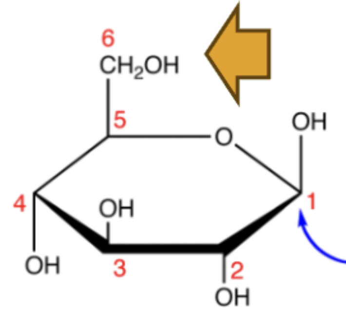
Glucose, Beta Anomer

Fructose, Alpha Anomer

Fructose, Beta Anomer
Pyranose
6-membered carbon ring with oxygen (Glucose)
Furanose
5-membered ring with oxygen (Fructose)
Ribose

Galactose

Fructose Fischer
Remember left = Up, right = Down

Glucose Fischer
Remember left = Up, right = Down

Epimer calculation
2^n chiral centers
Alpha-Pyranose conformation
Alpha is trans, other groups are equatorial
Alpha < Beta stability

Beta-Pyranose conformation
C1 is Equatorial or Axial Up
Beta > Alpha stability

Sugar Acids
Detoxify Liver via “Glucuronidation”
Inactivate hormones
Structural components in carbs

Sugar alcohols
(both sides)
Deoxy sugars
Sugars with -OH replaced with -H, including DNA
Amino Sugars
Have Amino/Amide Group
Found in structural components

Reducing Sugars
Sugars which become acidic as reducing agents (oxidized)

Glycotransferase
Enzyme which polymerizes monoshaccarides
Has no stop mechanism
Homopolysaccharides
Contain only 1 sugar unit,
ex: Glycogen and Starch, energy storage
Starch Structure
A1-4 “Glycosidic” bond
Hydrogen bonding between Glucoses

Heteropolysaccarides
Contain multiple sugar units
Often branched, Protein/Lipid Associated
Found in Extracellular Matrices, Connective Tissue, Mucus
Cellulose/Chitin Structure
Varies at R group (Carbon 2)

Glycoproteins
N-linked (Asn) or O-linked (Ser/Thr)
Protein with carbohydrates attached
Protein heavier than carbs
Many roles, ex: membrane proteins
Proteoglycan
Protein attached to “Glycoaminoglycan” (GAG)
Carb heavier than protein
Structural/Lubricant
Mucin/Mucoprotein
Hydrophobic protein attached to carb by “N-Acetylgalactoamine”
Often lubricants
Glyolipids
Play critical role in:
-Cell signalling
-Immunity
-Nervous System function
Usually has few sugars in carb
Lectins
Bind to sugars, are proteins
Cell-Cell recognition
Non-Enzymatic
Highly Specific
Multivalent (Binds to multiple sugar/glycoproteins at once)

Selectins
Transmembrane Glycoprotein with Sugar head
L- Leucytes (White Blood Cells)
E- Endothelial Cells
P- Platlets (blood clotting)
Fucosylation
Done by Fucosyltransferase, adds fucose to glycans, detected by Selectins
Sialylation
Done by Siayltransferases, adds sialic acid to glycans, detected by Selectins
Furanose puckering
C2 puckers for DNA
C3 puckers for RNA
Adenine
Connects to sugar at NH

Guanine
Connects to sugar at NH

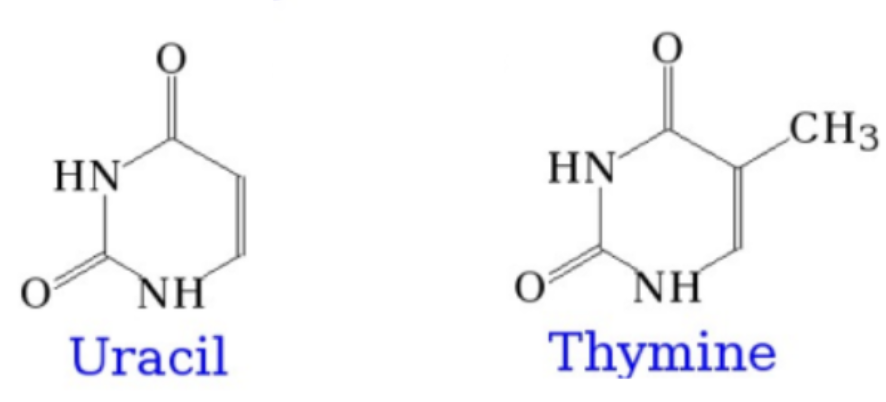
Thymine/Uracil
Connects to sugar at NH
Cytosine
Connects to sugar at NH

Nucleoside
Nucleotide without Phsophate
RNA Hydrolysis
Oxygen on C2 attacks Phosphate

GAGs
GlycoAminoGlycans
Act as Lubricants
Chondritan Sulfate
Connective and Heart Tissue, flexibility

Riboses
5-carbon sugars with Aldehyde group
Nucleotide Conformation
Beta, D isomer (-OH on right)
(B1-4)
T-A vs. C-G
T-A has 2 hydro bonds
C-G has 3 hydro bonds
Hairpin
“Palindrome” can also form hairpin

Palindrome, Mirror Repeats

Cruciform
DNA only

Wattson-Crick vs. Hoogstein
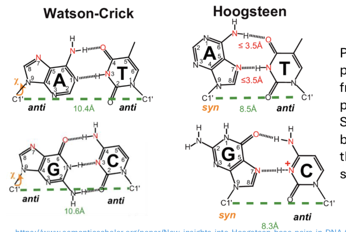
Triplex DNA
Forms at low pH, lots of C/G

Tetraplex
Forms from Guanine
Forms around Potassium
Stack on one another
Long RNAs
Ribosomal RNA (rRNA)
Enhancer RNA (eRNA)
Long Intergenic RNA (lincRNA)
DNA variants
A-form: anti-anti, dehydrated
B-form: anti-anti, most common
Z-form: anti-syn, high G-C content, left handed
t-RNA
D-loop: Aminoacyl-tRNA recognition site
Anticodon Loop: Binds only to corresponding anticodon
T-loop: Allows tRNA to dock

Hyper/Hypochromic Effect
Hypo - Close base packing in DNA decreases UV absorption
Hyper - Close base packing in DNA increases UV absorption
t(m)
Temperature at which half of DNA is double stranded, half is single
Deanimation
Loss of amino group becomes O= bond on Amino Acid, ex: Cytosine to Urocil

Depurination
N glycocidic bond lost with sugar

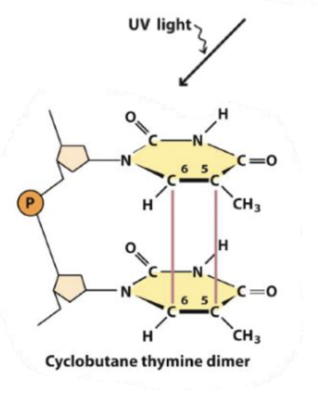
Thymine dimers
Two thymines merge together at 5 and 6 carbons
DNA Methylation
Usually happens to Adenine/Cytosine, controls expression

NAD+
Glycolysis (Oxidative Phosphorylation), Citric Acid Cycle

Coenzyme A
Used in:
Kreb’s Cycle
Fatty Acid Oxidation/Synthesis
Amino Acid Metabolism
Thiol (-SH) is active group
cGMP
Relaxes muscles/blood cells
Maintains ion balance

cAMP
Stimulates glucose/fat breakdown
Regulates kidney water balance
