Test Four
 Call Kai
 Learn
 Practice Test
 Spaced Repetition
 Match
 Flashcards
 Knowt Play
1/58

There's no tags or description
Looks like no tags are added yet.
59 Terms

Hematophagous Trophozoites
Trophozoites of Entamoeba histolytica shown with engulfed red blood cells.

1-3mm
Colonic ulcers caused by Entamoeba histolytica
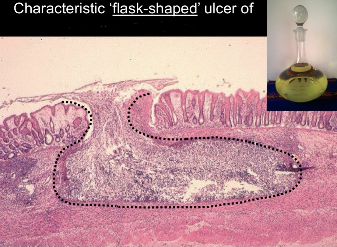
Flask Shaped Ulcer
Characteristic of intestinal amebiasis caused by Entamoeba histolytica

“Anchovy Paste” pus
Amebic liver abscess comprised of reddish brown pus, necrotic tissue, immune cells, and parasites. Characteristic of Entamoeba histolytica.

Trophozoite and Cyst
Balantidium coli

Cilia and Cytosome
Balantidium coli. Only ciliate known to infect humans.

Large and Ovoid Intestinal Cocci
Cystoisospora belli

Mid sized intestinal cocci
Cyclospora cayetanensis

Tiny Intestinal Cocci
Cryptospridium hominis (most medically relevant)

Basophilic Stippling “knobs”
On the surface of P. Falciparum infected erythrocytes

Cellular “knobs”
Occur on P. falciparum infected red blood cells. Promoted agglutination of red blood cells.

Male and Female Ganetophytes
P. Falciparum reproductive phase. Occurs within the mosquito

Volcano Sign
Characteristic painless lesion with rolled edges. Characteristic of leishmaniasis major

Maltese Crosses
Charateric of babesiosis

Giemsa Stain
Thick blood smear detects parasites. Thin blood smears identify species. Gold standard for detection of P. falciparum

Ring forms
Trophozoites of F. falciparum within red blood cells

Blood Smear
Numerous ring form P. falciparum

Microscopy
P. falciparum. Shows the large gametophyte, basophilic Stippling (top left), and rings (bottom)

Lung X-Ray of 9 year old boy
Hilar lynphadenopathy characteristic of Tuberculosis in children.

Lung X-ray of a 5 month old
White spots are miliary tuberculosis lesions

Lungs from a cadaver
Yellow arrows show millet granulomas

What is this?
Primary granuloma formed from a chain reaction macrophages and other phagocytic cells undergoing Tuberculosis induced apoptosis and being engulfed by other phagocytic cells. Forms a “jail” around the tuberculosis organisms and renders them dormant

Cadaver lung
Blue arrows indicate calcified lymph nodes and the green arrows indicate calcified periferal lung lesions. The infection travels from the peripheral lesion and spreads to the node. These combined form the Ranke Complex.

X-ray of lungs
Shows a Ranke Complex. Indicates past, spontaneously resolved tuberculosis infection.

X-ray and Cadaver lung
Shows liquified granulomas consistent with reactivated (cavitary) tuberculosis

Patient’s back
Deformation of the back, consistent with extrapulmonary tuberculosis consistent with Pott Disease. Pott Disease is a form of extrapulmonary Tuberculosis that infects the bones and joints

What is this?
Swelling of the skin as a result of Tuberculosis skin testing

What is this?
Growth of Tuberculosis on Lowensten-Jensen (LJ) agar. Highly selective for TB but organism takes around 4 weeks to form visible colonies. Brown, granular appearance

What is this?
Acid fast stain on mycobacterium tuberculosis

What is this?
Auramine-Rhodamine stain of Mycobacterium tuberculosis. Bacteria flouresce and show up as a bright orange or yellow stain. Cheaper but less specific than acid fast/Ziehl-Neelsen

What is this?
Shows M. Leprae next to Shwann cell membrane. M. Leprae replicates in macrophages and Shwann cells. It results in demylination of the Shwann cell resulting in cell death and permanent nerve damage.

Skin lesion
Superficial pale, flat lesion characteristic of Tuberculoid leprosy.

Patient
Patient shows hair loss in eyebrows and earlobe nodules consistent with Lepromatous leprosy

Patient
Severe, chronic M. Leprae infection has led to permanent neuropathy and resorption of distal phalanges leading to multiple distl amputations

Patient
Leprosy lesions highlighted by healthy skin that is stained dark from anti-leprosy drug clofazimine

1
Active hypopigmented anesthetic plaque
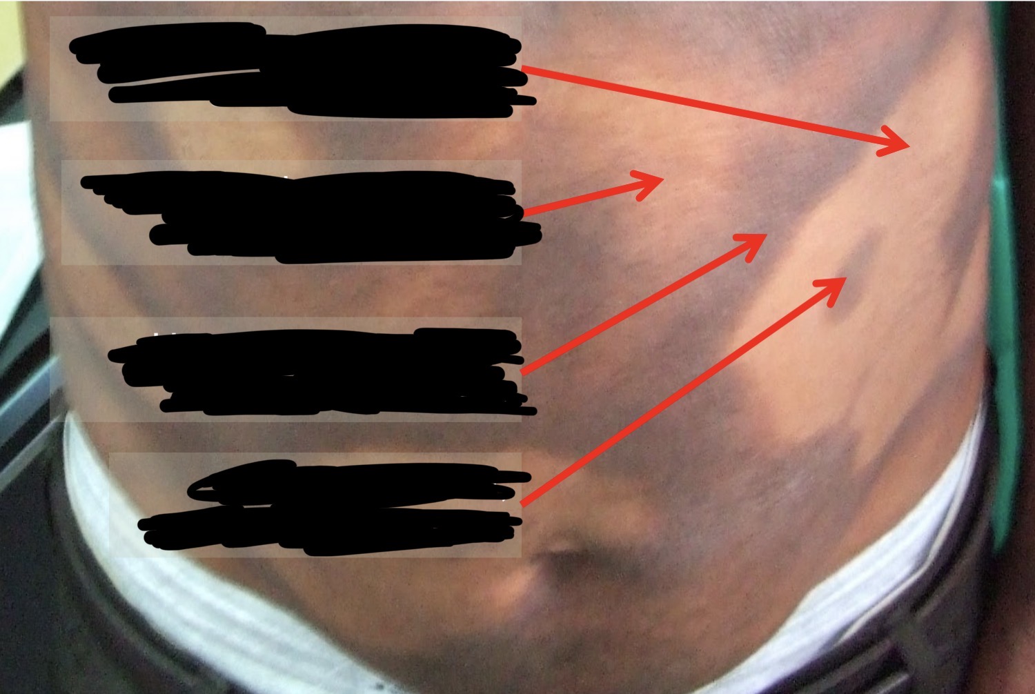
2
Drug staining of healthy skin (light staining)

3
Heavy staining of the border of the lesion

4
Central healed “punched out” lesion

Patient
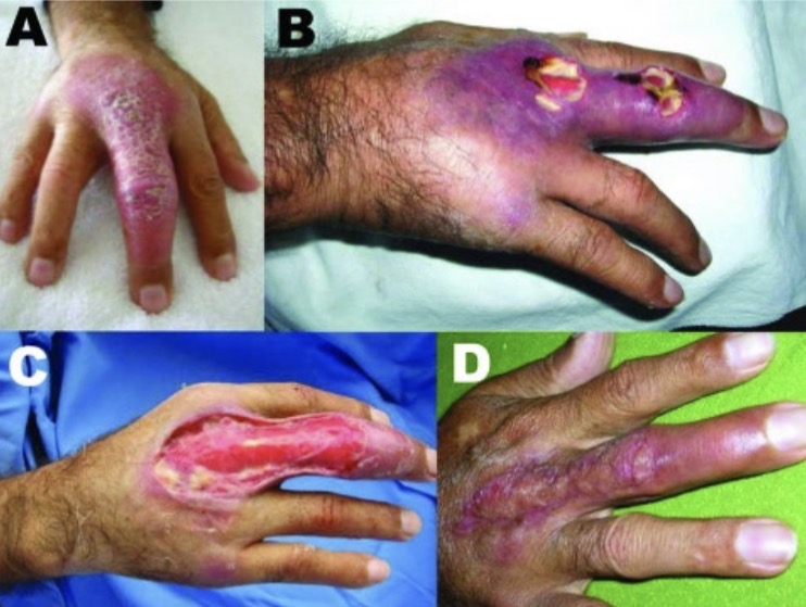
Buruli ulcer

Patient
Buruli ulcer
Patient
Buruli ulcer

Microscopy
Cell cycle arrest, apoptosis, necrosis caused by micobacterium ulcerans

Stain
Acid fast bacalli: mycobacterium ulcerans

X-ray and CT scan
Well defined, no calcified nodules, tissue necrosis (cavitation) caused by cryptospridium neoformans
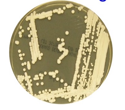
Agar plate
Sabourad agar plate of cryptospridium neoformans

Stain
H&E stain showing capule of cryptospridium neoformans

Stain
India ink stain show capsule (white halo) of cryptospridium neoformans

What organism is this?
Top: candida albicans
Botton: candida albicans stains gram positive

What form of what organism is this?
Yeast form of candida albicans, least virulent

What form of what organism is this
Psuedohyphae of candida albicans. Medium virulence
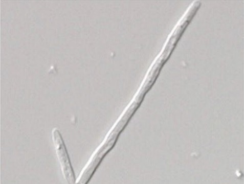
What form of what organism is this?
Hyphae form of candida albicans. Most virulent

Patient
Diaper rash caused by candida albicans

Patient
Oral candidiasis

Patient
Candida esopagitis in recipient of cadaveric renal transplant

Chest X-ray
Pulmonary histoplasmosis caused by histoplasma capsulatum

Patient
Rash from disseminated histoplasmosis

Patient
Rash from disseminated histoplasmosis

Stain
Intracellular yeast of histoplasma capsulatum