Ch 5: Membranes and an Introduction to Signal Transduction

0.0(0)
Studied by 0 people
call kaiCall Kai
learnLearn
examPractice Test
spaced repetitionSpaced Repetition
heart puzzleMatch
flashcardsFlashcards
GameKnowt Play
Card Sorting

1/60

encourage image

There's no tags or description

Looks like no tags are added yet.

61 Terms

1
New cards

What Are Cell Membranes?

Cell membranes surround all cells.

They have several important functions:

  • Separate the inside of the cell from the outside environment.

  • Control what enters and leaves the cell.

  • Help cells communicate with their surroundings.

  • Provide a location for important proteins involved in cellular activities.

Eukaryotic cells also have membranes surrounding their organelles.

For example:

  • Mitochondria.

  • Endoplasmic reticulum.

  • Golgi apparatus.

  • Nucleus.


2
New cards

The lipid bilayer

The main structure of a cell membrane is called the lipid bilayer. It consists of two layers of lipids arranged together. The membrane has two leaflets:

  • Outer leaflet: Faces the outside of the cell.

  • Inner leaflet: Faces the inside of the cell (cytosol).

The lipid bilayer also contains proteins, cholesterol, and carbohydrate groups.

<p><span>The</span> <span>main</span> <span>structure</span> <span>of</span> <span>a</span> <span>cell</span> <span>membrane</span> <span>is</span> <span>called</span> <span>the</span> <span style="line-height: inherit; font-size: inherit;"><strong>lipid bilayer</strong></span><span>. It</span> <span>consists</span> <span>of</span> <span>two</span> <span>layers</span> <span>of</span> <span>lipids</span> <span>arranged</span> <span>together. The</span> <span>membrane</span> <span>has</span> <span>two</span> <span>leaflets:</span></p><ul><li><p class="x1ghz6dp x1717udv x1wty727 x1pd3egz x4gjf66 x15eiy0f x1lziwak x15472z4 x1qlqyl8 x15bjb6t x1lliihq x14rh7hd xg07evd"><span style="line-height: inherit; font-size: inherit;"><strong>Outer leaflet:</strong></span> <span>Faces</span> <span>the</span> <span>outside</span> <span>of</span> <span>the</span> <span>cell.</span></p></li><li><p class="x1ghz6dp x1717udv x1wty727 x1pd3egz x4gjf66 x15eiy0f x1lziwak x15472z4 x1qlqyl8 x15bjb6t x1lliihq x14rh7hd xg07evd"><span style="line-height: inherit; font-size: inherit;"><strong>Inner leaflet:</strong></span> <span>Faces</span> <span>the</span> <span>inside</span> <span>of</span> <span>the</span> <span>cell</span> <span>(cytosol).</span></p></li></ul><p class="x1ghz6dp x1717udv x1wty727 x1pd3egz x4gjf66 x1wwbdsi x86bw1a x1lziwak x15472z4 x1qlqyl8 x15bjb6t x1lliihq x14rh7hd"><span>The</span> <span>lipid</span> <span>bilayer</span> <span>also</span> <span>contains</span> <span>proteins,</span> <span>cholesterol,</span> <span>and</span> <span>carbohydrate</span> <span>groups.</span></p>
3
New cards

What Are Lipids?

Lipids are different from proteins and nucleic acids because they are not defined by one common molecular structure. Instead, lipids are classified based on a physical property: Lipids are molecules that are soluble in nonpolar solvents.

Why are lipids nonpolar?

Most lipids contain large hydrocarbon regions.

Hydrocarbons contain:

  • Carbon atoms.

  • Hydrogen atoms.

Carbon-carbon bonds are nonpolar, and carbon-hydrogen bonds are relatively nonpolar. Therefore, molecules containing large hydrocarbon regions tend to be hydrophobic.

Remember: Like dissolves like

  • Polar substances dissolve in polar solvents.

  • Nonpolar substances dissolve in nonpolar solvents.

This explains why many lipids dissolve in nonpolar solvents.

Cholesterol is classified as a lipid because most of its structure is nonpolar and hydrocarbon-based. It contains:

  • A large nonpolar hydrocarbon structure.

  • One hydroxyl (-OH) group.

Cholesterol is an important component of cell membranes.

<p><span>Lipids</span> <span>are</span> <span>different</span> <span>from</span> <span>proteins</span> <span>and</span> <span>nucleic</span> <span>acids</span> <span>because</span> <span>they</span> <span>are</span> <span>not</span> <span>defined</span> <span>by</span> <span>one</span> <span>common</span> <span>molecular</span> <span>structure. Instead,</span> <span>lipids</span> <span>are</span> <span>classified</span> <span>based</span> <span>on</span> <span>a</span> <span>physical</span> <span>property: </span><span style="line-height: inherit; font-size: inherit;"><strong>Lipids are molecules that are soluble in nonpolar solvents.</strong></span></p><p><span><strong><u>Why</u></strong></span><strong><u> </u></strong><span><strong><u>are</u></strong></span><strong><u> </u></strong><span><strong><u>lipids</u></strong></span><strong><u> </u></strong><span><strong><u>nonpolar?</u></strong></span></p><p><span>Most</span> <span>lipids</span> <span>contain</span> <span>large</span> <span>hydrocarbon</span> <span>regions.</span></p><p class="x1ghz6dp x1717udv x1wty727 x1pd3egz x4gjf66 x1wwbdsi x86bw1a x1lziwak x15472z4 x1qlqyl8 x15bjb6t x1lliihq x14rh7hd"><span>Hydrocarbons</span> <span>contain:</span></p><ul><li><p class="x1ghz6dp x1717udv x1wty727 x1pd3egz x4gjf66 x15eiy0f x1lziwak x15472z4 x1qlqyl8 x15bjb6t x1lliihq x14rh7hd xg07evd"><span>Carbon</span> <span>atoms.</span></p></li><li><p class="x1ghz6dp x1717udv x1wty727 x1pd3egz x4gjf66 x15eiy0f x1lziwak x15472z4 x1qlqyl8 x15bjb6t x1lliihq x14rh7hd xg07evd"><span>Hydrogen</span> <span>atoms.</span></p></li></ul><p class="x1ghz6dp x1717udv x1wty727 x1pd3egz x4gjf66 x1wwbdsi x86bw1a x1lziwak x15472z4 x1qlqyl8 x15bjb6t x1lliihq x14rh7hd"><span>Carbon-carbon</span> <span>bonds</span> <span>are</span> <span>nonpolar,</span> <span>and</span> <span>carbon-hydrogen</span> <span>bonds</span> <span>are</span> <span>relatively</span> <span>nonpolar. Therefore,</span> <span>molecules</span> <span>containing</span> <span>large</span> <span>hydrocarbon</span> <span>regions</span> <span>tend</span> <span>to</span> <span>be</span> <span>hydrophobic. </span></p><p class="x1ghz6dp x1717udv x1wty727 x1pd3egz x4gjf66 x1wwbdsi x86bw1a x1lziwak x15472z4 x1qlqyl8 x15bjb6t x1lliihq x14rh7hd"><span><strong>Remember:</strong></span><strong> </strong><span><strong>Like</strong></span><strong> </strong><span><strong>dissolves</strong></span><strong> </strong><span><strong>like</strong></span></p><ul><li><p class="x1ghz6dp x1717udv x1wty727 x1pd3egz x4gjf66 x15eiy0f x1lziwak x15472z4 x1qlqyl8 x15bjb6t x1lliihq x14rh7hd xg07evd"><span>Polar</span> <span>substances</span> <span>dissolve</span> <span>in</span> <span>polar</span> <span>solvents.</span></p></li><li><p class="x1ghz6dp x1717udv x1wty727 x1pd3egz x4gjf66 x15eiy0f x1lziwak x15472z4 x1qlqyl8 x15bjb6t x1lliihq x14rh7hd xg07evd"><span>Nonpolar</span> <span>substances</span> <span>dissolve</span> <span>in</span> <span>nonpolar</span> <span>solvents.</span></p></li></ul><p class="x1ghz6dp x1717udv x1wty727 x1pd3egz x4gjf66 x1wwbdsi x86bw1a x1lziwak x15472z4 x1qlqyl8 x15bjb6t x1lliihq x14rh7hd"><span>This</span> <span>explains</span> <span>why</span> <span>many</span> <span>lipids</span> <span>dissolve</span> <span>in</span> <span>nonpolar</span> <span>solvents.</span></p><p class="x1ghz6dp x1717udv x1wty727 x1pd3egz x4gjf66 x1wwbdsi x86bw1a x1lziwak x15472z4 x1qlqyl8 x15bjb6t x1lliihq x14rh7hd"><span>Cholesterol</span> <span>is</span> <span>classified</span> <span>as</span> <span>a</span> <span>lipid</span> <span>because</span> <span>most</span> <span>of</span> <span>its</span> <span>structure</span> <span>is</span> <span>nonpolar</span> <span>and</span> <span>hydrocarbon-based. It</span> <span>contains:</span></p><ul><li><p class="x1ghz6dp x1717udv x1wty727 x1pd3egz x4gjf66 x15eiy0f x1lziwak x15472z4 x1qlqyl8 x15bjb6t x1lliihq x14rh7hd xg07evd"><span>A</span> <span>large</span> <span>nonpolar</span> <span>hydrocarbon</span> <span>structure.</span></p></li><li><p class="x1ghz6dp x1717udv x1wty727 x1pd3egz x4gjf66 x15eiy0f x1lziwak x15472z4 x1qlqyl8 x15bjb6t x1lliihq x14rh7hd xg07evd"><span>One</span> <span>hydroxyl</span> <span>(-OH)</span> <span>group.</span></p></li></ul><p class="x1ghz6dp x1717udv x1wty727 x1pd3egz x4gjf66 x1wwbdsi x86bw1a x1lziwak x15472z4 x1qlqyl8 x15bjb6t x1lliihq x14rh7hd"><span>Cholesterol</span> <span>is</span> <span>an</span> <span>important</span> <span>component</span> <span>of</span> <span>cell</span> <span>membranes.</span></p>
4
New cards

What Are Phospholipids?

Phospholipids are major components of cell membranes.

They contain:

  • A hydrophilic (water-loving) head.

  • Hydrophobic (water-fearing) fatty acid tails.

This makes phospholipids amphipathic molecules. Amphipathic means a molecule has both hydrophilic and hydrophobic regions.

Why are the two parts different?

Hydrophilic head:

  • Contains a phosphate group.

  • Has charged or polar components.

  • Interacts well with water.

Hydrophobic tails:

  • Contain long hydrocarbon chains.

  • Are nonpolar.

  • Do not interact well with water.


<p><span style="background-color: transparent;"><strong><u>Phospholipids are major components of cell membranes.</u></strong></span></p><p><span style="background-color: transparent;">They contain:</span></p><ul><li><p><span style="background-color: transparent;">A hydrophilic (water-loving) head.</span></p></li><li><p><span style="background-color: transparent;">Hydrophobic (water-fearing) fatty acid tails.</span></p></li></ul><p><span style="background-color: transparent;">This makes phospholipids amphipathic molecules. Amphipathic means a molecule has both hydrophilic and hydrophobic regions.</span></p><p><span style="background-color: transparent;"><strong>Why are the two parts different?</strong></span></p><p><span style="background-color: transparent;">Hydrophilic head:</span></p><ul><li><p><span style="background-color: transparent;">Contains a phosphate group.</span></p></li><li><p><span style="background-color: transparent;">Has charged or polar components.</span></p></li><li><p><span style="background-color: transparent;">Interacts well with water.</span></p></li></ul><p><span style="background-color: transparent;">Hydrophobic tails:</span></p><ul><li><p><span style="background-color: transparent;">Contain long hydrocarbon chains.</span></p></li><li><p><span style="background-color: transparent;">Are nonpolar.</span></p></li><li><p><span style="background-color: transparent;">Do not interact well with water.</span><br></p></li></ul><p></p>
5
New cards

Glycerol: The Backbone of Glycerophospholipids

A major type of membrane phospholipid is called a glycerophospholipid. It contains a glycerol backbone.

What is glycerol?

Glycerol is a three-carbon polyalcohol. It contains: 

  • Three carbon atoms connected in a row.

  • One hydroxyl (-OH) group attached to each carbon. CH2OH−CHOH−CH2OH

Memorize: Glycerol is a three-carbon alcohol with three hydroxyl groups.

<p><span style="background-color: transparent;">A major type of membrane phospholipid is called a glycerophospholipid. It contains a glycerol backbone.</span></p><p><span style="background-color: transparent;"><strong>What is glycerol?</strong></span></p><p><span style="background-color: transparent;">Glycerol is a three-carbon polyalcohol. It contains:&nbsp;</span></p><ul><li><p><span style="background-color: transparent;">Three carbon atoms connected in a row.</span></p></li><li><p><span style="background-color: transparent;">One hydroxyl (-OH) group attached to each carbon. </span><span>CH2OH−CHOH−CH2OH</span></p></li></ul><p><span><strong>Memorize:</strong> Glycerol is a three-carbon alcohol with three hydroxyl groups.</span></p>
6
New cards

Fatty Acids

A fatty acid is defined as: A long-chain carboxylic acid. This is an important definition to memorize. Fatty acids contain:

  • A carboxylic acid group.

  • A long hydrocarbon chain.

Common fatty acid chain lengths include 12, 14, 16, and 18 carbon atoms. The long hydrocarbon chain is responsible for the hydrophobic nature of the fatty acid.

What is an ester bond?

An ester forms when: 

  • A carboxylic acid reacts with an alcohol.

  • A dehydration reaction occurs.

  • Water is removed.

In glycerophospholipids:

  • The fatty acid provides the carboxylic acid group.

  • Glycerol provides the hydroxyl group.

  • An ester bond forms between them.

Two fatty acids attach to the glycerol backbone through ester bonds.

<p><span>A fatty acid is defined as: A long-chain carboxylic acid. This is an important definition to memorize. Fatty acids contain:</span></p><ul><li><p><span>A carboxylic acid group.</span></p></li><li><p><span>A long hydrocarbon chain.</span></p></li></ul><p><span>Common fatty acid chain lengths include 12, 14, 16, and 18 carbon atoms. The long hydrocarbon chain is responsible for the hydrophobic nature of the fatty acid.</span></p><p><span><strong>What is an ester bond?</strong></span></p><p><span>An ester forms when:&nbsp;</span></p><ul><li><p><span>A carboxylic acid reacts with an alcohol.</span></p></li><li><p><span>A dehydration reaction occurs.</span></p></li><li><p><span>Water is removed.</span></p></li></ul><p><span><strong>In glycerophospholipids:</strong></span></p><ul><li><p><span>The fatty acid provides the carboxylic acid group.</span></p></li><li><p><span>Glycerol provides the hydroxyl group.</span></p></li><li><p><span>An ester bond forms between them.</span></p></li></ul><p><span>Two fatty acids attach to the glycerol backbone through ester bonds.</span></p>
7
New cards

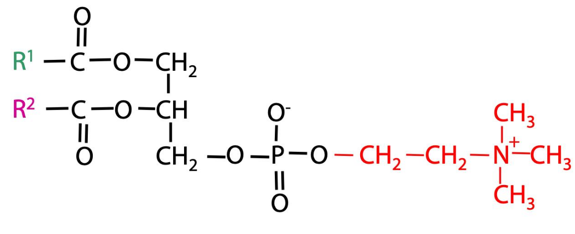
Phosphatidylcholine: An Example of a Glycerophospholipid

Phosphatidylcholine contains:

  • Glycerol backbone.

  • Two fatty acid chains.

  • Phosphate group.

  • Choline group.

Understanding the phosphate group

The phosphate group connects the glycerol backbone to another molecule through phosphoester bonds. The molecule attached to the phosphate helps determine the name of the phospholipid.

What is choline?

Choline is a two-carbon compound containing a nitrogen atom with three methyl groups attached to it. The nitrogen is positively charged. When choline is attached to the phosphate group of a phospholipid, the resulting molecule is called phosphatidylcholine.

The lecture also mentions that serine, threonine, and tyrosine are amino acids containing hydroxyl groups. These hydroxyl groups can participate in phosphoester bond formation.

Exam tip: Know that the attached head group determines the phospholipid's name.
<p><span><strong>Phosphatidylcholine contains:</strong></span></p><ul><li><p><span>Glycerol backbone.</span></p></li><li><p><span>Two fatty acid chains.</span></p></li><li><p><span>Phosphate group.</span></p></li><li><p><span>Choline group.</span></p></li></ul><p><span><strong>Understanding the phosphate group</strong></span></p><p><span>The phosphate group connects the glycerol backbone to another molecule through phosphoester bonds. The molecule attached to the phosphate helps determine the name of the phospholipid.</span></p><p><span><strong>What is choline?</strong></span></p><p><span>Choline is a two-carbon compound containing a nitrogen atom with three methyl groups attached to it. The nitrogen is positively charged. When choline is attached to the phosphate group of a phospholipid, the resulting molecule is called phosphatidylcholine.</span></p><p><span>The lecture also mentions tha<strong>t serine, threonine, and tyrosine are amino acids containing hydroxyl groups</strong>. <u>These hydroxyl groups can participate in phosphoester bond formation</u>.</span></p><p><span><strong>Exam tip</strong>: Know that the attached head group determines the phospholipid's name.</span></p>
8
New cards

Why Do Phospholipids Form Bilayers?

Phospholipids spontaneously form lipid bilayers when present above a sufficient concentration in an aqueous environment. Why? Because they have both hydrophilic and hydrophobic regions.

When phospholipids are surrounded by water:

  • The hydrophilic heads face the water.

  • The hydrophobic tails turn inward, away from water.

  • The fatty acid tails interact with one another.

This arrangement forms a stable lipid bilayer.

Important: The membrane forms spontaneously because this arrangement is energetically favorable.

<p><span>Phospholipids spontaneously form lipid bilayers when present above a sufficient concentration in an aqueous environment. Why? Because they have both hydrophilic and hydrophobic regions.</span></p><p><span><strong>When phospholipids are surrounded by water:</strong></span></p><ul><li><p><span>The hydrophilic heads face the water.</span></p></li><li><p><span>The hydrophobic tails turn inward, away from water.</span></p></li><li><p><span>The fatty acid tails interact with one another.</span></p></li></ul><p><span>This arrangement forms a stable lipid bilayer.</span></p><p><span><strong>Important</strong>: The membrane forms spontaneously because this arrangement is energetically favorable.</span></p>
9
New cards

What Does Cholesterol Do in the Membrane?

Cholesterol is another important component of cell membranes. It fits between the fatty acid tails of phospholipids.

What happens without cholesterol?

The fatty acid tails of neighboring phospholipids interact with each other through weak attractive forces. These include:

  • Van der Waals forces.

  • London dispersion forces.

What happens when cholesterol is present?

Cholesterol inserts itself between the fatty acid tails. This affects how closely the fatty acid chains can pack together and influences membrane fluidity and permeability. Main idea: Cholesterol helps regulate membrane fluidity.

<p><span>Cholesterol is another important component of cell membranes. It fits between the fatty acid tails of phospholipids.</span></p><p><span><strong>What happens without cholesterol?</strong></span></p><p><span>The fatty acid tails of neighboring phospholipids interact with each other through weak attractive forces. These include:</span></p><ul><li><p><span>Van der Waals forces.</span></p></li><li><p><span>London dispersion forces.</span></p></li></ul><p><span><strong>What happens when cholesterol is present?</strong></span></p><p><span>Cholesterol inserts itself between the fatty acid tails. This affects how closely the fatty acid chains can pack together and influences membrane fluidity and permeability. Main idea: Cholesterol helps regulate membrane fluidity.</span></p>
10
New cards

Membranes Are Dynamic Structures

The membrane is not a rigid structure. Phospholipids and other membrane components can move. There are several types of movement.

Four types of membrane movement:

  • Lateral Diffusion: Lipids move side to side within the same leaflet

  • Rotation: Lipids rotate around their own axis

  • Flexion: Fatty acid tails bend and move

  • Flip-flop: Lipids move from one leaflet to the other.

Flip-flop movement

Flip-flop is when a phospholipid moves from one side of the bilayer to the other. This movement is difficult because the hydrophilic head would have to pass through the hydrophobic interior of the membrane. Therefore, enzymes help this process occur. These enzymes are called flippases.

Memorize: Flippases move phospholipids between the two leaflets of a membrane.

<p><span>The membrane is not a rigid structure. Phospholipids and other membrane components can move. There are several types of movement.</span></p><p><span><strong>Four types of membrane movement:</strong></span></p><ul><li><p><span>Lateral Diffusion: Lipids move side to side within the same leaflet</span></p></li><li><p><span>Rotation: Lipids rotate around their own axis</span></p></li><li><p><span>Flexion: Fatty acid tails bend and move</span></p></li><li><p><span>Flip-flop: Lipids move from one leaflet to the other.</span></p></li></ul><p><span><strong>Flip-flop movement</strong></span></p><p><span>Flip-flop is when a phospholipid moves from one side of the bilayer to the other. This movement is difficult because the hydrophilic head would have to pass through the hydrophobic interior of the membrane. Therefore, enzymes help this process occur. These enzymes are called <strong>flippases.</strong></span></p><p><span><strong>Memorize</strong>: Flippases move phospholipids between the two leaflets of a membrane.</span></p>
11
New cards

Membrane Proteins

Different Ways Proteins Associate With Membranes

Proteins can be associated with membranes in several different ways. There are three major types discussed in the lecture:

  1. Integral membrane proteins.

  2. Peripheral membrane proteins.

  3. Lipid-anchored proteins.

Integral Membrane Proteins

  • Definition: Proteins that are permanently embedded directly within the hydrophobic core of the lipid bilayer.

  • Structure: They are amphipathic (having both hydrophobic and hydrophilic regions). Their hydrophobic amino acids interact with the lipid tails inside the membrane, while their hydrophilic regions stick out into the aqueous (water-based) environment inside or outside the cell.

  • Key Subtype: Transmembrane proteins span completely across the membrane from one side to the other.

  • Removal: They cannot be easily removed without using harsh agents like detergents that disrupt the lipid bilayer itself.

  • Function: Act as transport channels, carriers, receptors, and pumps.

Peripheral Membrane Proteins

  • Definition: Proteins that are located on the outside or inside surface of the membrane rather than being embedded in the lipid core.

  • Structure: They sit on the periphery and do not penetrate the hydrophobic layer.

  • Attachment: They bind loosely and non-covalently to the polar head groups of phospholipids or to the exposed parts of integral proteins via ionic or hydrogen bonds.

  • Removal: They can be easily washed off or detached using gentle methods like changing the pH or salt/ionic strength without harming the membrane.

  • Function: Serve as enzymes, structural supports (linking the membrane to the cytoskeleton), or signaling molecules.

Lipid-Anchored Proteins

  • Definition: Proteins that reside on the surface of the membrane but are permanently anchored to it via a covalent bond to a lipid molecule.

  • Structure: The protein itself does not touch or enter the lipid bilayer. Instead, a fatty acid chain, prenyl group, or glycolipid (such as a GPI anchor) acts as a tether, dipping directly into the membrane's lipid layer.

  • Removal: Because the lipid anchor is chemically (covalently) bound, releasing the protein usually requires specialized enzymes (like phospholipases) to cleave the link.

  • Function: Involved in cell signaling, enzymatic reactions, and anchoring proteins to specific membrane microdomains.

Integral Membrane Protein: Embedded in the lipid bilayer.

Transmembrane/ditopic: Passes completely through the bilayer.

Monotopic: Only partially enters the bilayer.

<p><strong>Different Ways Proteins Associate With Membranes</strong></p><p class="x1ghz6dp x1717udv x1wty727 x1pd3egz x4gjf66 x1kw3rcc x104jwf5 x1lziwak x15472z4 x1qlqyl8 x15bjb6t x1lliihq x14rh7hd">Proteins can be associated with membranes in several different ways. There are three major types discussed in the lecture:</p><ol><li><p class="x1ghz6dp x1717udv x1wty727 x1pd3egz x4gjf66 x15eiy0f x1lziwak x15472z4 x1qlqyl8 x15bjb6t x1lliihq x14rh7hd xg07evd">Integral membrane proteins.</p></li><li><p class="x1ghz6dp x1717udv x1wty727 x1pd3egz x4gjf66 x15eiy0f x1lziwak x15472z4 x1qlqyl8 x15bjb6t x1lliihq x14rh7hd xg07evd">Peripheral membrane proteins.</p></li><li><p class="x1ghz6dp x1717udv x1wty727 x1pd3egz x4gjf66 x15eiy0f x1lziwak x15472z4 x1qlqyl8 x15bjb6t x1lliihq x14rh7hd xg07evd">Lipid-anchored proteins.</p></li></ol><p><strong><u>Integral Membrane Proteins</u></strong></p><ul><li><p><strong>Definition:</strong> Proteins that are permanently embedded directly within the hydrophobic core of the lipid bilayer.</p></li><li><p><strong>Structure:</strong> They are amphipathic (having both hydrophobic and hydrophilic regions). Their hydrophobic amino acids interact with the lipid tails inside the membrane, while their hydrophilic regions stick out into the aqueous (water-based) environment inside or outside the cell.</p></li><li><p><strong>Key Subtype:</strong> <strong>Transmembrane proteins</strong> span completely across the membrane from one side to the other.</p></li><li><p><strong>Removal:</strong> They cannot be easily removed without using harsh agents like detergents that disrupt the lipid bilayer itself.</p></li><li><p><strong>Function:</strong> Act as transport channels, carriers, receptors, and pumps.</p></li></ul><p><strong><u>Peripheral Membrane Proteins</u></strong></p><ul><li><p><strong>Definition:</strong> Proteins that are located on the outside or inside surface of the membrane rather than being embedded in the lipid core.</p></li><li><p><strong>Structure:</strong> They sit on the periphery and do not penetrate the hydrophobic layer.</p></li><li><p><strong>Attachment:</strong> They bind loosely and non-covalently to the polar head groups of phospholipids or to the exposed parts of integral proteins via ionic or hydrogen bonds.</p></li><li><p><strong>Removal:</strong> They can be easily washed off or detached using gentle methods like changing the pH or salt/ionic strength without harming the membrane.</p></li><li><p><strong>Function:</strong> Serve as enzymes, structural supports (linking the membrane to the cytoskeleton), or signaling molecules.</p></li></ul><p><strong><u>Lipid-Anchored Proteins</u></strong></p><ul><li><p><strong>Definition:</strong> Proteins that reside on the surface of the membrane but are permanently anchored to it via a covalent bond to a lipid molecule.</p></li><li><p><strong>Structure:</strong> The protein itself does not touch or enter the lipid bilayer. Instead, a fatty acid chain, prenyl group, or glycolipid (such as a GPI anchor) acts as a tether, dipping directly into the membrane's lipid layer.</p></li><li><p><strong>Removal:</strong> Because the lipid anchor is chemically (covalently) bound, releasing the protein usually requires specialized enzymes (like phospholipases) to cleave the link.</p></li><li><p><strong>Function:</strong> Involved in cell signaling, enzymatic reactions, and anchoring proteins to specific membrane microdomains.</p></li></ul><p>Integral Membrane Protein: Embedded in the lipid bilayer.</p><p>Transmembrane/ditopic: Passes completely through the bilayer.</p><p>Monotopic: Only partially enters the bilayer.</p>
12
New cards

Integral Membrane Proteins

Integral membrane proteins are proteins embedded within the lipid bilayer. Some extend completely across the membrane, while others only partially enter the membrane.

13
New cards

Transmembrane proteins

A transmembrane protein passes completely through the membrane. It has regions exposed to both sides of the membrane. These are also called ditopic integral membrane proteins.

<p><span>A</span> <span>transmembrane</span> <span>protein</span> <span>passes</span> <span>completely</span> <span>through</span> <span>the</span> <span>membrane. It</span> <span>has</span> <span>regions</span> <span>exposed</span> <span>to</span> <span>both</span> <span>sides</span> <span>of</span> <span>the</span> <span>membrane. These</span> <span>are</span> <span>also</span> <span>called</span> <span style="line-height: inherit; font-size: inherit;"><strong>ditopic integral membrane proteins</strong></span><span>.</span></p>
14
New cards

Monotopic integral membrane proteins

These proteins are embedded in the membrane but do not pass completely through it. They only protrude into one side of the membrane.

15
New cards

What amino acids are found in different regions?

This is important because the membrane contains both hydrophobic and hydrophilic environments. Inside the membrane:

  • Mostly hydrophobic amino acid residues.

  • These interact with the hydrophobic fatty acid tails.

Outside the membrane:

  • Mostly hydrophilic amino acid residues.

  • These interact with the watery environment.

Remember: Protein structure is related to its function and location.

16
New cards

Peripheral Membrane Proteins

Peripheral membrane proteins are attached to the surface of the membrane. They do NOT penetrate the hydrophobic interior of the lipid bilayer. They attach through relatively weak interactions, such as:

  • Hydrogen bonds.

  • Salt bridges (ionic interactions).

  • London dispersion forces.

They can also attach to integral membrane proteins rather than directly to phospholipids. Key difference: Peripheral proteins sit on the surface, while integral proteins are embedded in the membrane.

<p><span>Peripheral</span> <span>membrane</span> <span>proteins</span> <span>are</span> <span>attached</span> <span>to</span> <span>the</span> <span>surface</span> <span>of</span> <span>the</span> <span>membrane. They</span> <span>do</span> <span>NOT</span> <span>penetrate</span> <span>the</span> <span>hydrophobic</span> <span>interior</span> <span>of</span> <span>the</span> <span>lipid</span> <span>bilayer. They</span> <span>attach</span> <span>through</span> <span>relatively</span> <span>weak</span> <span>interactions,</span> <span>such</span> <span>as:</span></p><ul><li><p class="x1ghz6dp x1717udv x1wty727 x1pd3egz x4gjf66 x15eiy0f x1lziwak x15472z4 x1qlqyl8 x15bjb6t x1lliihq x14rh7hd xg07evd"><span>Hydrogen</span> <span>bonds.</span></p></li><li><p class="x1ghz6dp x1717udv x1wty727 x1pd3egz x4gjf66 x15eiy0f x1lziwak x15472z4 x1qlqyl8 x15bjb6t x1lliihq x14rh7hd xg07evd"><span>Salt</span> <span>bridges</span> <span>(ionic</span> <span>interactions).</span></p></li><li><p class="x1ghz6dp x1717udv x1wty727 x1pd3egz x4gjf66 x15eiy0f x1lziwak x15472z4 x1qlqyl8 x15bjb6t x1lliihq x14rh7hd xg07evd"><span>London</span> <span>dispersion</span> <span>forces.</span></p></li></ul><p class="x1ghz6dp x1717udv x1wty727 x1pd3egz x4gjf66 x1wwbdsi x86bw1a x1lziwak x15472z4 x1qlqyl8 x15bjb6t x1lliihq x14rh7hd"><span>They</span> <span>can</span> <span>also</span> <span>attach</span> <span>to</span> <span>integral</span> <span>membrane</span> <span>proteins</span> <span>rather</span> <span>than</span> <span>directly</span> <span>to</span> <span>phospholipids. </span><span style="line-height: inherit; font-size: inherit;"><strong>Key difference:</strong></span> <span>Peripheral</span> <span>proteins</span> <span>sit</span> <span>on</span> <span>the</span> <span>surface,</span> <span>while</span> <span>integral</span> <span>proteins</span> <span>are</span> <span>embedded</span> <span>in</span> <span>the</span> <span>membrane.</span></p>
17
New cards

Lipid-Anchored Proteins

These are proteins that attach to membranes through a lipid molecule. This happens through a post-translational modification. A lipid, such as a fatty acid, is chemically attached to the protein. The fatty acid portion inserts into the hydrophobic interior of the membrane. This keeps the protein attached to the membrane.

Remember: Lipid-anchored proteins are attached through a lipid modification.

<p><span>These</span> <span>are</span> <span>proteins</span> <span>that</span> <span>attach</span> <span>to</span> <span>membranes</span> <span>through</span> <span>a</span> <span>lipid</span> <span>molecule. This</span> <span>happens</span> <span>through</span> <span>a</span> <span>post-translational</span> <span>modification. A</span> <span>lipid,</span> <span>such</span> <span>as</span> <span>a</span> <span>fatty</span> <span>acid,</span> <span>is</span> <span>chemically</span> <span>attached</span> <span>to</span> <span>the</span> <span>protein. The</span> <span>fatty</span> <span>acid</span> <span>portion</span> <span>inserts</span> <span>into</span> <span>the</span> <span>hydrophobic</span> <span>interior</span> <span>of</span> <span>the</span> <span>membrane. This</span> <span>keeps</span> <span>the</span> <span>protein</span> <span>attached</span> <span>to</span> <span>the</span> <span>membrane.</span></p><p class="x1ghz6dp x1717udv x1wty727 x1pd3egz x4gjf66 x1wwbdsi x86bw1a x1lziwak x15472z4 x1qlqyl8 x15bjb6t x1lliihq x14rh7hd"><span style="line-height: inherit; font-size: inherit;"><strong>Remember:</strong></span> <span>Lipid-anchored</span> <span>proteins</span> <span>are</span> <span>attached</span> <span>through</span> <span>a</span> <span>lipid</span> <span>modification.</span></p>
18
New cards

Summary of membrane protein types

(see image)

<p>(see image)</p>
19
New cards

Membrane Transport Proteins

Some integral membrane proteins function as transporters. They help move substances across the cell membrane. There are three important types:

  1. Uniporters

  2. Symporters

  3. Antiporters


20
New cards

Uniporters

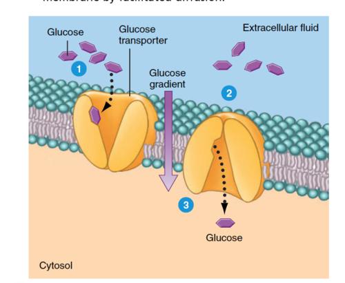
A uniporter transports one type of molecule in one direction across the membrane. Example:

- Glucose transporter proteins move glucose across the cell membrane.

<p><span>A uniporter transports one type of molecule in one direction across the membrane. Example:</span></p><p class="x1ghz6dp x1717udv x1wty727 x1pd3egz x4gjf66 x1wwbdsi x86bw1a x1lziwak x15472z4 x1qlqyl8 x15bjb6t x1lliihq x14rh7hd"><span>-</span> <span>Glucose</span> <span>transporter</span> <span>proteins</span> <span>move</span> <span>glucose</span> <span>across</span> <span>the</span> <span>cell</span> <span>membrane.</span></p>
21
New cards

Symporters

A symporter moves two different substances in the same direction across the membrane. For example:

  • Substance A moves into the cell.

  • Substance B also moves into the cell.


<p><span>A symporter moves two different substances in the same direction across the membrane. For</span> <span>example:</span></p><ul><li><p class="x1ghz6dp x1717udv x1wty727 x1pd3egz x4gjf66 x15eiy0f x1lziwak x15472z4 x1qlqyl8 x15bjb6t x1lliihq x14rh7hd xg07evd"><span>Substance</span> <span>A</span> <span>moves</span> <span>into</span> <span>the</span> <span>cell.</span></p></li><li><p class="x1ghz6dp x1717udv x1wty727 x1pd3egz x4gjf66 x15eiy0f x1lziwak x15472z4 x1qlqyl8 x15bjb6t x1lliihq x14rh7hd xg07evd"><span>Substance</span> <span>B</span> <span>also</span> <span>moves</span> <span>into</span> <span>the</span> <span>cell.</span></p></li></ul><p></p>
22
New cards

Antiporters

An antiporter moves two different substances in opposite directions across the membrane. For example:

  • Substance A moves into the cell.

  • Substance B moves out of the cell.

Easy way to remember:

  • Uni = one.

  • Sym = same.

  • Anti = opposite.


<p><span>An antiporter moves two different substances in opposite directions across the membrane. For</span> <span>example:</span></p><ul><li><p class="x1ghz6dp x1717udv x1wty727 x1pd3egz x4gjf66 x15eiy0f x1lziwak x15472z4 x1qlqyl8 x15bjb6t x1lliihq x14rh7hd xg07evd"><span>Substance</span> <span>A</span> <span>moves</span> <span>into</span> <span>the</span> <span>cell.</span></p></li><li><p class="x1ghz6dp x1717udv x1wty727 x1pd3egz x4gjf66 x15eiy0f x1lziwak x15472z4 x1qlqyl8 x15bjb6t x1lliihq x14rh7hd xg07evd"><span>Substance</span> <span>B</span> <span>moves</span> <span>out</span> <span>of</span> <span>the</span> <span>cell.</span></p></li></ul><p><span style="line-height: inherit; font-size: inherit;"><strong>Easy way to remember:</strong></span></p><ul><li><p class="x1ghz6dp x1717udv x1wty727 x1pd3egz x4gjf66 x15eiy0f x1lziwak x15472z4 x1qlqyl8 x15bjb6t x1lliihq x14rh7hd xg07evd"><span>Uni</span> <span>=</span> <span>one.</span></p></li><li><p class="x1ghz6dp x1717udv x1wty727 x1pd3egz x4gjf66 x15eiy0f x1lziwak x15472z4 x1qlqyl8 x15bjb6t x1lliihq x14rh7hd xg07evd"><span>Sym</span> <span>=</span> <span>same.</span></p></li><li><p class="x1ghz6dp x1717udv x1wty727 x1pd3egz x4gjf66 x15eiy0f x1lziwak x15472z4 x1qlqyl8 x15bjb6t x1lliihq x14rh7hd xg07evd"><span>Anti</span> <span>=</span> <span>opposite.</span></p></li></ul><p></p>
23
New cards

Active vs. Passive Transport

Transport proteins can also be classified based on whether they require energy.

24
New cards

Passive transport

Passive transport does NOT require energy from ATP. It typically moves substances down their concentration gradient. This means:

  • From high concentration.

  • To low concentration.

Example: Glucose transport through glucose transporter proteins.
<p><span>Passive</span> <span>transport</span> <span>does</span> <span>NOT</span> <span>require</span> <span>energy</span> <span>from</span> <span>ATP. It</span> <span>typically</span> <span>moves</span> <span>substances</span> <span>down</span> <span>their</span> <span>concentration</span> <span>gradient. This</span> <span>means:</span></p><ul><li><p class="x1ghz6dp x1717udv x1wty727 x1pd3egz x4gjf66 x15eiy0f x1lziwak x15472z4 x1qlqyl8 x15bjb6t x1lliihq x14rh7hd xg07evd"><span>From</span> <span>high</span> <span>concentration.</span></p></li><li><p class="x1ghz6dp x1717udv x1wty727 x1pd3egz x4gjf66 x15eiy0f x1lziwak x15472z4 x1qlqyl8 x15bjb6t x1lliihq x14rh7hd xg07evd"><span>To</span> <span>low</span> <span>concentration.</span></p></li></ul><p class="x1ghz6dp x1717udv x1wty727 x1pd3egz x4gjf66 x1wwbdsi x86bw1a x1lziwak x15472z4 x1qlqyl8 x15bjb6t x1lliihq x14rh7hd"><span>Example:</span> <span>Glucose</span> <span>transport</span> <span>through</span> <span>glucose</span> <span>transporter</span> <span>proteins.</span></p>
25
New cards
<p><span>Active</span> <span>transport</span></p>

Active transport

Active transport requires energy to move substances across the membrane. The energy often comes from ATP hydrolysis. It can move substances against their concentration gradient. This means:

  • From low concentration.

  • To high concentration.

(see image for comparison)

Exam tip: Remember that the glucose transporter discussed in the lecture is an example of passive transport.

<p><span>Active</span> <span>transport</span> <span>requires</span> <span>energy</span> <span>to</span> <span>move</span> <span>substances</span> <span>across</span> <span>the</span> <span>membrane. The</span> <span>energy</span> <span>often</span> <span>comes</span> <span>from</span> <span>ATP</span> <span>hydrolysis. It</span> <span>can</span> <span>move</span> <span>substances</span> <span>against</span> <span>their</span> <span>concentration</span> <span>gradient. This</span> <span>means:</span></p><ul><li><p class="x1ghz6dp x1717udv x1wty727 x1pd3egz x4gjf66 x15eiy0f x1lziwak x15472z4 x1qlqyl8 x15bjb6t x1lliihq x14rh7hd xg07evd"><span>From</span> <span>low</span> <span>concentration.</span></p></li><li><p class="x1ghz6dp x1717udv x1wty727 x1pd3egz x4gjf66 x15eiy0f x1lziwak x15472z4 x1qlqyl8 x15bjb6t x1lliihq x14rh7hd xg07evd"><span>To</span> <span>high</span> <span>concentration.</span></p></li></ul><p>(see image for comparison)</p><p><span style="line-height: inherit;"><strong>Exam tip:</strong></span><span> Remember that the glucose transporter discussed in the lecture is an example of passive transport.</span></p>
26
New cards

Glycosylation of Membrane Components

Glycosylation is the addition of sugar molecules to another molecule. In membranes, sugars can be attached to:

  • Proteins.

  • Lipids.

Glycoproteins: These are proteins with carbohydrate (sugar) groups attached.

Glycolipids: These are lipids with carbohydrate (sugar) groups attached. These modifications contribute to the structure and function of the cell membrane.

Important: Glycosylation can occur on both proteins and lipids.

<p><span><strong>Glycosylation</strong></span><strong> </strong><span>is</span> <span>the</span> <span>addition</span> <span>of</span> <span>sugar</span> <span>molecules</span> <span>to</span> <span>another</span> <span>molecule. In</span> <span>membranes,</span> <span>sugars</span> <span>can</span> <span>be</span> <span>attached</span> <span>to:</span></p><ul><li><p class="x1ghz6dp x1717udv x1wty727 x1pd3egz x4gjf66 x15eiy0f x1lziwak x15472z4 x1qlqyl8 x15bjb6t x1lliihq x14rh7hd xg07evd"><span>Proteins.</span></p></li><li><p class="x1ghz6dp x1717udv x1wty727 x1pd3egz x4gjf66 x15eiy0f x1lziwak x15472z4 x1qlqyl8 x15bjb6t x1lliihq x14rh7hd xg07evd"><span>Lipids.</span></p></li></ul><p><span><strong>Glycoproteins: </strong>These</span> <span>are</span> <span>proteins</span> <span>with</span> <span>carbohydrate</span> <span>(sugar)</span> <span>groups</span> <span>attached.</span></p><p><span><strong>Glycolipids: </strong>These</span> <span>are</span> <span>lipids</span> <span>with</span> <span>carbohydrate</span> <span>(sugar)</span> <span>groups</span> <span>attached. These</span> <span>modifications</span> <span>contribute</span> <span>to</span> <span>the</span> <span>structure</span> <span>and</span> <span>function</span> <span>of</span> <span>the</span> <span>cell</span> <span>membrane.</span></p><p class="x1ghz6dp x1717udv x1wty727 x1pd3egz x4gjf66 x1wwbdsi x86bw1a x1lziwak x15472z4 x1qlqyl8 x15bjb6t x1lliihq x14rh7hd"><span style="line-height: inherit; font-size: inherit;"><strong>Important:</strong></span> <span>Glycosylation</span> <span>can</span> <span>occur</span> <span>on</span> <span>both</span> <span>proteins</span> <span>and</span> <span>lipids.</span></p>
27
New cards

Membrane Lipid Composition

Not every membrane in a cell has the same lipid composition. For example, the plasma membrane contains different proportions of:

  • Cholesterol.

  • Phosphatidylcholine.

  • Phosphatidylethanolamine.

  • Phosphatidylserine.

  • Sphingomyelin.

  • Cardiolipin.

(see image)

Important concepts

  • Different membranes have different lipid compositions.

  • Different membranes also have different protein compositions.

  • These differences exist because membranes perform different functions.

For example:

  • The plasma membrane has its own specialized composition.

  • The inner mitochondrial membrane has a different composition because it performs different functions.

Structure-function relationship

Whenever two biological structures have different compositions or structures, consider whether they perform different functions. You do NOT need to memorize the exact percentages of each lipid. Just understand that membrane composition varies depending on the membrane's function.

28
New cards

Membrane Fusion

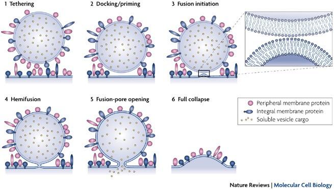
Membrane fusion occurs when two separate lipid bilayers join together to form one continuous membrane. This process requires specific proteins that help the membranes come together and merge.
<p><span>Membrane fusion occurs when two separate lipid bilayers join together to form one continuous membrane. This process requires specific proteins that help the membranes come together and merge.</span></p>
29
New cards

Membrane budding

Budding is essentially the opposite process. A portion of a membrane separates and forms a vesicle. This process also requires specialized proteins. Main idea: Membranes can fuse together or separate through budding. These processes involve many proteins, not just phospholipids.

<p><span>Budding</span> <span>is</span> <span>essentially</span> <span>the</span> <span>opposite</span> <span>process. A</span> <span>portion</span> <span>of</span> <span>a</span> <span>membrane</span> <span>separates</span> <span>and</span> <span>forms</span> <span>a</span> <span>vesicle. </span>This process also requires specialized proteins. Main idea: Membranes can fuse together or separate through budding. These processes involve many proteins, not just phospholipids.</p>
30
New cards

Signal Transduction

Signal transduction is the process by which a signal from outside a cell is transmitted to the inside of the cell. The purpose is to tell the cell to perform a particular action. For example:

  • Make a protein.

  • Change cellular activity.

  • Divide.

  • Move.

  • Undergo cell death.

Imagine someone standing outside a room trying to communicate with someone inside. They need a way to send the message through the door.

Similarly, a cell needs a way to receive information from its environment and communicate that information to its internal machinery. That process is called signal transduction.

31
New cards

The Two Main Types of Signal Transduction

Both methods begin with a signaling molecule outside the cell binding to a receptor protein. These receptors are typically ditopic integral membrane proteins.

<p><span>Both</span> <span>methods</span> <span>begin</span> <span>with</span> <span>a</span> <span>signaling</span> <span>molecule</span> <span>outside</span> <span>the</span> <span>cell</span> <span>binding</span> <span>to</span> <span>a</span> <span>receptor</span> <span>protein. These</span> <span>receptors</span> <span>are</span> <span>typically</span> <span>ditopic</span> <span>integral</span> <span>membrane</span> <span>proteins. </span></p>
32
New cards

Pathway 1: Signal transduction involving a second messenger

Steps:

  1. A signaling molecule outside the cell binds to a receptor.

  2. The receptor undergoes a conformational change.

  3. The receptor activates or interacts with another molecule inside the cell.

  4. An enzyme produces a second messenger.

  5. The second messenger binds to another protein, such as a kinase.

  6. The kinase becomes activated.

  7. The kinase phosphorylates a target protein.

  8. The target protein produces a cellular response.


<p><span>Steps:</span></p><ol><li><p class="x1ghz6dp x1717udv x1wty727 x1pd3egz x4gjf66 x15eiy0f x1lziwak x15472z4 x1qlqyl8 x15bjb6t x1lliihq x14rh7hd xg07evd"><span>A</span> <span>signaling</span> <span>molecule</span> <span>outside</span> <span>the</span> <span>cell</span> <span>binds</span> <span>to</span> <span>a</span> <span>receptor.</span></p></li><li><p class="x1ghz6dp x1717udv x1wty727 x1pd3egz x4gjf66 x15eiy0f x1lziwak x15472z4 x1qlqyl8 x15bjb6t x1lliihq x14rh7hd xg07evd"><span>The</span> <span>receptor</span> <span>undergoes</span> <span>a</span> <span>conformational</span> <span>change.</span></p></li><li><p class="x1ghz6dp x1717udv x1wty727 x1pd3egz x4gjf66 x15eiy0f x1lziwak x15472z4 x1qlqyl8 x15bjb6t x1lliihq x14rh7hd xg07evd"><span>The</span> <span>receptor</span> <span>activates</span> <span>or</span> <span>interacts</span> <span>with</span> <span>another</span> <span>molecule</span> <span>inside</span> <span>the</span> <span>cell.</span></p></li><li><p class="x1ghz6dp x1717udv x1wty727 x1pd3egz x4gjf66 x15eiy0f x1lziwak x15472z4 x1qlqyl8 x15bjb6t x1lliihq x14rh7hd xg07evd"><span>An</span> <span>enzyme</span> <span>produces</span> <span>a</span> <span>second</span> <span>messenger.</span></p></li><li><p class="x1ghz6dp x1717udv x1wty727 x1pd3egz x4gjf66 x15eiy0f x1lziwak x15472z4 x1qlqyl8 x15bjb6t x1lliihq x14rh7hd xg07evd"><span>The</span> <span>second</span> <span>messenger</span> <span>binds</span> <span>to</span> <span>another</span> <span>protein,</span> <span>such</span> <span>as</span> <span>a</span> <span>kinase.</span></p></li><li><p class="x1ghz6dp x1717udv x1wty727 x1pd3egz x4gjf66 x15eiy0f x1lziwak x15472z4 x1qlqyl8 x15bjb6t x1lliihq x14rh7hd xg07evd"><span>The</span> <span>kinase</span> <span>becomes</span> <span>activated.</span></p></li><li><p class="x1ghz6dp x1717udv x1wty727 x1pd3egz x4gjf66 x15eiy0f x1lziwak x15472z4 x1qlqyl8 x15bjb6t x1lliihq x14rh7hd xg07evd"><span>The</span> <span>kinase</span> <span>phosphorylates</span> <span>a</span> <span>target</span> <span>protein.</span></p></li><li><p class="x1ghz6dp x1717udv x1wty727 x1pd3egz x4gjf66 x15eiy0f x1lziwak x15472z4 x1qlqyl8 x15bjb6t x1lliihq x14rh7hd xg07evd"><span>The</span> <span>target</span> <span>protein</span> <span>produces</span> <span>a</span> <span>cellular</span> <span>response.</span></p></li></ol><p></p>
33
New cards

Pathway 2: Enzyme cascade

This pathway does not require a second messenger to be produced.

Instead:

  1. A signaling molecule binds to a receptor.

  2. The receptor undergoes a conformational change.

  3. An enzyme becomes activated.

  4. That enzyme activates another enzyme.

  5. Additional enzymes become activated.

  6. Eventually, a cellular response occurs.

Key difference:

  • Pathway 1 produces a second messenger.

  • Pathway 2 involves an enzyme cascade without a second messenger being produced.

(An enzyme cascade (or enzymatic cascade) is a sequence of consecutive, linked biochemical reactions where the product of one enzyme-catalyzed reaction immediately serves as the substrate for the next enzyme in the pathway).

<p><span>This</span> <span>pathway</span> <span>does</span> <span>not</span> <span>require</span> <span>a</span> <span>second</span> <span>messenger</span> <span>to</span> <span>be</span> <span>produced.</span></p><p class="x1ghz6dp x1717udv x1wty727 x1pd3egz x4gjf66 x1wwbdsi x86bw1a x1lziwak x15472z4 x1qlqyl8 x15bjb6t x1lliihq x14rh7hd"><span>Instead:</span></p><ol><li><p class="x1ghz6dp x1717udv x1wty727 x1pd3egz x4gjf66 x15eiy0f x1lziwak x15472z4 x1qlqyl8 x15bjb6t x1lliihq x14rh7hd xg07evd"><span>A</span> <span>signaling</span> <span>molecule</span> <span>binds</span> <span>to</span> <span>a</span> <span>receptor.</span></p></li><li><p class="x1ghz6dp x1717udv x1wty727 x1pd3egz x4gjf66 x15eiy0f x1lziwak x15472z4 x1qlqyl8 x15bjb6t x1lliihq x14rh7hd xg07evd"><span>The</span> <span>receptor</span> <span>undergoes</span> <span>a</span> <span>conformational</span> <span>change.</span></p></li><li><p class="x1ghz6dp x1717udv x1wty727 x1pd3egz x4gjf66 x15eiy0f x1lziwak x15472z4 x1qlqyl8 x15bjb6t x1lliihq x14rh7hd xg07evd"><span>An</span> <span>enzyme</span> <span>becomes</span> <span>activated.</span></p></li><li><p class="x1ghz6dp x1717udv x1wty727 x1pd3egz x4gjf66 x15eiy0f x1lziwak x15472z4 x1qlqyl8 x15bjb6t x1lliihq x14rh7hd xg07evd"><span>That</span> <span>enzyme</span> <span>activates</span> <span>another</span> <span>enzyme.</span></p></li><li><p class="x1ghz6dp x1717udv x1wty727 x1pd3egz x4gjf66 x15eiy0f x1lziwak x15472z4 x1qlqyl8 x15bjb6t x1lliihq x14rh7hd xg07evd"><span>Additional</span> <span>enzymes</span> <span>become</span> <span>activated.</span></p></li><li><p class="x1ghz6dp x1717udv x1wty727 x1pd3egz x4gjf66 x15eiy0f x1lziwak x15472z4 x1qlqyl8 x15bjb6t x1lliihq x14rh7hd xg07evd"><span>Eventually,</span> <span>a</span> <span>cellular</span> <span>response</span> <span>occurs.</span></p></li></ol><p><span style="line-height: inherit; font-size: inherit;"><strong>Key difference:</strong></span></p><ul><li><p class="x1ghz6dp x1717udv x1wty727 x1pd3egz x4gjf66 x15eiy0f x1lziwak x15472z4 x1qlqyl8 x15bjb6t x1lliihq x14rh7hd xg07evd"><span>Pathway</span> <span>1</span> <span>produces</span> <span>a</span> <span>second</span> <span>messenger.</span></p></li><li><p class="x1ghz6dp x1717udv x1wty727 x1pd3egz x4gjf66 x15eiy0f x1lziwak x15472z4 x1qlqyl8 x15bjb6t x1lliihq x14rh7hd xg07evd"><span>Pathway</span> <span>2</span> <span>involves</span> <span>an</span> <span>enzyme</span> <span>cascade</span> <span>without</span> <span>a</span> <span>second</span> <span>messenger</span> <span>being</span> <span>produced.</span></p></li></ul><p>(An enzyme cascade (or enzymatic cascade) is a sequence of consecutive, linked biochemical reactions where the product of one enzyme-catalyzed reaction immediately serves as the substrate for the next enzyme in the pathway).</p>
34
New cards

First messenger

The first messenger is the signaling molecule outside the cell. It binds to the receptor and starts the signaling process. Example: Epinephrine.

<p><span>The</span> <span>first</span> <span>messenger</span> <span>is</span> <span>the</span> <span>signaling</span> <span>molecule</span> <span>outside</span> <span>the</span> <span>cell. It</span> <span>binds</span> <span>to</span> <span>the</span> <span>receptor</span> <span>and</span> <span>starts</span> <span>the</span> <span>signaling</span> <span>process. Example:</span> <span>Epinephrine.</span></p>
35
New cards

Second messenger

The second messenger is a small molecule produced inside the cell in response to the first messenger. It carries the signal further into the cell. Example: Cyclic AMP (cAMP).

Remember:

  • First messenger = outside the cell.

  • Second messenger = produced inside the cell.

<p><span>The</span> <span>second</span> <span>messenger</span> <span>is</span> <span>a</span> <span>small</span> <span>molecule</span> <span>produced</span> <span>inside</span> <span>the</span> <span>cell</span> <span>in</span> <span>response</span> <span>to</span> <span>the</span> <span>first</span> <span>messenger. It</span> <span>carries</span> <span>the</span> <span>signal</span> <span>further</span> <span>into</span> <span>the</span> <span>cell. Example:</span> <span>Cyclic</span> <span>AMP</span> <span>(cAMP).</span></p><p><span style="line-height: inherit; font-size: inherit;"><strong>Remember:</strong></span></p><ul><li><p class="x1ghz6dp x1717udv x1wty727 x1pd3egz x4gjf66 x15eiy0f x1lziwak x15472z4 x1qlqyl8 x15bjb6t x1lliihq x14rh7hd xg07evd"><span>First</span> <span>messenger</span> <span>=</span> <span>outside</span> <span>the</span> <span>cell.</span></p></li><li><p class="x1ghz6dp x1717udv x1wty727 x1pd3egz x4gjf66 x15eiy0f x1lziwak x15472z4 x1qlqyl8 x15bjb6t x1lliihq x14rh7hd xg07evd"><span>Second</span> <span>messenger</span> <span>=</span> <span>produced</span> <span>inside</span> <span>the</span> <span>cell.</span></p></li></ul><p></p>
36
New cards

Protein Kinase

Cells receive signals from outside via receptors. When a ligand binds to its receptor, or a signal is transmitted as part of the signaling cascade, which may alter the function of the cell. A central element of these signaling cascades are protein kinases. Protein kinases are enzymes that transfer a phosphate group onto a target protein, thereby changing protein function from inactive to active.

Protein Kinases usually use ATP as a source of phosphate. Protein kinase binds ATP as well as the target protein and then transfers a phosphate group. The phosphorylated protein and the remaining Adenosine diphosphate (ATP missing one of its 3 phosphates) are subsequently released by protein kinase. Phosphorylation can active a proteins function. But other things can happen

  • Protein function can be activated

  • Deactivated

  • The transferred phosphate group can be a signal to other proteins to interact with the protein.

The signal is turned off if another group of of enzymes called the phosphatases are able to remove the phosphate group from the target protein, therefore turning the signal off. Phosphorylation is therefore a reversible protein modification that is regulated by a balance of kinase and phosphatase activity.

37
New cards

What Is a Kinase?

A kinase is an enzyme that catalyzes the ATP-dependent phosphorylation of a substrate. This is a definition you should memorize.

What does phosphorylation mean?

Phosphorylation is the addition of a phosphate group to a molecule. Kinases use ATP as the phosphate source. They transfer a phosphate group from ATP to a substrate.

Phosphorylation can change a protein's:

  • Activity.

  • Structure.

  • Function.

  • Interactions with other molecules.

Kinases can activate or deactivate proteins depending on the specific protein and phosphorylation site.

Protein Kinases usually act on 1/3 residues:

  1. Tyrosine

  2. Serine

  3. Threonine

However, Kinases that are able to transfer phosphate groups to serine residues are also able to do so for threonine residues, therefore they are often grouped together as serine/threonine kinases.

38
New cards

Protein Kinase A (PKA) Signaling Pathway

What Is the PKA Signaling Pathway?

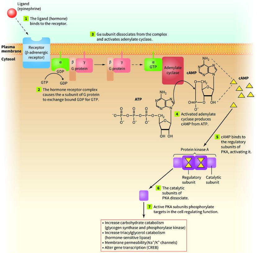
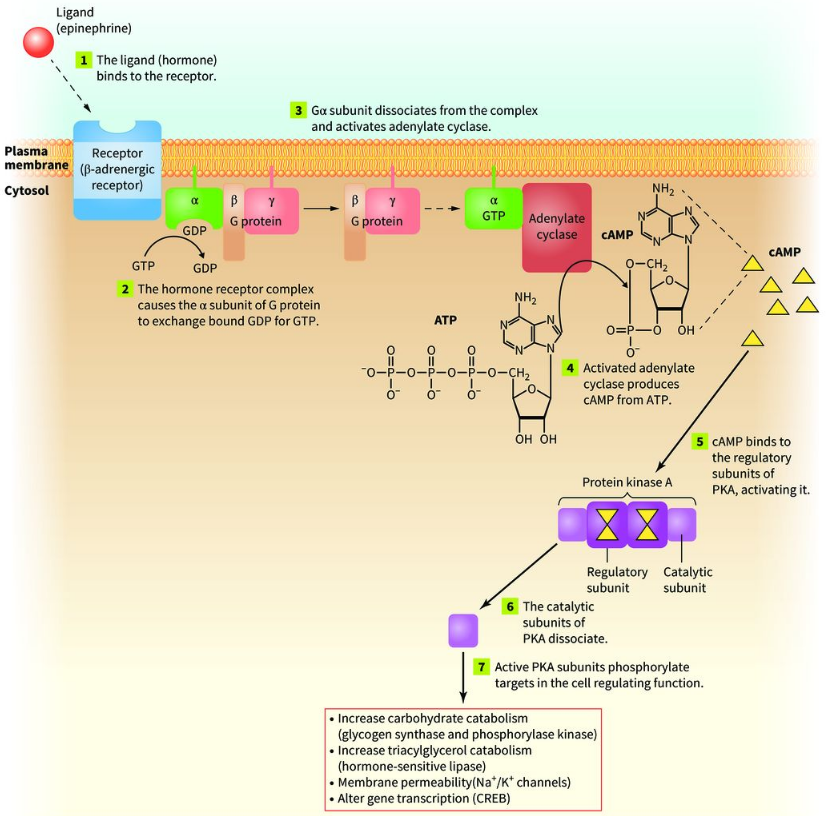
The protein kinase A (PKA) signaling pathway is a specific example of signal transduction that uses a second messenger. The second messenger is cyclic AMP (cAMP). This pathway is activated by epinephrine (adrenaline).

Epinephrine (Adrenaline)

Epinephrine is a hormone released by the adrenal glands. The adrenal glands sit on top of the kidneys. Epinephrine is released during stressful situations. This is part of the body's fight-or-flight response. Epinephrine prepares the body to respond to danger.

The first messenger in the PKA pathway

Epinephrine is the first messenger. It travels through the bloodstream and binds to a receptor on the surface of target cells. The specific receptor discussed in the lecture is the beta-adrenergic receptor.

Step 1: Epinephrine Binds to the Beta-Adrenergic Receptor

The beta-adrenergic receptor is a ditopic integral membrane protein. This means it spans the entire membrane. When epinephrine binds to the receptor:

  • The molecules have structural complementarity.

  • The receptor undergoes a conformational change.

Important: Ligand binding commonly causes a conformational change in a protein.

Step 2: Activation of the G Protein

On the inside of the cell, the beta-adrenergic receptor is associated with a G protein. This G protein has three subunits:

  • Alpha (α).

  • Beta (β).

  • Gamma (γ).

This is called a heterotrimeric G protein.

What happens when the receptor changes shape?

The receptor causes the alpha subunit to exchange GDP for GTP. Initially: GDP is attached to the alpha subunit. After receptor activation:

  • GDP is released.

  • GTP from the cytosol binds to the alpha subunit.

Very important: GDP does NOT get phosphorylated to become GTP. Instead, GDP is released and replaced by a separate GTP molecule. This is called a nucleotide exchange.

Step 3: The Alpha Subunit Separates

After GTP binds to the alpha subunit:

  • The alpha subunit undergoes a conformational change.

  • The alpha subunit separates from the beta and gamma subunits.

The alpha subunit remains associated with the inner surface of the membrane. It moves along the membrane until it encounters another protein called adenylate cyclase.

Step 4: Activation of Adenylate Cyclase

Adenylate cyclase is an enzyme associated with the inner surface of the cell membrane. When the activated alpha subunit binds to adenylate cyclase:

  • Adenylate cyclase undergoes a conformational change.

  • It becomes enzymatically active.

What reaction does adenylate cyclase catalyze?

Its substrate is ATP. It converts ATP into cyclic AMP (cAMP).

ATP —> cAMP + PPi

Where:

  • ATP = adenosine triphosphate.

  • cAMP = cyclic adenosine monophosphate.

  • PPi= pyrophosphate.

Understanding cyclic AMP

AMP stands for adenosine monophosphate.

  • Adenosine = adenine + ribose sugar.

  • Monophosphate = one phosphate group.

In cyclic AMP:

  • The phosphate is attached to the 5' carbon of the ribose.

  • The same phosphate is also connected to the 3' carbon.

This creates a cyclic structure. Therefore, the specific product is 3',5'-cyclic AMP.

What is pyrophosphate?

When ATP is converted into cyclic AMP, two phosphate groups are released together. These two linked phosphate groups are called pyrophosphate (PPiPPi​). Memorize: Adenylate cyclase converts ATP into cAMP and pyrophosphate.

Step 5: cAMP Acts as the Second Messenger

The cyclic AMP produced inside the cell is the second messenger.

Remember:

  • Epinephrine = first messenger.

  • cAMP = second messenger.

The cAMP then binds to another protein called protein kinase A (PKA). This continues the signaling pathway.
<p><span><strong>What</strong></span><strong> </strong><span><strong>Is</strong></span><strong> </strong><span><strong>the</strong></span><strong> </strong><span><strong>PKA</strong></span><strong> </strong><span><strong>Signaling</strong></span><strong> </strong><span><strong>Pathway?</strong></span></p><p><span>The</span> <span>protein</span> <span>kinase</span> <span>A</span> <span>(PKA)</span> <span>signaling</span> <span>pathway</span> <span>is</span> <span>a</span> <span>specific</span> <span>example</span> <span>of</span> <span>signal</span> <span>transduction</span> <span>that</span> <span>uses</span> <span>a</span> <span>second</span> <span>messenger. The</span> <span>second</span> <span>messenger</span> <span>is</span> <span style="line-height: inherit; font-size: inherit;"><strong>cyclic AMP (cAMP)</strong></span><span>. This</span> <span>pathway</span> <span>is</span> <span>activated</span> <span>by</span> <span>epinephrine</span> <span>(adrenaline).</span></p><p><span><strong>Epinephrine</strong></span><strong> </strong><span><strong>(Adrenaline)</strong></span></p><p class="x1ghz6dp x1717udv x1wty727 x1pd3egz x4gjf66 x1kw3rcc x104jwf5 x1lziwak x15472z4 x1qlqyl8 x15bjb6t x1lliihq x14rh7hd"><span>Epinephrine</span> <span>is</span> <span>a</span> <span>hormone</span> <span>released</span> <span>by</span> <span>the</span> <span>adrenal</span> <span>glands. The</span> <span>adrenal</span> <span>glands</span> <span>sit</span> <span>on</span> <span>top</span> <span>of</span> <span>the</span> <span>kidneys. Epinephrine</span> <span>is</span> <span>released</span> <span>during</span> <span>stressful</span> <span>situations. This</span> <span>is</span> <span>part</span> <span>of</span> <span>the</span> <span>body's</span> <span style="line-height: inherit; font-size: inherit;"><strong>fight-or-flight response</strong></span><span>. Epinephrine</span> <span>prepares</span> <span>the</span> <span>body</span> <span>to</span> <span>respond</span> <span>to</span> <span>danger.</span></p><p class="x1ghz6dp x1717udv x1wty727 x1pd3egz x4gjf66 x1kw3rcc x104jwf5 x1lziwak x15472z4 x1qlqyl8 x15bjb6t x1lliihq x14rh7hd"><span><strong>The</strong></span><strong> </strong><span><strong>first</strong></span><strong> </strong><span><strong>messenger</strong></span><strong> </strong><span><strong>in</strong></span><strong> </strong><span><strong>the</strong></span><strong> </strong><span><strong>PKA</strong></span><strong> </strong><span><strong>pathway</strong></span></p><p class="x1ghz6dp x1717udv x1wty727 x1pd3egz x4gjf66 x1kw3rcc x104jwf5 x1lziwak x15472z4 x1qlqyl8 x15bjb6t x1lliihq x14rh7hd"><span>Epinephrine</span> <span>is</span> <span>the</span> <span>first</span> <span>messenger. It</span> <span>travels</span> <span>through</span> <span>the</span> <span>bloodstream</span> <span>and</span> <span>binds</span> <span>to</span> <span>a</span> <span>receptor</span> <span>on</span> <span>the</span> <span>surface</span> <span>of</span> <span>target</span> <span>cells. The</span> <span>specific</span> <span>receptor</span> <span>discussed</span> <span>in</span> <span>the</span> <span>lecture</span> <span>is</span> <span>the</span> <span style="line-height: inherit; font-size: inherit;"><strong>beta-adrenergic receptor</strong></span><span>.</span></p><p class="x1ghz6dp x1717udv x1wty727 x1pd3egz x4gjf66 x1kw3rcc x104jwf5 x1lziwak x15472z4 x1qlqyl8 x15bjb6t x1lliihq x14rh7hd"><span><strong><u>Step</u></strong></span><strong><u> </u></strong><span><strong><u>1:</u></strong></span><strong><u> </u></strong><span><strong><u>Epinephrine</u></strong></span><strong><u> </u></strong><span><strong><u>Binds</u></strong></span><strong><u> </u></strong><span><strong><u>to</u></strong></span><strong><u> </u></strong><span><strong><u>the</u></strong></span><strong><u> </u></strong><span><strong><u>Beta-Adrenergic</u></strong></span><strong><u> </u></strong><span><strong><u>Receptor</u></strong></span></p><p class="x1ghz6dp x1717udv x1wty727 x1pd3egz x4gjf66 x1kw3rcc x104jwf5 x1lziwak x15472z4 x1qlqyl8 x15bjb6t x1lliihq x14rh7hd"><span>The</span> <span>beta-adrenergic</span> <span>receptor</span> <span>is</span> <span>a</span> <span>ditopic</span> <span>integral</span> <span>membrane</span> <span>protein. This</span> <span>means</span> <span>it</span> <span>spans</span> <span>the</span> <span>entire</span> <span>membrane. When</span> <span>epinephrine</span> <span>binds</span> <span>to</span> <span>the</span> <span>receptor:</span></p><ul><li><p class="x1ghz6dp x1717udv x1wty727 x1pd3egz x4gjf66 x15eiy0f x1lziwak x15472z4 x1qlqyl8 x15bjb6t x1lliihq x14rh7hd xg07evd"><span>The</span> <span>molecules</span> <span>have</span> <span>structural</span> <span>complementarity.</span></p></li><li><p class="x1ghz6dp x1717udv x1wty727 x1pd3egz x4gjf66 x15eiy0f x1lziwak x15472z4 x1qlqyl8 x15bjb6t x1lliihq x14rh7hd xg07evd"><span>The</span> <span>receptor</span> <span>undergoes</span> <span>a</span> <span>conformational</span> <span>change.</span></p></li></ul><p><span style="line-height: inherit;"><strong>Important:</strong></span><span> Ligand binding commonly causes a conformational change in a protein.</span></p><p><span><strong><u>Step</u></strong></span><strong><u> </u></strong><span><strong><u>2:</u></strong></span><strong><u> </u></strong><span><strong><u>Activation</u></strong></span><strong><u> </u></strong><span><strong><u>of</u></strong></span><strong><u> </u></strong><span><strong><u>the</u></strong></span><strong><u> </u></strong><span><strong><u>G</u></strong></span><strong><u> </u></strong><span><strong><u>Protein</u></strong></span></p><p><span>On</span> <span>the</span> <span>inside</span> <span>of</span> <span>the</span> <span>cell,</span> <span>the</span> <span>beta-adrenergic</span> <span>receptor</span> <span>is</span> <span>associated</span> <span>with</span> <span>a</span> <span>G</span> <span>protein. This</span> <span>G</span> <span>protein</span> <span>has</span> <span>three</span> <span>subunits:</span></p><ul><li><p class="x1ghz6dp x1717udv x1wty727 x1pd3egz x4gjf66 x15eiy0f x1lziwak x15472z4 x1qlqyl8 x15bjb6t x1lliihq x14rh7hd xg07evd"><span>Alpha</span> <span>(α).</span></p></li><li><p class="x1ghz6dp x1717udv x1wty727 x1pd3egz x4gjf66 x15eiy0f x1lziwak x15472z4 x1qlqyl8 x15bjb6t x1lliihq x14rh7hd xg07evd"><span>Beta</span> <span>(β).</span></p></li><li><p class="x1ghz6dp x1717udv x1wty727 x1pd3egz x4gjf66 x15eiy0f x1lziwak x15472z4 x1qlqyl8 x15bjb6t x1lliihq x14rh7hd xg07evd"><span>Gamma</span> <span>(γ).</span></p></li></ul><p><span>This is called a heterotrimeric G protein.</span></p><p><span><strong>What</strong></span><strong> </strong><span><strong>happens</strong></span><strong> </strong><span><strong>when</strong></span><strong> </strong><span><strong>the</strong></span><strong> </strong><span><strong>receptor</strong></span><strong> </strong><span><strong>changes</strong></span><strong> </strong><span><strong>shape?</strong></span></p><p><span>The</span> <span>receptor</span> <span>causes</span> <span>the</span> <span>alpha</span> <span>subunit</span> <span>to</span> <span>exchange</span> <span>GDP</span> <span>for</span> <span>GTP. Initially:</span> <span>GDP</span> <span>is</span> <span>attached</span> <span>to</span> <span>the</span> <span>alpha</span> <span>subunit. After</span> <span>receptor</span> <span>activation:</span></p><ul><li><p class="x1ghz6dp x1717udv x1wty727 x1pd3egz x4gjf66 x15eiy0f x1lziwak x15472z4 x1qlqyl8 x15bjb6t x1lliihq x14rh7hd xg07evd"><span>GDP</span> <span>is</span> <span>released.</span></p></li><li><p class="x1ghz6dp x1717udv x1wty727 x1pd3egz x4gjf66 x15eiy0f x1lziwak x15472z4 x1qlqyl8 x15bjb6t x1lliihq x14rh7hd xg07evd"><span>GTP</span> <span>from</span> <span>the</span> <span>cytosol</span> <span>binds</span> <span>to</span> <span>the</span> <span>alpha</span> <span>subunit.</span></p></li></ul><p class="x1ghz6dp x1717udv x1wty727 x1pd3egz x4gjf66 x1wwbdsi x86bw1a x1lziwak x15472z4 x1qlqyl8 x15bjb6t x1lliihq x14rh7hd"><span style="line-height: inherit; font-size: inherit;"><strong>Very important:</strong></span> <span>GDP</span> <span>does</span> <span>NOT</span> <span>get</span> <span>phosphorylated</span> <span>to</span> <span>become</span> <span>GTP. Instead,</span> <span>GDP</span> <span>is</span> <span>released</span> <span>and</span> <span>replaced</span> <span>by</span> <span>a</span> <span>separate</span> <span>GTP</span> <span>molecule. This</span> <span>is</span> <span>called</span> <span>a</span> <span>nucleotide</span> <span>exchange.</span></p><p class="x1ghz6dp x1717udv x1wty727 x1pd3egz x4gjf66 x1wwbdsi x86bw1a x1lziwak x15472z4 x1qlqyl8 x15bjb6t x1lliihq x14rh7hd"><span><strong><u>Step</u></strong></span><strong><u> </u></strong><span><strong><u>3:</u></strong></span><strong><u> </u></strong><span><strong><u>The</u></strong></span><strong><u> </u></strong><span><strong><u>Alpha</u></strong></span><strong><u> </u></strong><span><strong><u>Subunit</u></strong></span><strong><u> </u></strong><span><strong><u>Separates</u></strong></span></p><p class="x1ghz6dp x1717udv x1wty727 x1pd3egz x4gjf66 x1wwbdsi x86bw1a x1lziwak x15472z4 x1qlqyl8 x15bjb6t x1lliihq x14rh7hd"><span>After</span> <span>GTP</span> <span>binds</span> <span>to</span> <span>the</span> <span>alpha</span> <span>subunit:</span></p><ul><li><p class="x1ghz6dp x1717udv x1wty727 x1pd3egz x4gjf66 x15eiy0f x1lziwak x15472z4 x1qlqyl8 x15bjb6t x1lliihq x14rh7hd xg07evd"><span>The</span> <span>alpha</span> <span>subunit</span> <span>undergoes</span> <span>a</span> <span>conformational</span> <span>change.</span></p></li><li><p class="x1ghz6dp x1717udv x1wty727 x1pd3egz x4gjf66 x15eiy0f x1lziwak x15472z4 x1qlqyl8 x15bjb6t x1lliihq x14rh7hd xg07evd"><span>The</span> <span>alpha</span> <span>subunit</span> <span>separates</span> <span>from</span> <span>the</span> <span>beta</span> <span>and</span> <span>gamma</span> <span>subunits.</span></p></li></ul><p><span>The</span> <span>alpha</span> <span>subunit</span> <span>remains</span> <span>associated</span> <span>with</span> <span>the</span> <span>inner</span> <span>surface</span> <span>of</span> <span>the</span> <span>membrane. It</span> <span>moves</span> <span>along</span> <span>the</span> <span>membrane</span> <span>until</span> <span>it</span> <span>encounters</span> <span>another</span> <span>protein</span> <span>called</span> <span style="line-height: inherit; font-size: inherit;"><strong>adenylate cyclase</strong></span><span>.</span></p><p><span><strong><u>Step</u></strong></span><strong><u> </u></strong><span><strong><u>4:</u></strong></span><strong><u> </u></strong><span><strong><u>Activation</u></strong></span><strong><u> </u></strong><span><strong><u>of</u></strong></span><strong><u> </u></strong><span><strong><u>Adenylate</u></strong></span><strong><u> </u></strong><span><strong><u>Cyclase</u></strong></span></p><p><span>Adenylate</span> <span>cyclase</span> <span>is</span> <span>an</span> <span>enzyme</span> <span>associated</span> <span>with</span> <span>the</span> <span>inner</span> <span>surface</span> <span>of</span> <span>the</span> <span>cell</span> <span>membrane. When</span> <span>the</span> <span>activated</span> <span>alpha</span> <span>subunit</span> <span>binds</span> <span>to</span> <span>adenylate</span> <span>cyclase:</span></p><ul><li><p class="x1ghz6dp x1717udv x1wty727 x1pd3egz x4gjf66 x15eiy0f x1lziwak x15472z4 x1qlqyl8 x15bjb6t x1lliihq x14rh7hd xg07evd"><span>Adenylate</span> <span>cyclase</span> <span>undergoes</span> <span>a</span> <span>conformational</span> <span>change.</span></p></li><li><p class="x1ghz6dp x1717udv x1wty727 x1pd3egz x4gjf66 x15eiy0f x1lziwak x15472z4 x1qlqyl8 x15bjb6t x1lliihq x14rh7hd xg07evd"><span>It</span> <span>becomes</span> <span>enzymatically</span> <span>active.</span></p></li></ul><p><span><strong>What</strong></span><strong> </strong><span><strong>reaction</strong></span><strong> </strong><span><strong>does</strong></span><strong> </strong><span><strong>adenylate</strong></span><strong> </strong><span><strong>cyclase</strong></span><strong> </strong><span><strong>catalyze?</strong></span></p><p class="x1ghz6dp x1717udv x1wty727 x1pd3egz x4gjf66 x1kw3rcc x104jwf5 x1lziwak x15472z4 x1qlqyl8 x15bjb6t x1lliihq x14rh7hd"><span>Its</span> <span>substrate</span> <span>is</span> <span>ATP. It</span> <span>converts</span> <span>ATP</span> <span>into</span> <span>cyclic</span> <span>AMP</span> <span>(cAMP).</span></p><p class="x1ghz6dp x1717udv x1wty727 x1pd3egz x4gjf66 x1kw3rcc x104jwf5 x1lziwak x15472z4 x1qlqyl8 x15bjb6t x1lliihq x14rh7hd"><span>ATP —&gt; cAMP + PPi</span></p><p class="x1ghz6dp x1717udv x1wty727 x1pd3egz x4gjf66 x1kw3rcc x104jwf5 x1lziwak x15472z4 x1qlqyl8 x15bjb6t x1lliihq x14rh7hd"><span>Where:</span></p><ul><li><p class="x1ghz6dp x1717udv x1wty727 x1pd3egz x4gjf66 x15eiy0f x1lziwak x15472z4 x1qlqyl8 x15bjb6t x1lliihq x14rh7hd xg07evd"><span>ATP</span> <span>=</span> <span>adenosine</span> <span>triphosphate.</span></p></li><li><p class="x1ghz6dp x1717udv x1wty727 x1pd3egz x4gjf66 x15eiy0f x1lziwak x15472z4 x1qlqyl8 x15bjb6t x1lliihq x14rh7hd xg07evd"><span>cAMP</span> <span>=</span> <span>cyclic</span> <span>adenosine</span> <span>monophosphate.</span></p></li><li><p class="x1ghz6dp x1717udv x1wty727 x1pd3egz x4gjf66 x15eiy0f x1lziwak x15472z4 x1qlqyl8 x15bjb6t x1lliihq x14rh7hd xg07evd"><span style="font-family: KaTeX_Main, &quot;Times New Roman&quot;, serif; line-height: 1.2; font-size: 1.21em;">PPi</span><span>=</span> <span>pyrophosphate.</span></p></li></ul><p><span><strong>Understanding</strong></span><strong> </strong><span><strong>cyclic</strong></span><strong> </strong><span><strong>AMP</strong></span></p><p class="x1ghz6dp x1717udv x1wty727 x1pd3egz x4gjf66 x1kw3rcc x104jwf5 x1lziwak x15472z4 x1qlqyl8 x15bjb6t x1lliihq x14rh7hd"><span>AMP</span> <span>stands</span> <span>for</span> <span>adenosine</span> <span>monophosphate.</span></p><ul><li><p class="x1ghz6dp x1717udv x1wty727 x1pd3egz x4gjf66 x15eiy0f x1lziwak x15472z4 x1qlqyl8 x15bjb6t x1lliihq x14rh7hd xg07evd"><span>Adenosine</span> <span>=</span> <span>adenine</span> <span>+</span> <span>ribose</span> <span>sugar.</span></p></li><li><p class="x1ghz6dp x1717udv x1wty727 x1pd3egz x4gjf66 x15eiy0f x1lziwak x15472z4 x1qlqyl8 x15bjb6t x1lliihq x14rh7hd xg07evd"><span>Monophosphate</span> <span>=</span> <span>one</span> <span>phosphate</span> <span>group.</span></p></li></ul><p class="x1ghz6dp x1717udv x1wty727 x1pd3egz x4gjf66 x1wwbdsi x86bw1a x1lziwak x15472z4 x1qlqyl8 x15bjb6t x1lliihq x14rh7hd"><span>In</span> <span>cyclic</span> <span>AMP:</span></p><ul><li><p class="x1ghz6dp x1717udv x1wty727 x1pd3egz x4gjf66 x15eiy0f x1lziwak x15472z4 x1qlqyl8 x15bjb6t x1lliihq x14rh7hd xg07evd"><span>The</span> <span>phosphate</span> <span>is</span> <span>attached</span> <span>to</span> <span>the</span> <span>5'</span> <span>carbon</span> <span>of</span> <span>the</span> <span>ribose.</span></p></li><li><p class="x1ghz6dp x1717udv x1wty727 x1pd3egz x4gjf66 x15eiy0f x1lziwak x15472z4 x1qlqyl8 x15bjb6t x1lliihq x14rh7hd xg07evd"><span>The</span> <span>same</span> <span>phosphate</span> <span>is</span> <span>also</span> <span>connected</span> <span>to</span> <span>the</span> <span>3'</span> <span>carbon.</span></p></li></ul><p class="x1ghz6dp x1717udv x1wty727 x1pd3egz x4gjf66 x1wwbdsi x86bw1a x1lziwak x15472z4 x1qlqyl8 x15bjb6t x1lliihq x14rh7hd"><span>This</span> <span>creates</span> <span>a</span> <span>cyclic</span> <span>structure. Therefore,</span> <span>the</span> <span>specific</span> <span>product</span> <span>is</span> <span style="line-height: inherit; font-size: inherit;"><strong>3',5'-cyclic AMP</strong></span><span>.</span></p><p><span><strong>What</strong></span><strong> </strong><span><strong>is</strong></span><strong> </strong><span><strong>pyrophosphate?</strong></span></p><p class="x1ghz6dp x1717udv x1wty727 x1pd3egz x4gjf66 x1kw3rcc x104jwf5 x1lziwak x15472z4 x1qlqyl8 x15bjb6t x1lliihq x14rh7hd"><span>When</span> <span>ATP</span> <span>is</span> <span>converted</span> <span>into</span> <span>cyclic</span> <span>AMP,</span> <span>two</span> <span>phosphate</span> <span>groups</span> <span>are</span> <span>released</span> <span>together. These</span> <span>two</span> <span>linked</span> <span>phosphate</span> <span>groups</span> <span>are</span> <span>called</span> <span>pyrophosphate</span> <span>(</span><span style="font-family: KaTeX_Main, &quot;Times New Roman&quot;, serif; line-height: 1.2; font-size: 1.21em;">PPi<em>PPi</em>​</span><span>). </span><span style="line-height: inherit; font-size: inherit;"><strong>Memorize:</strong></span> <span>Adenylate</span> <span>cyclase</span> <span>converts</span> <span>ATP</span> <span>into</span> <span>cAMP</span> <span>and</span> <span>pyrophosphate.</span></p><p class="x1ghz6dp x1717udv x1wty727 x1pd3egz x4gjf66 x1kw3rcc x104jwf5 x1lziwak x15472z4 x1qlqyl8 x15bjb6t x1lliihq x14rh7hd"><span><strong><u>Step</u></strong></span><strong><u> </u></strong><span><strong><u>5:</u></strong></span><strong><u> </u></strong><span><strong><u>cAMP</u></strong></span><strong><u> </u></strong><span><strong><u>Acts</u></strong></span><strong><u> </u></strong><span><strong><u>as</u></strong></span><strong><u> </u></strong><span><strong><u>the</u></strong></span><strong><u> </u></strong><span><strong><u>Second</u></strong></span><strong><u> </u></strong><span><strong><u>Messenger</u></strong></span></p><p class="x1ghz6dp x1717udv x1wty727 x1pd3egz x4gjf66 x1kw3rcc x104jwf5 x1lziwak x15472z4 x1qlqyl8 x15bjb6t x1lliihq x14rh7hd"><span>The</span> <span>cyclic</span> <span>AMP</span> <span>produced</span> <span>inside</span> <span>the</span> <span>cell</span> <span>is</span> <span>the</span> <span>second</span> <span>messenger.</span></p><p class="x1ghz6dp x1717udv x1wty727 x1pd3egz x4gjf66 x1wwbdsi x86bw1a x1lziwak x15472z4 x1qlqyl8 x15bjb6t x1lliihq x14rh7hd"><span>Remember:</span></p><ul><li><p class="x1ghz6dp x1717udv x1wty727 x1pd3egz x4gjf66 x15eiy0f x1lziwak x15472z4 x1qlqyl8 x15bjb6t x1lliihq x14rh7hd xg07evd"><span>Epinephrine</span> <span>=</span> <span>first</span> <span>messenger.</span></p></li><li><p class="x1ghz6dp x1717udv x1wty727 x1pd3egz x4gjf66 x15eiy0f x1lziwak x15472z4 x1qlqyl8 x15bjb6t x1lliihq x14rh7hd xg07evd"><span>cAMP</span> <span>=</span> <span>second</span> <span>messenger.</span></p></li></ul><p class="x1ghz6dp x1717udv x1wty727 x1pd3egz x4gjf66 x1wwbdsi x86bw1a x1lziwak x15472z4 x1qlqyl8 x15bjb6t x1lliihq x14rh7hd"><span>The</span> <span>cAMP</span> <span>then</span> <span>binds</span> <span>to</span> <span>another</span> <span>protein</span> <span>called</span> <span>protein</span> <span>kinase</span> <span>A</span> <span>(PKA). This</span> <span>continues</span> <span>the</span> <span>signaling</span> <span>pathway.</span></p>
39
New cards

Signal transduction

Signal transduction is the process by which a cell receives a signal and converts it into a response inside the cell.

There are two important terms:

  • First messenger: The signaling molecule (ligand) that binds to a receptor.

  • Second messenger: A small molecule produced inside the cell that carries the signal further.

In the first pathway discussed, epinephrine is the first messenger, and cyclic AMP (cAMP) is the second messenger.

Epinephrine signaling

  • Epinephrine (adrenaline) is released during stressful situations.

  • It binds to a receptor on the cell membrane.

  • This activates a series of events inside the cell.

  • The cell responds by making more energy available.
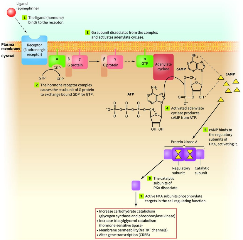

<p><span style="line-height: inherit; font-size: inherit;"><strong>Signal transduction</strong></span> <span>is</span> <span>the</span> <span>process</span> <span>by</span> <span>which</span> <span>a</span> <span>cell</span> <span>receives</span> <span>a</span> <span>signal</span> <span>and</span> <span>converts</span> <span>it</span> <span>into</span> <span>a</span> <span>response</span> <span>inside</span> <span>the</span> <span>cell.</span></p><p class="x1ghz6dp x1717udv x1wty727 x1pd3egz x4gjf66 x1wwbdsi x86bw1a x1lziwak x15472z4 x1qlqyl8 x15bjb6t x1lliihq x14rh7hd"><span>There</span> <span>are</span> <span>two</span> <span>important</span> <span>terms:</span></p><ul><li><p class="x1ghz6dp x1717udv x1wty727 x1pd3egz x4gjf66 x15eiy0f x1lziwak x15472z4 x1qlqyl8 x15bjb6t x1lliihq x14rh7hd xg07evd"><span style="line-height: inherit; font-size: inherit;"><strong>First messenger:</strong></span> <span>The</span> <span>signaling</span> <span>molecule</span> <span>(ligand)</span> <span>that</span> <span>binds</span> <span>to</span> <span>a</span> <span>receptor.</span></p></li><li><p class="x1ghz6dp x1717udv x1wty727 x1pd3egz x4gjf66 x15eiy0f x1lziwak x15472z4 x1qlqyl8 x15bjb6t x1lliihq x14rh7hd xg07evd"><span style="line-height: inherit; font-size: inherit;"><strong>Second messenger:</strong></span> <span>A</span> <span>small</span> <span>molecule</span> <span>produced</span> <span>inside</span> <span>the</span> <span>cell</span> <span>that</span> <span>carries</span> <span>the</span> <span>signal</span> <span>further.</span></p></li></ul><p class="x1ghz6dp x1717udv x1wty727 x1pd3egz x4gjf66 x1wwbdsi x86bw1a x1lziwak x15472z4 x1qlqyl8 x15bjb6t x1lliihq x14rh7hd"><span>In</span> <span>the</span> <span>first</span> <span>pathway</span> <span>discussed,</span> <span>epinephrine</span> <span>is</span> <span>the</span> <span>first</span> <span>messenger,</span> <span>and</span> <span>cyclic</span> <span>AMP</span> <span>(cAMP)</span> <span>is</span> <span>the</span> <span>second</span> <span>messenger.</span></p><p class="x1ghz6dp x1717udv x1wty727 x1pd3egz x4gjf66 x1wwbdsi x86bw1a x1lziwak x15472z4 x1qlqyl8 x15bjb6t x1lliihq x14rh7hd"><span><strong><u>Epinephrine</u></strong></span><strong><u> </u></strong><span><strong><u>signaling</u></strong></span></p><ul><li><p class="x1ghz6dp x1717udv x1wty727 x1pd3egz x4gjf66 x15eiy0f x1lziwak x15472z4 x1qlqyl8 x15bjb6t x1lliihq x14rh7hd xg07evd"><span>Epinephrine</span> <span>(adrenaline)</span> <span>is</span> <span>released</span> <span>during</span> <span>stressful</span> <span>situations.</span></p></li><li><p class="x1ghz6dp x1717udv x1wty727 x1pd3egz x4gjf66 x15eiy0f x1lziwak x15472z4 x1qlqyl8 x15bjb6t x1lliihq x14rh7hd xg07evd"><span>It</span> <span>binds</span> <span>to</span> <span>a</span> <span>receptor</span> <span>on</span> <span>the</span> <span>cell</span> <span>membrane.</span></p></li><li><p class="x1ghz6dp x1717udv x1wty727 x1pd3egz x4gjf66 x15eiy0f x1lziwak x15472z4 x1qlqyl8 x15bjb6t x1lliihq x14rh7hd xg07evd"><span>This</span> <span>activates</span> <span>a</span> <span>series</span> <span>of</span> <span>events</span> <span>inside</span> <span>the</span> <span>cell.</span></p></li><li><p class="x1ghz6dp x1717udv x1wty727 x1pd3egz x4gjf66 x15eiy0f x1lziwak x15472z4 x1qlqyl8 x15bjb6t x1lliihq x14rh7hd xg07evd"><span>The</span> <span>cell</span> <span>responds</span> <span>by</span> <span>making</span> <span>more</span> <span>energy</span> <span>available.</span></p></li></ul><p></p>
40
New cards

The cAMP – Protein Kinase A (PKA) Pathway

This pathway involves a G-protein-coupled receptor (GPCR), a G protein, cAMP, and protein kinase A. Step-by-step process:

Step 1: Epinephrine binds to the receptor

  • Epinephrine (first messenger) binds to a receptor on the cell membrane.

  • The receptor undergoes a conformational change (change in shape).

  • This activates the G protein associated with the receptor.

Step 2: G protein becomes activated

The G protein consists of three subunits:

  • Alpha (α)

  • Beta (β)

  • Gamma (γ)

Normally, the alpha subunit is bound to GDP. When the receptor is activated:

  • GDP is released.

  • GTP binds to the alpha subunit.

  • The alpha subunit undergoes a conformational change.

  • The alpha subunit separates from the beta and gamma subunits.

Important: The alpha subunit bound to GTP is the active form.

Step 3: Adenylate cyclase is activated

  • The activated alpha subunit interacts with adenylate cyclase.

  • Adenylate cyclase is an enzyme located in the cell membrane.

  • It converts ATP into cyclic AMP (cAMP). ATP → cAMP.

Remember:

  • Adenylate cyclase produces cAMP.

  • cAMP is the second messenger.

Step 4: cAMP activates Protein Kinase A (PKA)

PKA is made up of multiple subunits:

  • 2 regulatory subunits

  • 2 catalytic subunits

When cAMP binds to the regulatory subunits:

  1. The regulatory subunits undergo a conformational change.

  2. The catalytic subunits are released.

  3. The catalytic subunits become active.

  4. The active catalytic subunits can now phosphorylate other proteins.

Step 5: PKA phosphorylates other proteins

A kinase is an enzyme that catalyzes the ATP-dependent phosphorylation of a substrate. In other words, it transfers a phosphate group from ATP onto another molecule. A protein kinase specifically phosphorylates proteins. PKA phosphorylates many different proteins, including other enzymes. This changes their activity and produces different responses inside the cell.
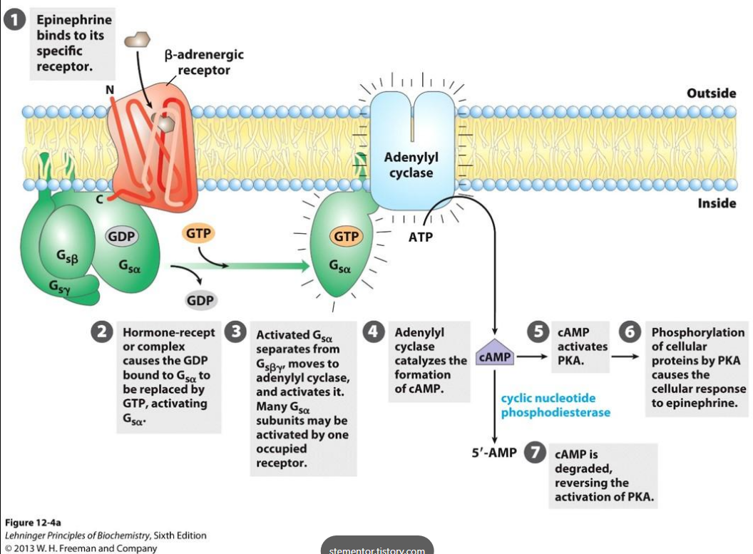
<p><span>This</span> <span>pathway</span> <span>involves</span> <span>a</span> <span>G-protein-coupled</span> <span>receptor</span> <span>(GPCR),</span> <span>a</span> <span>G</span> <span>protein,</span> <span>cAMP,</span> <span>and</span> <span>protein</span> <span>kinase</span> <span>A. Step-by-step process:</span></p><p class="x1ghz6dp x1717udv x1wty727 x1pd3egz x4gjf66 x1kw3rcc x104jwf5 x1lziwak x15472z4 x1qlqyl8 x15bjb6t x1lliihq x14rh7hd"><span style="line-height: inherit; font-size: inherit;"><strong><u>Step 1: Epinephrine binds to the receptor</u></strong></span></p><ul><li><p class="x1ghz6dp x1717udv x1wty727 x1pd3egz x4gjf66 x15eiy0f x1lziwak x15472z4 x1qlqyl8 x15bjb6t x1lliihq x14rh7hd xg07evd"><span>Epinephrine</span> <span>(first</span> <span>messenger)</span> <span>binds</span> <span>to</span> <span>a</span> <span>receptor</span> <span>on</span> <span>the</span> <span>cell</span> <span>membrane.</span></p></li><li><p class="x1ghz6dp x1717udv x1wty727 x1pd3egz x4gjf66 x15eiy0f x1lziwak x15472z4 x1qlqyl8 x15bjb6t x1lliihq x14rh7hd xg07evd"><span>The</span> <span>receptor</span> <span>undergoes</span> <span>a</span> <span>conformational</span> <span>change</span> <span>(change</span> <span>in</span> <span>shape).</span></p></li><li><p class="x1ghz6dp x1717udv x1wty727 x1pd3egz x4gjf66 x15eiy0f x1lziwak x15472z4 x1qlqyl8 x15bjb6t x1lliihq x14rh7hd xg07evd"><span>This</span> <span>activates</span> <span>the</span> <span>G</span> <span>protein</span> <span>associated</span> <span>with</span> <span>the</span> <span>receptor.</span></p></li></ul><p><span style="line-height: inherit; font-size: inherit;"><strong><u>Step 2: G protein becomes activated</u></strong></span></p><p class="x1ghz6dp x1717udv x1wty727 x1pd3egz x4gjf66 x1wwbdsi x86bw1a x1lziwak x15472z4 x1qlqyl8 x15bjb6t x1lliihq x14rh7hd"><span>The</span> <span>G</span> <span>protein</span> <span>consists</span> <span>of</span> <span>three</span> <span>subunits:</span></p><ul><li><p class="x1ghz6dp x1717udv x1wty727 x1pd3egz x4gjf66 x15eiy0f x1lziwak x15472z4 x1qlqyl8 x15bjb6t x1lliihq x14rh7hd xg07evd"><span>Alpha</span> <span>(α)</span></p></li><li><p class="x1ghz6dp x1717udv x1wty727 x1pd3egz x4gjf66 x15eiy0f x1lziwak x15472z4 x1qlqyl8 x15bjb6t x1lliihq x14rh7hd xg07evd"><span>Beta</span> <span>(β)</span></p></li><li><p class="x1ghz6dp x1717udv x1wty727 x1pd3egz x4gjf66 x15eiy0f x1lziwak x15472z4 x1qlqyl8 x15bjb6t x1lliihq x14rh7hd xg07evd"><span>Gamma</span> <span>(γ)</span></p></li></ul><p><span>Normally,</span> <span>the</span> <span>alpha</span> <span>subunit</span> <span>is</span> <span>bound</span> <span>to</span> <span>GDP. When</span> <span>the</span> <span>receptor</span> <span>is</span> <span>activated:</span></p><ul><li><p class="x1ghz6dp x1717udv x1wty727 x1pd3egz x4gjf66 x15eiy0f x1lziwak x15472z4 x1qlqyl8 x15bjb6t x1lliihq x14rh7hd xg07evd"><span>GDP</span> <span>is</span> <span>released.</span></p></li><li><p class="x1ghz6dp x1717udv x1wty727 x1pd3egz x4gjf66 x15eiy0f x1lziwak x15472z4 x1qlqyl8 x15bjb6t x1lliihq x14rh7hd xg07evd"><span>GTP</span> <span>binds</span> <span>to</span> <span>the</span> <span>alpha</span> <span>subunit.</span></p></li><li><p class="x1ghz6dp x1717udv x1wty727 x1pd3egz x4gjf66 x15eiy0f x1lziwak x15472z4 x1qlqyl8 x15bjb6t x1lliihq x14rh7hd xg07evd"><span>The</span> <span>alpha</span> <span>subunit</span> <span>undergoes</span> <span>a</span> <span>conformational</span> <span>change.</span></p></li><li><p class="x1ghz6dp x1717udv x1wty727 x1pd3egz x4gjf66 x15eiy0f x1lziwak x15472z4 x1qlqyl8 x15bjb6t x1lliihq x14rh7hd xg07evd"><span>The</span> <span>alpha</span> <span>subunit</span> <span>separates</span> <span>from</span> <span>the</span> <span>beta</span> <span>and</span> <span>gamma</span> <span>subunits.</span></p></li></ul><p><span><strong>Important:</strong> The alpha subunit bound to GTP is the active form.</span></p><p><span style="line-height: inherit; font-size: inherit;"><strong>Step 3: Adenylate cyclase is activated</strong></span></p><ul><li><p class="x1ghz6dp x1717udv x1wty727 x1pd3egz x4gjf66 x15eiy0f x1lziwak x15472z4 x1qlqyl8 x15bjb6t x1lliihq x14rh7hd xg07evd"><span>The</span> <span>activated</span> <span>alpha</span> <span>subunit</span> <span>interacts</span> <span>with</span> <span>adenylate</span> <span>cyclase.</span></p></li><li><p class="x1ghz6dp x1717udv x1wty727 x1pd3egz x4gjf66 x15eiy0f x1lziwak x15472z4 x1qlqyl8 x15bjb6t x1lliihq x14rh7hd xg07evd"><span>Adenylate</span> <span>cyclase</span> <span>is</span> <span>an</span> <span>enzyme</span> <span>located</span> <span>in</span> <span>the</span> <span>cell</span> <span>membrane.</span></p></li><li><p class="x1ghz6dp x1717udv x1wty727 x1pd3egz x4gjf66 x15eiy0f x1lziwak x15472z4 x1qlqyl8 x15bjb6t x1lliihq x14rh7hd xg07evd"><span>It</span> <span>converts</span> <span>ATP</span> <span>into</span> <span>cyclic</span> <span>AMP</span> <span>(cAMP). ATP</span> <span>→</span> <span>cAMP. </span></p></li></ul><p class="x1ghz6dp x1717udv x1wty727 x1pd3egz x4gjf66 x1wwbdsi x86bw1a x1lziwak x15472z4 x1qlqyl8 x15bjb6t x1lliihq x14rh7hd"><span style="line-height: inherit; font-size: inherit;"><strong>Remember:</strong></span></p><ul><li><p class="x1ghz6dp x1717udv x1wty727 x1pd3egz x4gjf66 x15eiy0f x1lziwak x15472z4 x1qlqyl8 x15bjb6t x1lliihq x14rh7hd xg07evd"><span>Adenylate</span> <span>cyclase</span> <span>produces</span> <span>cAMP.</span></p></li><li><p class="x1ghz6dp x1717udv x1wty727 x1pd3egz x4gjf66 x15eiy0f x1lziwak x15472z4 x1qlqyl8 x15bjb6t x1lliihq x14rh7hd xg07evd"><span>cAMP</span> <span>is</span> <span>the</span> <span>second</span> <span>messenger.</span></p></li></ul><p><span style="line-height: inherit; font-size: inherit;"><strong><u>Step 4: cAMP activates Protein Kinase A (PKA)</u></strong></span></p><p class="x1ghz6dp x1717udv x1wty727 x1pd3egz x4gjf66 x1wwbdsi x86bw1a x1lziwak x15472z4 x1qlqyl8 x15bjb6t x1lliihq x14rh7hd"><span>PKA</span> <span>is</span> <span>made</span> <span>up</span> <span>of</span> <span>multiple</span> <span>subunits:</span></p><ul><li><p class="x1ghz6dp x1717udv x1wty727 x1pd3egz x4gjf66 x15eiy0f x1lziwak x15472z4 x1qlqyl8 x15bjb6t x1lliihq x14rh7hd xg07evd"><span>2</span> <span>regulatory</span> <span>subunits</span></p></li><li><p class="x1ghz6dp x1717udv x1wty727 x1pd3egz x4gjf66 x15eiy0f x1lziwak x15472z4 x1qlqyl8 x15bjb6t x1lliihq x14rh7hd xg07evd"><span>2</span> <span>catalytic</span> <span>subunits</span></p></li></ul><p class="x1ghz6dp x1717udv x1wty727 x1pd3egz x4gjf66 x1wwbdsi x86bw1a x1lziwak x15472z4 x1qlqyl8 x15bjb6t x1lliihq x14rh7hd"><span>When</span> <span>cAMP</span> <span>binds</span> <span>to</span> <span>the</span> <span>regulatory</span> <span>subunits:</span></p><ol><li><p class="x1ghz6dp x1717udv x1wty727 x1pd3egz x4gjf66 x15eiy0f x1lziwak x15472z4 x1qlqyl8 x15bjb6t x1lliihq x14rh7hd xg07evd"><span>The</span> <span>regulatory</span> <span>subunits</span> <span>undergo</span> <span>a</span> <span>conformational</span> <span>change.</span></p></li><li><p class="x1ghz6dp x1717udv x1wty727 x1pd3egz x4gjf66 x15eiy0f x1lziwak x15472z4 x1qlqyl8 x15bjb6t x1lliihq x14rh7hd xg07evd"><span>The</span> <span>catalytic</span> <span>subunits</span> <span>are</span> <span>released.</span></p></li><li><p class="x1ghz6dp x1717udv x1wty727 x1pd3egz x4gjf66 x15eiy0f x1lziwak x15472z4 x1qlqyl8 x15bjb6t x1lliihq x14rh7hd xg07evd"><span>The</span> <span>catalytic</span> <span>subunits</span> <span>become</span> <span>active.</span></p></li><li><p class="x1ghz6dp x1717udv x1wty727 x1pd3egz x4gjf66 x15eiy0f x1lziwak x15472z4 x1qlqyl8 x15bjb6t x1lliihq x14rh7hd xg07evd"><span>The</span> <span>active</span> <span>catalytic</span> <span>subunits</span> <span>can</span> <span>now</span> <span>phosphorylate</span> <span>other</span> <span>proteins.</span></p></li></ol><p><span><strong><u>Step 5: PKA phosphorylates other proteins</u></strong></span></p><p><span>A</span> <span>kinase</span> <span>is</span> <span>an</span> <span>enzyme</span> <span>that</span> <span>catalyzes</span> <span>the</span> <span>ATP-dependent</span> <span>phosphorylation</span> <span>of</span> <span>a</span> <span>substrate. In</span> <span>other</span> <span>words,</span> <span>it</span> <span>transfers</span> <span>a</span> <span>phosphate</span> <span>group</span> <span>from</span> <span>ATP</span> <span>onto</span> <span>another</span> <span>molecule. A</span> <span>protein</span> <span>kinase</span> <span>specifically</span> <span>phosphorylates</span> <span>proteins. PKA</span> <span>phosphorylates</span> <span>many</span> <span>different</span> <span>proteins,</span> <span>including</span> <span>other</span> <span>enzymes. This</span> <span>changes</span> <span>their</span> <span>activity</span> <span>and</span> <span>produces</span> <span>different</span> <span>responses</span> <span>inside</span> <span>the</span> <span>cell.</span></p>
41
New cards

Effects of activating PKA

The activation of PKA can cause several cellular responses (see image). Important vocabulary:

  • Catabolism = breakdown of molecules.

  • Anabolism = building molecules.

  • Triglycerides (triacylglycerols) = the stored form of fat, made of one glycerol and three fatty acids.

Why does epinephrine increase fat and carbohydrate breakdown?

Because the body needs energy quickly during stressful situations. For example, when you need to run away from danger, your muscles need a rapid supply of energy.

<p><span>The activation of PKA can cause several cellular responses (see image). </span><span style="line-height: inherit; font-size: inherit;"><strong>Important vocabulary:</strong></span></p><ul><li><p class="x1ghz6dp x1717udv x1wty727 x1pd3egz x4gjf66 x15eiy0f x1lziwak x15472z4 x1qlqyl8 x15bjb6t x1lliihq x14rh7hd xg07evd"><span>Catabolism</span> <span>=</span> <span>breakdown</span> <span>of</span> <span>molecules.</span></p></li><li><p class="x1ghz6dp x1717udv x1wty727 x1pd3egz x4gjf66 x15eiy0f x1lziwak x15472z4 x1qlqyl8 x15bjb6t x1lliihq x14rh7hd xg07evd"><span>Anabolism</span> <span>=</span> <span>building</span> <span>molecules.</span></p></li><li><p class="x1ghz6dp x1717udv x1wty727 x1pd3egz x4gjf66 x15eiy0f x1lziwak x15472z4 x1qlqyl8 x15bjb6t x1lliihq x14rh7hd xg07evd"><span>Triglycerides</span> <span>(triacylglycerols)</span> <span>=</span> <span>the</span> <span>stored</span> <span>form</span> <span>of</span> <span>fat,</span> <span>made</span> <span>of</span> <span>one</span> <span>glycerol</span> <span>and</span> <span>three</span> <span>fatty</span> <span>acids.</span></p></li></ul><p class="x1ghz6dp x1717udv x1wty727 x1pd3egz x4gjf66 x1wwbdsi x86bw1a x1lziwak x15472z4 x1qlqyl8 x15bjb6t x1lliihq x14rh7hd"><span style="line-height: inherit; font-size: inherit;"><strong>Why does epinephrine increase fat and carbohydrate breakdown?</strong></span></p><p class="x1ghz6dp x1717udv x1wty727 x1pd3egz x4gjf66 x1wwbdsi x86bw1a x1lziwak x15472z4 x1qlqyl8 x15bjb6t x1lliihq x14rh7hd"><span>Because</span> <span>the</span> <span>body</span> <span>needs</span> <span>energy</span> <span>quickly</span> <span>during</span> <span>stressful</span> <span>situations. For example, when you need to run away from danger, your muscles need a rapid supply of energy.</span></p>
42
New cards

How Does the Cell Turn OFF the PKA Pathway?

The signaling pathway cannot stay activated forever. It needs to be reversible so the cell can:

  • Turn the pathway on when needed.

  • Turn it off when the signal is no longer needed.

  • Activate it again later.

There are several ways to shut down this pathway.

Method 1: Remove epinephrine from the receptor

  • Epinephrine can detach from the receptor.

  • Without the ligand bound, the receptor is no longer activated.

  • The downstream signaling pathway stops being stimulated.

Method 2: An antagonist binds to the receptor

An antagonist is a molecule that binds to a receptor and blocks its normal activation.

  • Epinephrine normally activates the receptor.

  • An antagonist can bind instead.

  • This prevents epinephrine from activating the receptor.

Method 3: Remove the receptor from the cell membrane

  • The receptor and ligand can be taken into the cell through internalization.

  • The receptor is removed from the cell surface.

  • This prevents it from continuing to receive signals from outside the cell.

Method 4: GTP is converted back into GDP

This is one of the most important ways to shut down the pathway. Remember that the alpha subunit of the G protein becomes active when GTP binds to it. However, the alpha subunit has an inherent GTPase activity. GTPase activity allows it to hydrolyze GTP into GDP.

GTP → GDP + Pi

What happens next?

  1. The alpha subunit hydrolyzes GTP into GDP.

  2. It undergoes a conformational change.

  3. It becomes inactive.

  4. It stops interacting with adenylate cyclase.

  5. It reassociates with the beta and gamma subunits.

  6. The G protein returns to its inactive state.

GTPase activity acts like a built-in timer.

The alpha subunit naturally turns itself off by converting GTP into GDP. This ensures that the signaling pathway is only active for a limited amount of time.

Can the pathway happen multiple times?

Yes! The G protein can be activated again if another signal activates the receptor.

Method 5: Break down cAMP

Another way to stop the pathway is to remove the second messenger.

An enzyme called phosphodiesterase (PDE) breaks down cAMP.

  • cAMP is converted into 5'-AMP.

  • AMP cannot activate the regulatory subunits of PKA.

  • PKA becomes inactive.

cAMP → 5'-AMP. This prevents the catalytic subunits of PKA from remaining activated.

Important distinction: Phosphatases

A student suggested using a phosphatase to remove phosphate groups from the proteins phosphorylated by PKA. A phosphatase does remove phosphate groups from proteins. However, this is different from directly shutting down the signal transduction pathway.

  • Phosphatases can reverse phosphorylation of downstream proteins.

  • They help regulate the activity of the enzymes affected by PKA.

  • They do not directly remove cAMP or deactivate the G protein.

(see image for summary)

<p><span>The</span> <span>signaling</span> <span>pathway</span> <span>cannot</span> <span>stay</span> <span>activated</span> <span>forever. It</span> <span>needs</span> <span>to</span> <span>be</span> <span>reversible</span> <span>so</span> <span>the</span> <span>cell</span> <span>can:</span></p><ul><li><p class="x1ghz6dp x1717udv x1wty727 x1pd3egz x4gjf66 x15eiy0f x1lziwak x15472z4 x1qlqyl8 x15bjb6t x1lliihq x14rh7hd xg07evd"><span>Turn</span> <span>the</span> <span>pathway</span> <span>on</span> <span>when</span> <span>needed.</span></p></li><li><p class="x1ghz6dp x1717udv x1wty727 x1pd3egz x4gjf66 x15eiy0f x1lziwak x15472z4 x1qlqyl8 x15bjb6t x1lliihq x14rh7hd xg07evd"><span>Turn</span> <span>it</span> <span>off</span> <span>when</span> <span>the</span> <span>signal</span> <span>is</span> <span>no</span> <span>longer</span> <span>needed.</span></p></li><li><p class="x1ghz6dp x1717udv x1wty727 x1pd3egz x4gjf66 x15eiy0f x1lziwak x15472z4 x1qlqyl8 x15bjb6t x1lliihq x14rh7hd xg07evd"><span>Activate</span> <span>it</span> <span>again</span> <span>later.</span></p></li></ul><p class="x1ghz6dp x1717udv x1wty727 x1pd3egz x4gjf66 x1wwbdsi x86bw1a x1lziwak x15472z4 x1qlqyl8 x15bjb6t x1lliihq x14rh7hd"><span>There</span> <span>are</span> <span>several</span> <span>ways</span> <span>to</span> <span>shut</span> <span>down</span> <span>this</span> <span>pathway. </span></p><p class="x1ghz6dp x1717udv x1wty727 x1pd3egz x4gjf66 x1wwbdsi x86bw1a x1lziwak x15472z4 x1qlqyl8 x15bjb6t x1lliihq x14rh7hd"><span><strong><u>Method</u></strong></span><strong><u> </u></strong><span><strong><u>1:</u></strong></span><strong><u> </u></strong><span><strong><u>Remove</u></strong></span><strong><u> </u></strong><span><strong><u>epinephrine</u></strong></span><strong><u> </u></strong><span><strong><u>from</u></strong></span><strong><u> </u></strong><span><strong><u>the</u></strong></span><strong><u> </u></strong><span><strong><u>receptor</u></strong></span></p><ul><li><p class="x1ghz6dp x1717udv x1wty727 x1pd3egz x4gjf66 x15eiy0f x1lziwak x15472z4 x1qlqyl8 x15bjb6t x1lliihq x14rh7hd xg07evd"><span>Epinephrine</span> <span>can</span> <span>detach</span> <span>from</span> <span>the</span> <span>receptor.</span></p></li><li><p class="x1ghz6dp x1717udv x1wty727 x1pd3egz x4gjf66 x15eiy0f x1lziwak x15472z4 x1qlqyl8 x15bjb6t x1lliihq x14rh7hd xg07evd"><span>Without</span> <span>the</span> <span>ligand</span> <span>bound,</span> <span>the</span> <span>receptor</span> <span>is</span> <span>no</span> <span>longer</span> <span>activated.</span></p></li><li><p class="x1ghz6dp x1717udv x1wty727 x1pd3egz x4gjf66 x15eiy0f x1lziwak x15472z4 x1qlqyl8 x15bjb6t x1lliihq x14rh7hd xg07evd"><span>The</span> <span>downstream</span> <span>signaling</span> <span>pathway</span> <span>stops</span> <span>being</span> <span>stimulated.</span></p></li></ul><p><span><strong><u>Method</u></strong></span><strong><u> </u></strong><span><strong><u>2:</u></strong></span><strong><u> </u></strong><span><strong><u>An</u></strong></span><strong><u> </u></strong><span><strong><u>antagonist</u></strong></span><strong><u> </u></strong><span><strong><u>binds</u></strong></span><strong><u> </u></strong><span><strong><u>to</u></strong></span><strong><u> </u></strong><span><strong><u>the</u></strong></span><strong><u> </u></strong><span><strong><u>receptor</u></strong></span></p><p class="x1ghz6dp x1717udv x1wty727 x1pd3egz x4gjf66 x1kw3rcc x104jwf5 x1lziwak x15472z4 x1qlqyl8 x15bjb6t x1lliihq x14rh7hd"><span>An</span> <span style="line-height: inherit; font-size: inherit;"><strong>antagonist</strong></span> <span>is</span> <span>a</span> <span>molecule</span> <span>that</span> <span>binds</span> <span>to</span> <span>a</span> <span>receptor</span> <span>and</span> <span>blocks</span> <span>its</span> <span>normal</span> <span>activation.</span></p><ul><li><p class="x1ghz6dp x1717udv x1wty727 x1pd3egz x4gjf66 x15eiy0f x1lziwak x15472z4 x1qlqyl8 x15bjb6t x1lliihq x14rh7hd xg07evd"><span>Epinephrine</span> <span>normally</span> <span>activates</span> <span>the</span> <span>receptor.</span></p></li><li><p class="x1ghz6dp x1717udv x1wty727 x1pd3egz x4gjf66 x15eiy0f x1lziwak x15472z4 x1qlqyl8 x15bjb6t x1lliihq x14rh7hd xg07evd"><span>An</span> <span>antagonist</span> <span>can</span> <span>bind</span> <span>instead.</span></p></li><li><p class="x1ghz6dp x1717udv x1wty727 x1pd3egz x4gjf66 x15eiy0f x1lziwak x15472z4 x1qlqyl8 x15bjb6t x1lliihq x14rh7hd xg07evd"><span>This</span> <span>prevents</span> <span>epinephrine</span> <span>from</span> <span>activating</span> <span>the</span> <span>receptor.</span></p></li></ul><p><span><strong><u>Method</u></strong></span><strong><u> </u></strong><span><strong><u>3:</u></strong></span><strong><u> </u></strong><span><strong><u>Remove</u></strong></span><strong><u> </u></strong><span><strong><u>the</u></strong></span><strong><u> </u></strong><span><strong><u>receptor</u></strong></span><strong><u> </u></strong><span><strong><u>from</u></strong></span><strong><u> </u></strong><span><strong><u>the</u></strong></span><strong><u> </u></strong><span><strong><u>cell</u></strong></span><strong><u> </u></strong><span><strong><u>membrane</u></strong></span></p><ul><li><p class="x1ghz6dp x1717udv x1wty727 x1pd3egz x4gjf66 x15eiy0f x1lziwak x15472z4 x1qlqyl8 x15bjb6t x1lliihq x14rh7hd xg07evd"><span>The</span> <span>receptor</span> <span>and</span> <span>ligand</span> <span>can</span> <span>be</span> <span>taken</span> <span>into</span> <span>the</span> <span>cell</span> <span>through</span> <span>internalization.</span></p></li><li><p class="x1ghz6dp x1717udv x1wty727 x1pd3egz x4gjf66 x15eiy0f x1lziwak x15472z4 x1qlqyl8 x15bjb6t x1lliihq x14rh7hd xg07evd"><span>The</span> <span>receptor</span> <span>is</span> <span>removed</span> <span>from</span> <span>the</span> <span>cell</span> <span>surface.</span></p></li><li><p class="x1ghz6dp x1717udv x1wty727 x1pd3egz x4gjf66 x15eiy0f x1lziwak x15472z4 x1qlqyl8 x15bjb6t x1lliihq x14rh7hd xg07evd"><span>This</span> <span>prevents</span> <span>it</span> <span>from</span> <span>continuing</span> <span>to</span> <span>receive</span> <span>signals</span> <span>from</span> <span>outside</span> <span>the</span> <span>cell.</span></p></li></ul><p><span><strong><u>Method</u></strong></span><strong><u> </u></strong><span><strong><u>4:</u></strong></span><strong><u> </u></strong><span><strong><u>GTP</u></strong></span><strong><u> </u></strong><span><strong><u>is</u></strong></span><strong><u> </u></strong><span><strong><u>converted</u></strong></span><strong><u> </u></strong><span><strong><u>back</u></strong></span><strong><u> </u></strong><span><strong><u>into</u></strong></span><strong><u> </u></strong><span><strong><u>GDP</u></strong></span></p><p class="x1ghz6dp x1717udv x1wty727 x1pd3egz x4gjf66 x1kw3rcc x104jwf5 x1lziwak x15472z4 x1qlqyl8 x15bjb6t x1lliihq x14rh7hd"><span>This</span> <span>is</span> <span>one</span> <span>of</span> <span>the</span> <span>most</span> <span>important</span> <span>ways</span> <span>to</span> <span>shut</span> <span>down</span> <span>the</span> <span>pathway. Remember</span> <span>that</span> <span>the</span> <span>alpha</span> <span>subunit</span> <span>of</span> <span>the</span> <span>G</span> <span>protein</span> <span>becomes</span> <span>active</span> <span>when</span> <span>GTP</span> <span>binds</span> <span>to</span> <span>it. However,</span> <span>the</span> <span>alpha</span> <span>subunit</span> <span>has</span> <span>an</span> <span style="line-height: inherit; font-size: inherit;"><strong>inherent GTPase activity</strong></span><span>. GTPase</span> <span>activity</span> <span>allows</span> <span>it</span> <span>to</span> <span>hydrolyze</span> <span>GTP</span> <span>into</span> <span>GDP.</span></p><p class="x1ghz6dp x1717udv x1wty727 x1pd3egz x4gjf66 x1wwbdsi x86bw1a x1lziwak x15472z4 x1qlqyl8 x15bjb6t x1lliihq x14rh7hd"><span>GTP</span> <span>→</span> <span>GDP</span> <span>+</span> <span>Pi</span></p><p class="x1ghz6dp x1717udv x1wty727 x1pd3egz x4gjf66 x1wwbdsi x86bw1a x1lziwak x15472z4 x1qlqyl8 x15bjb6t x1lliihq x14rh7hd"><span>What</span> <span>happens</span> <span>next?</span></p><ol><li><p class="x1ghz6dp x1717udv x1wty727 x1pd3egz x4gjf66 x15eiy0f x1lziwak x15472z4 x1qlqyl8 x15bjb6t x1lliihq x14rh7hd xg07evd"><span>The</span> <span>alpha</span> <span>subunit</span> <span>hydrolyzes</span> <span>GTP</span> <span>into</span> <span>GDP.</span></p></li><li><p class="x1ghz6dp x1717udv x1wty727 x1pd3egz x4gjf66 x15eiy0f x1lziwak x15472z4 x1qlqyl8 x15bjb6t x1lliihq x14rh7hd xg07evd"><span>It</span> <span>undergoes</span> <span>a</span> <span>conformational</span> <span>change.</span></p></li><li><p class="x1ghz6dp x1717udv x1wty727 x1pd3egz x4gjf66 x15eiy0f x1lziwak x15472z4 x1qlqyl8 x15bjb6t x1lliihq x14rh7hd xg07evd"><span>It</span> <span>becomes</span> <span>inactive.</span></p></li><li><p class="x1ghz6dp x1717udv x1wty727 x1pd3egz x4gjf66 x15eiy0f x1lziwak x15472z4 x1qlqyl8 x15bjb6t x1lliihq x14rh7hd xg07evd"><span>It</span> <span>stops</span> <span>interacting</span> <span>with</span> <span>adenylate</span> <span>cyclase.</span></p></li><li><p class="x1ghz6dp x1717udv x1wty727 x1pd3egz x4gjf66 x15eiy0f x1lziwak x15472z4 x1qlqyl8 x15bjb6t x1lliihq x14rh7hd xg07evd"><span>It</span> <span>reassociates</span> <span>with</span> <span>the</span> <span>beta</span> <span>and</span> <span>gamma</span> <span>subunits.</span></p></li><li><p class="x1ghz6dp x1717udv x1wty727 x1pd3egz x4gjf66 x15eiy0f x1lziwak x15472z4 x1qlqyl8 x15bjb6t x1lliihq x14rh7hd xg07evd"><span>The</span> <span>G</span> <span>protein</span> <span>returns</span> <span>to</span> <span>its</span> <span>inactive</span> <span>state.</span></p></li></ol><p><span style="line-height: inherit; font-size: inherit;"><strong>GTPase activity acts like a built-in timer.</strong></span></p><p class="x1ghz6dp x1717udv x1wty727 x1pd3egz x4gjf66 xdj266r x15eiy0f x1lziwak x15472z4 x1qlqyl8 x15bjb6t x1lliihq x14rh7hd"><span>The</span> <span>alpha</span> <span>subunit</span> <span>naturally</span> <span>turns</span> <span>itself</span> <span>off</span> <span>by</span> <span>converting</span> <span>GTP</span> <span>into</span> <span>GDP. This</span> <span>ensures</span> <span>that</span> <span>the</span> <span>signaling</span> <span>pathway</span> <span>is</span> <span>only</span> <span>active</span> <span>for</span> <span>a</span> <span>limited</span> <span>amount</span> <span>of</span> <span>time.</span></p><p class="x1ghz6dp x1717udv x1wty727 x1pd3egz x4gjf66 xdj266r x15eiy0f x1lziwak x15472z4 x1qlqyl8 x15bjb6t x1lliihq x14rh7hd"><span style="line-height: inherit; font-size: inherit;"><strong>Can the pathway happen multiple times?</strong></span></p><p class="x1ghz6dp x1717udv x1wty727 x1pd3egz x4gjf66 x1wwbdsi x86bw1a x1lziwak x15472z4 x1qlqyl8 x15bjb6t x1lliihq x14rh7hd"><span>Yes!</span> <span>The</span> <span>G</span> <span>protein</span> <span>can</span> <span>be</span> <span>activated</span> <span>again</span> <span>if</span> <span>another</span> <span>signal</span> <span>activates</span> <span>the</span> <span>receptor.</span></p><p class="x1ghz6dp x1717udv x1wty727 x1pd3egz x4gjf66 x1wwbdsi x86bw1a x1lziwak x15472z4 x1qlqyl8 x15bjb6t x1lliihq x14rh7hd"><span><strong><u>Method</u></strong></span><strong><u> </u></strong><span><strong><u>5:</u></strong></span><strong><u> </u></strong><span><strong><u>Break</u></strong></span><strong><u> </u></strong><span><strong><u>down</u></strong></span><strong><u> </u></strong><span><strong><u>cAMP</u></strong></span></p><p class="x1ghz6dp x1717udv x1wty727 x1pd3egz x4gjf66 x1kw3rcc x104jwf5 x1lziwak x15472z4 x1qlqyl8 x15bjb6t x1lliihq x14rh7hd"><span>Another</span> <span>way</span> <span>to</span> <span>stop</span> <span>the</span> <span>pathway</span> <span>is</span> <span>to</span> <span>remove</span> <span>the</span> <span>second</span> <span>messenger.</span></p><p class="x1ghz6dp x1717udv x1wty727 x1pd3egz x4gjf66 x1wwbdsi x86bw1a x1lziwak x15472z4 x1qlqyl8 x15bjb6t x1lliihq x14rh7hd"><span>An</span> <span>enzyme</span> <span>called</span> <span style="line-height: inherit; font-size: inherit;"><strong>phosphodiesterase (PDE)</strong></span> <span>breaks</span> <span>down</span> <span>cAMP.</span></p><ul><li><p class="x1ghz6dp x1717udv x1wty727 x1pd3egz x4gjf66 x15eiy0f x1lziwak x15472z4 x1qlqyl8 x15bjb6t x1lliihq x14rh7hd xg07evd"><span>cAMP</span> <span>is</span> <span>converted</span> <span>into</span> <span>5'-AMP.</span></p></li><li><p class="x1ghz6dp x1717udv x1wty727 x1pd3egz x4gjf66 x15eiy0f x1lziwak x15472z4 x1qlqyl8 x15bjb6t x1lliihq x14rh7hd xg07evd"><span>AMP</span> <span>cannot</span> <span>activate</span> <span>the</span> <span>regulatory</span> <span>subunits</span> <span>of</span> <span>PKA.</span></p></li><li><p class="x1ghz6dp x1717udv x1wty727 x1pd3egz x4gjf66 x15eiy0f x1lziwak x15472z4 x1qlqyl8 x15bjb6t x1lliihq x14rh7hd xg07evd"><span>PKA</span> <span>becomes</span> <span>inactive.</span></p></li></ul><p class="x1ghz6dp x1717udv x1wty727 x1pd3egz x4gjf66 x1wwbdsi x86bw1a x1lziwak x15472z4 x1qlqyl8 x15bjb6t x1lliihq x14rh7hd"><span>cAMP</span> <span>→</span> <span>5'-AMP. This</span> <span>prevents</span> <span>the</span> <span>catalytic</span> <span>subunits</span> <span>of</span> <span>PKA</span> <span>from</span> <span>remaining</span> <span>activated.</span></p><p class="x1ghz6dp x1717udv x1wty727 x1pd3egz x4gjf66 x1wwbdsi x86bw1a x1lziwak x15472z4 x1qlqyl8 x15bjb6t x1lliihq x14rh7hd"><span>Important</span> <span>distinction:</span> <span>Phosphatases</span></p><p class="x1ghz6dp x1717udv x1wty727 x1pd3egz x4gjf66 x1kw3rcc x104jwf5 x1lziwak x15472z4 x1qlqyl8 x15bjb6t x1lliihq x14rh7hd"><span>A</span> <span>student</span> <span>suggested</span> <span>using</span> <span>a</span> <span>phosphatase</span> <span>to</span> <span>remove</span> <span>phosphate</span> <span>groups</span> <span>from</span> <span>the</span> <span>proteins</span> <span>phosphorylated</span> <span>by</span> <span>PKA. A</span> <span>phosphatase</span> <span>does</span> <span>remove</span> <span>phosphate</span> <span>groups</span> <span>from</span> <span>proteins. However,</span> <span>this</span> <span>is</span> <span>different</span> <span>from</span> <span>directly</span> <span>shutting</span> <span>down</span> <span>the</span> <span>signal</span> <span>transduction</span> <span>pathway.</span></p><ul><li><p class="x1ghz6dp x1717udv x1wty727 x1pd3egz x4gjf66 x15eiy0f x1lziwak x15472z4 x1qlqyl8 x15bjb6t x1lliihq x14rh7hd xg07evd"><span>Phosphatases</span> <span>can</span> <span>reverse</span> <span>phosphorylation</span> <span>of</span> <span>downstream</span> <span>proteins.</span></p></li><li><p class="x1ghz6dp x1717udv x1wty727 x1pd3egz x4gjf66 x15eiy0f x1lziwak x15472z4 x1qlqyl8 x15bjb6t x1lliihq x14rh7hd xg07evd"><span>They</span> <span>help</span> <span>regulate</span> <span>the</span> <span>activity</span> <span>of</span> <span>the</span> <span>enzymes</span> <span>affected</span> <span>by</span> <span>PKA.</span></p></li><li><p class="x1ghz6dp x1717udv x1wty727 x1pd3egz x4gjf66 x15eiy0f x1lziwak x15472z4 x1qlqyl8 x15bjb6t x1lliihq x14rh7hd xg07evd"><span>They</span> <span>do</span> <span>not</span> <span>directly</span> <span>remove</span> <span>cAMP</span> <span>or</span> <span>deactivate</span> <span>the</span> <span>G</span> <span>protein.</span></p></li></ul><p>(see image for summary)</p>
43
New cards

Cholera: How Bacteria Hijack the PKA Pathway

Cholera is an intestinal disease caused by the bacterium Vibrio cholerae. It is commonly spread through contaminated water. The bacteria produce a protein called cholera toxin.

Structure of cholera toxin

Cholera toxin contains:

  • A subunit: Responsible for the toxin's enzymatic activity.

  • B subunits: Recognize and bind to receptors on the surface of cells.

The B subunits help the toxin attach to the cell, allowing it to enter. The A subunit then modifies the alpha subunit of the G protein.

What does the toxin do?

Normally, the alpha subunit has GTPase activity, which allows it to turn itself off. However, cholera toxin causes an irreversible activation of the alpha subunit through a covalent modification. This means the alpha subunit cannot properly turn itself off.

Step-by-step effects of cholera toxin

  1. Cholera toxin enters intestinal cells.

  2. Its A subunit modifies the alpha subunit of the G protein.

  3. The alpha subunit remains continuously activated.

  4. It continuously activates adenylate cyclase.

  5. Adenylate cyclase continuously produces cAMP.

  6. cAMP continuously activates PKA.

  7. PKA phosphorylates and activates CFTR.

  8. CFTR transports chloride ions (Cl⁻) out of intestinal cells and into the intestinal lumen.

What is CFTR?

CFTR (cystic fibrosis transmembrane conductance regulator) is a membrane transport protein that transports chloride ions across the cell membrane.

It is also the protein associated with cystic fibrosis.

  • In cystic fibrosis, mutations in the CFTR gene cause the transporter to function improperly.

  • In cholera, CFTR becomes excessively activated.

These are two different problems involving the same protein. Why does cholera cause severe diarrhea?

This is an important sequence to understand:

  1. CFTR continuously transports chloride ions into the intestinal lumen.

  2. Sodium ions (Na⁺) follow to maintain electrical balance.

  3. The concentration of salt in the intestinal lumen increases.

  4. Water moves into the intestinal lumen through osmosis.

  5. Large amounts of water accumulate in the intestine.

  6. The person develops severe watery diarrhea.

Osmosis: The movement of water across a selectively permeable membrane from an area of lower solute concentration to an area of higher solute concentration.

Why is cholera dangerous?

Severe diarrhea causes:

  • Dehydration

  • Loss of electrolytes

  • Disruption of fluid balance

  • Potentially life-threatening complications

Without treatment, severe cholera can be fatal. How is cholera treated? The main treatment is fluid and electrolyte replacement. This can be done through:

  • Oral rehydration solution (ORS).

  • Intravenous (IV) fluids in severe cases.

Antibiotics may also be used to help eliminate the bacteria and reduce the duration or severity of illness. Important: Replacing fluids does not immediately reverse the toxin's effect. It prevents dangerous dehydration while the infection is cleared and affected proteins are eventually degraded.

Why does the cholera bacterium produce the toxin?

The toxin gives the bacteria an evolutionary advantage. It causes severe watery diarrhea, which:

  1. Helps spread the bacteria into the environment.

  2. Allows the bacteria to contaminate water supplies.

  3. Increases the chance that other people will consume contaminated water.

  4. Helps the bacteria spread to new hosts.

The diarrhea can also flush out normal intestinal bacteria, reducing competition for resources. Main idea: The toxin helps the bacteria spread and survive by increasing transmission to new hosts.

<p><span>Cholera</span> <span>is</span> <span>an</span> <span>intestinal</span> <span>disease</span> <span>caused</span> <span>by</span> <span>the</span> <span>bacterium</span> <span style="line-height: inherit; font-size: inherit;"><em>Vibrio cholerae</em></span><span>. It</span> <span>is</span> <span>commonly</span> <span>spread</span> <span>through</span> <span>contaminated</span> <span>water. The</span> <span>bacteria</span> <span>produce</span> <span>a</span> <span>protein</span> <span>called</span> <span style="line-height: inherit; font-size: inherit;"><strong>cholera toxin</strong></span><span>.</span></p><p><span><strong>Structure of cholera toxin </strong></span></p><p><span>Cholera</span> <span>toxin</span> <span>contains:</span></p><ul><li><p class="x1ghz6dp x1717udv x1wty727 x1pd3egz x4gjf66 x15eiy0f x1lziwak x15472z4 x1qlqyl8 x15bjb6t x1lliihq x14rh7hd xg07evd"><span>A</span> <span>subunit:</span> <span>Responsible</span> <span>for</span> <span>the</span> <span>toxin's</span> <span>enzymatic</span> <span>activity.</span></p></li><li><p class="x1ghz6dp x1717udv x1wty727 x1pd3egz x4gjf66 x15eiy0f x1lziwak x15472z4 x1qlqyl8 x15bjb6t x1lliihq x14rh7hd xg07evd"><span>B</span> <span>subunits:</span> <span>Recognize</span> <span>and</span> <span>bind</span> <span>to</span> <span>receptors</span> <span>on</span> <span>the</span> <span>surface</span> <span>of</span> <span>cells.</span></p></li></ul><p class="x1ghz6dp x1717udv x1wty727 x1pd3egz x4gjf66 x1wwbdsi x86bw1a x1lziwak x15472z4 x1qlqyl8 x15bjb6t x1lliihq x14rh7hd"><span>The</span> <span>B</span> <span>subunits</span> <span>help</span> <span>the</span> <span>toxin</span> <span>attach</span> <span>to</span> <span>the</span> <span>cell,</span> <span>allowing</span> <span>it</span> <span>to</span> <span>enter. The</span> <span>A</span> <span>subunit</span> <span>then</span> <span>modifies</span> <span>the</span> <span>alpha</span> <span>subunit</span> <span>of</span> <span>the</span> <span>G</span> <span>protein. </span></p><p class="x1ghz6dp x1717udv x1wty727 x1pd3egz x4gjf66 x1wwbdsi x86bw1a x1lziwak x15472z4 x1qlqyl8 x15bjb6t x1lliihq x14rh7hd"><span><strong>What</strong></span><strong> </strong><span><strong>does</strong></span><strong> </strong><span><strong>the</strong></span><strong> </strong><span><strong>toxin</strong></span><strong> </strong><span><strong>do?</strong></span></p><p class="x1ghz6dp x1717udv x1wty727 x1pd3egz x4gjf66 x1kw3rcc x104jwf5 x1lziwak x15472z4 x1qlqyl8 x15bjb6t x1lliihq x14rh7hd"><span>Normally,</span> <span>the</span> <span>alpha</span> <span>subunit</span> <span>has</span> <span>GTPase</span> <span>activity,</span> <span>which</span> <span>allows</span> <span>it</span> <span>to</span> <span>turn</span> <span>itself</span> <span>off. However,</span> <span>cholera</span> <span>toxin</span> <span>causes</span> <span>an</span> <span>irreversible</span> <span>activation</span> <span>of</span> <span>the</span> <span>alpha</span> <span>subunit</span> <span>through</span> <span>a</span> <span>covalent</span> <span>modification. This</span> <span>means</span> <span>the</span> <span>alpha</span> <span>subunit</span> <span>cannot</span> <span>properly</span> <span>turn</span> <span>itself</span> <span>off.</span></p><p class="x1ghz6dp x1717udv x1wty727 x1pd3egz x4gjf66 x1kw3rcc x104jwf5 x1lziwak x15472z4 x1qlqyl8 x15bjb6t x1lliihq x14rh7hd"><span>Step-by-step</span> <span>effects</span> <span>of</span> <span>cholera</span> <span>toxin</span></p><ol><li><p class="x1ghz6dp x1717udv x1wty727 x1pd3egz x4gjf66 x15eiy0f x1lziwak x15472z4 x1qlqyl8 x15bjb6t x1lliihq x14rh7hd xg07evd"><span>Cholera</span> <span>toxin</span> <span>enters</span> <span>intestinal</span> <span>cells.</span></p></li><li><p class="x1ghz6dp x1717udv x1wty727 x1pd3egz x4gjf66 x15eiy0f x1lziwak x15472z4 x1qlqyl8 x15bjb6t x1lliihq x14rh7hd xg07evd"><span>Its</span> <span>A</span> <span>subunit</span> <span>modifies</span> <span>the</span> <span>alpha</span> <span>subunit</span> <span>of</span> <span>the</span> <span>G</span> <span>protein.</span></p></li><li><p class="x1ghz6dp x1717udv x1wty727 x1pd3egz x4gjf66 x15eiy0f x1lziwak x15472z4 x1qlqyl8 x15bjb6t x1lliihq x14rh7hd xg07evd"><span>The</span> <span>alpha</span> <span>subunit</span> <span>remains</span> <span>continuously</span> <span>activated.</span></p></li><li><p class="x1ghz6dp x1717udv x1wty727 x1pd3egz x4gjf66 x15eiy0f x1lziwak x15472z4 x1qlqyl8 x15bjb6t x1lliihq x14rh7hd xg07evd"><span>It</span> <span>continuously</span> <span>activates</span> <span>adenylate</span> <span>cyclase.</span></p></li><li><p class="x1ghz6dp x1717udv x1wty727 x1pd3egz x4gjf66 x15eiy0f x1lziwak x15472z4 x1qlqyl8 x15bjb6t x1lliihq x14rh7hd xg07evd"><span>Adenylate</span> <span>cyclase</span> <span>continuously</span> <span>produces</span> <span>cAMP.</span></p></li><li><p class="x1ghz6dp x1717udv x1wty727 x1pd3egz x4gjf66 x15eiy0f x1lziwak x15472z4 x1qlqyl8 x15bjb6t x1lliihq x14rh7hd xg07evd"><span>cAMP</span> <span>continuously</span> <span>activates</span> <span>PKA.</span></p></li><li><p class="x1ghz6dp x1717udv x1wty727 x1pd3egz x4gjf66 x15eiy0f x1lziwak x15472z4 x1qlqyl8 x15bjb6t x1lliihq x14rh7hd xg07evd"><span>PKA</span> <span>phosphorylates</span> <span>and</span> <span>activates</span> <span>CFTR.</span></p></li><li><p class="x1ghz6dp x1717udv x1wty727 x1pd3egz x4gjf66 x15eiy0f x1lziwak x15472z4 x1qlqyl8 x15bjb6t x1lliihq x14rh7hd xg07evd"><span>CFTR</span> <span>transports</span> <span>chloride</span> <span>ions</span> <span>(Cl⁻)</span> <span>out</span> <span>of</span> <span>intestinal</span> <span>cells</span> <span>and</span> <span>into</span> <span>the</span> <span>intestinal</span> <span>lumen.</span></p></li></ol><p><span><strong>What</strong></span><strong> </strong><span><strong>is</strong></span><strong> </strong><span><strong>CFTR?</strong></span></p><p class="x1ghz6dp x1717udv x1wty727 x1pd3egz x4gjf66 x1kw3rcc x104jwf5 x1lziwak x15472z4 x1qlqyl8 x15bjb6t x1lliihq x14rh7hd"><span style="line-height: inherit; font-size: inherit;"><strong>CFTR (cystic fibrosis transmembrane conductance regulator)</strong></span> <span>is</span> <span>a</span> <span>membrane</span> <span>transport</span> <span>protein</span> <span>that</span> <span>transports</span> <span>chloride</span> <span>ions</span> <span>across</span> <span>the</span> <span>cell</span> <span>membrane.</span></p><p class="x1ghz6dp x1717udv x1wty727 x1pd3egz x4gjf66 x1wwbdsi x86bw1a x1lziwak x15472z4 x1qlqyl8 x15bjb6t x1lliihq x14rh7hd"><span>It</span> <span>is</span> <span>also</span> <span>the</span> <span>protein</span> <span>associated</span> <span>with</span> <span>cystic</span> <span>fibrosis.</span></p><ul><li><p class="x1ghz6dp x1717udv x1wty727 x1pd3egz x4gjf66 x15eiy0f x1lziwak x15472z4 x1qlqyl8 x15bjb6t x1lliihq x14rh7hd xg07evd"><span>In</span> <span>cystic</span> <span>fibrosis,</span> <span>mutations</span> <span>in</span> <span>the</span> <span>CFTR</span> <span>gene</span> <span>cause</span> <span>the</span> <span>transporter</span> <span>to</span> <span>function</span> <span>improperly.</span></p></li><li><p class="x1ghz6dp x1717udv x1wty727 x1pd3egz x4gjf66 x15eiy0f x1lziwak x15472z4 x1qlqyl8 x15bjb6t x1lliihq x14rh7hd xg07evd"><span>In</span> <span>cholera,</span> <span>CFTR</span> <span>becomes</span> <span>excessively</span> <span>activated.</span></p></li></ul><p class="x1ghz6dp x1717udv x1wty727 x1pd3egz x4gjf66 x1wwbdsi x86bw1a x1lziwak x15472z4 x1qlqyl8 x15bjb6t x1lliihq x14rh7hd"><span>These</span> <span>are</span> <span>two</span> <span>different</span> <span>problems</span> <span>involving</span> <span>the</span> <span>same</span> <span>protein. Why</span> <span>does</span> <span>cholera</span> <span>cause</span> <span>severe</span> <span>diarrhea?</span></p><p class="x1ghz6dp x1717udv x1wty727 x1pd3egz x4gjf66 x1kw3rcc x104jwf5 x1lziwak x15472z4 x1qlqyl8 x15bjb6t x1lliihq x14rh7hd"><span>This</span> <span>is</span> <span>an</span> <span>important</span> <span>sequence</span> <span>to</span> <span>understand:</span></p><ol><li><p class="x1ghz6dp x1717udv x1wty727 x1pd3egz x4gjf66 x15eiy0f x1lziwak x15472z4 x1qlqyl8 x15bjb6t x1lliihq x14rh7hd xg07evd"><span>CFTR</span> <span>continuously</span> <span>transports</span> <span>chloride</span> <span>ions</span> <span>into</span> <span>the</span> <span>intestinal</span> <span>lumen.</span></p></li><li><p class="x1ghz6dp x1717udv x1wty727 x1pd3egz x4gjf66 x15eiy0f x1lziwak x15472z4 x1qlqyl8 x15bjb6t x1lliihq x14rh7hd xg07evd"><span>Sodium</span> <span>ions</span> <span>(Na⁺)</span> <span>follow</span> <span>to</span> <span>maintain</span> <span>electrical</span> <span>balance.</span></p></li><li><p class="x1ghz6dp x1717udv x1wty727 x1pd3egz x4gjf66 x15eiy0f x1lziwak x15472z4 x1qlqyl8 x15bjb6t x1lliihq x14rh7hd xg07evd"><span>The</span> <span>concentration</span> <span>of</span> <span>salt</span> <span>in</span> <span>the</span> <span>intestinal</span> <span>lumen</span> <span>increases.</span></p></li><li><p class="x1ghz6dp x1717udv x1wty727 x1pd3egz x4gjf66 x15eiy0f x1lziwak x15472z4 x1qlqyl8 x15bjb6t x1lliihq x14rh7hd xg07evd"><span>Water</span> <span>moves</span> <span>into</span> <span>the</span> <span>intestinal</span> <span>lumen</span> <span>through</span> <span>osmosis.</span></p></li><li><p class="x1ghz6dp x1717udv x1wty727 x1pd3egz x4gjf66 x15eiy0f x1lziwak x15472z4 x1qlqyl8 x15bjb6t x1lliihq x14rh7hd xg07evd"><span>Large</span> <span>amounts</span> <span>of</span> <span>water</span> <span>accumulate</span> <span>in</span> <span>the</span> <span>intestine.</span></p></li><li><p class="x1ghz6dp x1717udv x1wty727 x1pd3egz x4gjf66 x15eiy0f x1lziwak x15472z4 x1qlqyl8 x15bjb6t x1lliihq x14rh7hd xg07evd"><span>The</span> <span>person</span> <span>develops</span> <span>severe</span> <span>watery</span> <span>diarrhea.</span></p></li></ol><p><span style="line-height: inherit;"><strong>Osmosis:</strong></span><span> The movement of water across a selectively permeable membrane from an area of lower solute concentration to an area of higher solute concentration.</span></p><p><span><strong>Why</strong></span><strong> </strong><span><strong>is</strong></span><strong> </strong><span><strong>cholera</strong></span><strong> </strong><span><strong>dangerous?</strong></span></p><p class="x1ghz6dp x1717udv x1wty727 x1pd3egz x4gjf66 x1kw3rcc x104jwf5 x1lziwak x15472z4 x1qlqyl8 x15bjb6t x1lliihq x14rh7hd"><span>Severe</span> <span>diarrhea</span> <span>causes:</span></p><ul><li><p class="x1ghz6dp x1717udv x1wty727 x1pd3egz x4gjf66 x15eiy0f x1lziwak x15472z4 x1qlqyl8 x15bjb6t x1lliihq x14rh7hd xg07evd"><span>Dehydration</span></p></li><li><p class="x1ghz6dp x1717udv x1wty727 x1pd3egz x4gjf66 x15eiy0f x1lziwak x15472z4 x1qlqyl8 x15bjb6t x1lliihq x14rh7hd xg07evd"><span>Loss</span> <span>of</span> <span>electrolytes</span></p></li><li><p class="x1ghz6dp x1717udv x1wty727 x1pd3egz x4gjf66 x15eiy0f x1lziwak x15472z4 x1qlqyl8 x15bjb6t x1lliihq x14rh7hd xg07evd"><span>Disruption</span> <span>of</span> <span>fluid</span> <span>balance</span></p></li><li><p class="x1ghz6dp x1717udv x1wty727 x1pd3egz x4gjf66 x15eiy0f x1lziwak x15472z4 x1qlqyl8 x15bjb6t x1lliihq x14rh7hd xg07evd"><span>Potentially</span> <span>life-threatening</span> <span>complications</span></p></li></ul><p class="x1ghz6dp x1717udv x1wty727 x1pd3egz x4gjf66 x1wwbdsi x86bw1a x1lziwak x15472z4 x1qlqyl8 x15bjb6t x1lliihq x14rh7hd"><span>Without</span> <span>treatment,</span> <span>severe</span> <span>cholera</span> <span>can</span> <span>be</span> <span>fatal. How</span> <span>is</span> <span>cholera</span> <span>treated? The</span> <span>main</span> <span>treatment</span> <span>is</span> <span style="line-height: inherit; font-size: inherit;"><strong>fluid and electrolyte replacement</strong></span><span>. This</span> <span>can</span> <span>be</span> <span>done</span> <span>through:</span></p><ul><li><p class="x1ghz6dp x1717udv x1wty727 x1pd3egz x4gjf66 x15eiy0f x1lziwak x15472z4 x1qlqyl8 x15bjb6t x1lliihq x14rh7hd xg07evd"><span>Oral</span> <span>rehydration</span> <span>solution</span> <span>(ORS).</span></p></li><li><p class="x1ghz6dp x1717udv x1wty727 x1pd3egz x4gjf66 x15eiy0f x1lziwak x15472z4 x1qlqyl8 x15bjb6t x1lliihq x14rh7hd xg07evd"><span>Intravenous</span> <span>(IV)</span> <span>fluids</span> <span>in</span> <span>severe</span> <span>cases.</span></p></li></ul><p class="x1ghz6dp x1717udv x1wty727 x1pd3egz x4gjf66 x1wwbdsi x86bw1a x1lziwak x15472z4 x1qlqyl8 x15bjb6t x1lliihq x14rh7hd"><span>Antibiotics</span> <span>may</span> <span>also</span> <span>be</span> <span>used</span> <span>to</span> <span>help</span> <span>eliminate</span> <span>the</span> <span>bacteria</span> <span>and</span> <span>reduce</span> <span>the</span> <span>duration</span> <span>or</span> <span>severity</span> <span>of</span> <span>illness. </span><span style="line-height: inherit; font-size: inherit;"><strong>Important:</strong></span> <span>Replacing</span> <span>fluids</span> <span>does</span> <span>not</span> <span>immediately</span> <span>reverse</span> <span>the</span> <span>toxin's</span> <span>effect.</span> <span>It</span> <span>prevents</span> <span>dangerous</span> <span>dehydration</span> <span>while</span> <span>the</span> <span>infection</span> <span>is</span> <span>cleared</span> <span>and</span> <span>affected</span> <span>proteins</span> <span>are</span> <span>eventually</span> <span>degraded. </span></p><p class="x1ghz6dp x1717udv x1wty727 x1pd3egz x4gjf66 x1wwbdsi x86bw1a x1lziwak x15472z4 x1qlqyl8 x15bjb6t x1lliihq x14rh7hd"><span><strong>Why</strong></span><strong> </strong><span><strong>does</strong></span><strong> </strong><span><strong>the</strong></span><strong> </strong><span><strong>cholera</strong></span><strong> </strong><span><strong>bacterium</strong></span><strong> </strong><span><strong>produce</strong></span><strong> </strong><span><strong>the</strong></span><strong> </strong><span><strong>toxin?</strong></span></p><p class="x1ghz6dp x1717udv x1wty727 x1pd3egz x4gjf66 x1kw3rcc x104jwf5 x1lziwak x15472z4 x1qlqyl8 x15bjb6t x1lliihq x14rh7hd"><span>The</span> <span>toxin</span> <span>gives</span> <span>the</span> <span>bacteria</span> <span>an</span> <span>evolutionary</span> <span>advantage. It</span> <span>causes</span> <span>severe</span> <span>watery</span> <span>diarrhea,</span> <span>which:</span></p><ol><li><p class="x1ghz6dp x1717udv x1wty727 x1pd3egz x4gjf66 x15eiy0f x1lziwak x15472z4 x1qlqyl8 x15bjb6t x1lliihq x14rh7hd xg07evd"><span>Helps</span> <span>spread</span> <span>the</span> <span>bacteria</span> <span>into</span> <span>the</span> <span>environment.</span></p></li><li><p class="x1ghz6dp x1717udv x1wty727 x1pd3egz x4gjf66 x15eiy0f x1lziwak x15472z4 x1qlqyl8 x15bjb6t x1lliihq x14rh7hd xg07evd"><span>Allows</span> <span>the</span> <span>bacteria</span> <span>to</span> <span>contaminate</span> <span>water</span> <span>supplies.</span></p></li><li><p class="x1ghz6dp x1717udv x1wty727 x1pd3egz x4gjf66 x15eiy0f x1lziwak x15472z4 x1qlqyl8 x15bjb6t x1lliihq x14rh7hd xg07evd"><span>Increases</span> <span>the</span> <span>chance</span> <span>that</span> <span>other</span> <span>people</span> <span>will</span> <span>consume</span> <span>contaminated</span> <span>water.</span></p></li><li><p class="x1ghz6dp x1717udv x1wty727 x1pd3egz x4gjf66 x15eiy0f x1lziwak x15472z4 x1qlqyl8 x15bjb6t x1lliihq x14rh7hd xg07evd"><span>Helps</span> <span>the</span> <span>bacteria</span> <span>spread</span> <span>to</span> <span>new</span> <span>hosts.</span></p></li></ol><p class="x1ghz6dp x1717udv x1wty727 x1pd3egz x4gjf66 x1wwbdsi x86bw1a x1lziwak x15472z4 x1qlqyl8 x15bjb6t x1lliihq x14rh7hd"><span>The</span> <span>diarrhea</span> <span>can</span> <span>also</span> <span>flush</span> <span>out</span> <span>normal</span> <span>intestinal</span> <span>bacteria,</span> <span>reducing</span> <span>competition</span> <span>for</span> <span>resources. </span><span style="line-height: inherit; font-size: inherit;"><strong>Main idea:</strong></span> <span>The</span> <span>toxin</span> <span>helps</span> <span>the</span> <span>bacteria</span> <span>spread</span> <span>and</span> <span>survive</span> <span>by</span> <span>increasing</span> <span>transmission</span> <span>to</span> <span>new</span> <span>hosts.</span></p>
44
New cards

Insulin Signaling Pathway

Insulin signaling is another example of signal transduction. However, there is one major difference between this pathway and the PKA pathway. The insulin pathway does NOT produce a second messenger like cAMP.

What is insulin?

  • Insulin is a peptide hormone (a small protein).

  • It is produced by the pancreas.

  • It helps regulate blood glucose levels.

  • One of its major functions is stimulating glucose uptake from the blood into cells.

Insulin enters the bloodstream from the pancreas and circulates throughout the body, allowing it to interact with insulin receptors on different cells.

How Does the Insulin Receptor Work?

The insulin receptor is a cell surface receptor with an inherent tyrosine kinase activity. What is a tyrosine kinase? A tyrosine kinase is an enzyme that catalyzes the ATP-dependent phosphorylation of tyrosine residues on proteins.

Step-by-step process

Step 1: Insulin binds to its receptor

  • Insulin binds to the insulin receptor on the cell membrane.

  • The receptor undergoes a conformational change.

Step 2: The receptor phosphorylates itself

The insulin receptor has its own tyrosine kinase activity.

When insulin binds:

  1. The receptor becomes activated.

  2. It uses ATP to add phosphate groups to tyrosine residues on its intracellular portion.

  3. This is called autophosphorylation.

Autophosphorylation: When a protein adds phosphate groups to itself.\

The phosphorylation causes additional conformational changes that help activate the receptor and allow it to interact with other proteins.

Step 3: The receptor phosphorylates IRS

IRS stands for Insulin Receptor Substrate.

  • The activated insulin receptor phosphorylates IRS.

  • IRS helps transmit the signal to other proteins inside the cell.

The signal then branches into different pathways.

<p><span>Insulin</span> <span>signaling</span> <span>is</span> <span>another</span> <span>example</span> <span>of</span> <span>signal</span> <span>transduction. However,</span> <span>there</span> <span>is</span> <span>one</span> <span>major</span> <span>difference</span> <span>between</span> <span>this</span> <span>pathway</span> <span>and</span> <span>the</span> <span>PKA</span> <span>pathway. </span><span style="line-height: inherit; font-size: inherit;"><strong>The insulin pathway does NOT produce a second messenger like cAMP.</strong></span></p><p><span><strong><u>What</u></strong></span><strong><u> </u></strong><span><strong><u>is</u></strong></span><strong><u> </u></strong><span><strong><u>insulin?</u></strong></span></p><ul><li><p class="x1ghz6dp x1717udv x1wty727 x1pd3egz x4gjf66 x15eiy0f x1lziwak x15472z4 x1qlqyl8 x15bjb6t x1lliihq x14rh7hd xg07evd"><span>Insulin</span> <span>is</span> <span>a</span> <span>peptide</span> <span>hormone</span> <span>(a</span> <span>small</span> <span>protein).</span></p></li><li><p class="x1ghz6dp x1717udv x1wty727 x1pd3egz x4gjf66 x15eiy0f x1lziwak x15472z4 x1qlqyl8 x15bjb6t x1lliihq x14rh7hd xg07evd"><span>It</span> <span>is</span> <span>produced</span> <span>by</span> <span>the</span> <span>pancreas.</span></p></li><li><p class="x1ghz6dp x1717udv x1wty727 x1pd3egz x4gjf66 x15eiy0f x1lziwak x15472z4 x1qlqyl8 x15bjb6t x1lliihq x14rh7hd xg07evd"><span>It</span> <span>helps</span> <span>regulate</span> <span>blood</span> <span>glucose</span> <span>levels.</span></p></li><li><p class="x1ghz6dp x1717udv x1wty727 x1pd3egz x4gjf66 x15eiy0f x1lziwak x15472z4 x1qlqyl8 x15bjb6t x1lliihq x14rh7hd xg07evd"><span>One</span> <span>of</span> <span>its</span> <span>major</span> <span>functions</span> <span>is</span> <span>stimulating</span> <span>glucose</span> <span>uptake</span> <span>from</span> <span>the</span> <span>blood</span> <span>into</span> <span>cells.</span></p></li></ul><p class="x1ghz6dp x1717udv x1wty727 x1pd3egz x4gjf66 x1wwbdsi x86bw1a x1lziwak x15472z4 x1qlqyl8 x15bjb6t x1lliihq x14rh7hd"><span>Insulin</span> <span>enters</span> <span>the</span> <span>bloodstream</span> <span>from</span> <span>the</span> <span>pancreas</span> <span>and</span> <span>circulates</span> <span>throughout</span> <span>the</span> <span>body,</span> <span>allowing</span> <span>it</span> <span>to</span> <span>interact</span> <span>with</span> <span>insulin</span> <span>receptors</span> <span>on</span> <span>different</span> <span>cells.</span></p><p class="x1ghz6dp x1717udv x1wty727 x1pd3egz x4gjf66 x1wwbdsi x86bw1a x1lziwak x15472z4 x1qlqyl8 x15bjb6t x1lliihq x14rh7hd"><span><strong><u>How</u></strong></span><strong><u> </u></strong><span><strong><u>Does</u></strong></span><strong><u> </u></strong><span><strong><u>the</u></strong></span><strong><u> </u></strong><span><strong><u>Insulin</u></strong></span><strong><u> </u></strong><span><strong><u>Receptor</u></strong></span><strong><u> </u></strong><span><strong><u>Work?</u></strong></span></p><p class="x1ghz6dp x1717udv x1wty727 x1pd3egz x4gjf66 x1kw3rcc x104jwf5 x1lziwak x15472z4 x1qlqyl8 x15bjb6t x1lliihq x14rh7hd"><span>The</span> <span>insulin</span> <span>receptor</span> <span>is</span> <span>a</span> <span>cell</span> <span>surface</span> <span>receptor</span> <span>with</span> <span>an</span> <span>inherent</span> <span style="line-height: inherit; font-size: inherit;"><strong>tyrosine kinase activity</strong></span><span>. What</span> <span>is</span> <span>a</span> <span>tyrosine</span> <span>kinase? A</span> <span>tyrosine</span> <span>kinase</span> <span>is</span> <span>an</span> <span>enzyme</span> <span>that</span> <span>catalyzes</span> <span>the</span> <span>ATP-dependent</span> <span>phosphorylation</span> <span>of</span> <span>tyrosine</span> <span>residues</span> <span>on</span> <span>proteins.</span></p><p class="x1ghz6dp x1717udv x1wty727 x1pd3egz x4gjf66 x1kw3rcc x104jwf5 x1lziwak x15472z4 x1qlqyl8 x15bjb6t x1lliihq x14rh7hd"><span><strong><u>Step-by-step</u></strong></span><strong><u> </u></strong><span><strong><u>process</u></strong></span></p><p class="x1ghz6dp x1717udv x1wty727 x1pd3egz x4gjf66 x1kw3rcc x104jwf5 x1lziwak x15472z4 x1qlqyl8 x15bjb6t x1lliihq x14rh7hd"><span style="line-height: inherit; font-size: inherit;"><strong>Step 1: Insulin binds to its receptor</strong></span></p><ul><li><p class="x1ghz6dp x1717udv x1wty727 x1pd3egz x4gjf66 x15eiy0f x1lziwak x15472z4 x1qlqyl8 x15bjb6t x1lliihq x14rh7hd xg07evd"><span>Insulin</span> <span>binds</span> <span>to</span> <span>the</span> <span>insulin</span> <span>receptor</span> <span>on</span> <span>the</span> <span>cell</span> <span>membrane.</span></p></li><li><p class="x1ghz6dp x1717udv x1wty727 x1pd3egz x4gjf66 x15eiy0f x1lziwak x15472z4 x1qlqyl8 x15bjb6t x1lliihq x14rh7hd xg07evd"><span>The</span> <span>receptor</span> <span>undergoes</span> <span>a</span> <span>conformational</span> <span>change.</span></p></li></ul><p class="x1ghz6dp x1717udv x1wty727 x1pd3egz x4gjf66 x1wwbdsi x86bw1a x1lziwak x15472z4 x1qlqyl8 x15bjb6t x1lliihq x14rh7hd"><span style="line-height: inherit; font-size: inherit;"><strong>Step 2: The receptor phosphorylates itself</strong></span></p><p class="x1ghz6dp x1717udv x1wty727 x1pd3egz x4gjf66 x1wwbdsi x86bw1a x1lziwak x15472z4 x1qlqyl8 x15bjb6t x1lliihq x14rh7hd"><span>The</span> <span>insulin</span> <span>receptor</span> <span>has</span> <span>its</span> <span>own</span> <span>tyrosine</span> <span>kinase</span> <span>activity.</span></p><p class="x1ghz6dp x1717udv x1wty727 x1pd3egz x4gjf66 x1wwbdsi x86bw1a x1lziwak x15472z4 x1qlqyl8 x15bjb6t x1lliihq x14rh7hd"><span>When</span> <span>insulin</span> <span>binds:</span></p><ol><li><p class="x1ghz6dp x1717udv x1wty727 x1pd3egz x4gjf66 x15eiy0f x1lziwak x15472z4 x1qlqyl8 x15bjb6t x1lliihq x14rh7hd xg07evd"><span>The</span> <span>receptor</span> <span>becomes</span> <span>activated.</span></p></li><li><p class="x1ghz6dp x1717udv x1wty727 x1pd3egz x4gjf66 x15eiy0f x1lziwak x15472z4 x1qlqyl8 x15bjb6t x1lliihq x14rh7hd xg07evd"><span>It</span> <span>uses</span> <span>ATP</span> <span>to</span> <span>add</span> <span>phosphate</span> <span>groups</span> <span>to</span> <span>tyrosine</span> <span>residues</span> <span>on</span> <span>its</span> <span>intracellular</span> <span>portion.</span></p></li><li><p class="x1ghz6dp x1717udv x1wty727 x1pd3egz x4gjf66 x15eiy0f x1lziwak x15472z4 x1qlqyl8 x15bjb6t x1lliihq x14rh7hd xg07evd"><span>This</span> <span>is</span> <span>called</span> <span>autophosphorylation.</span></p></li></ol><p><span style="line-height: inherit;"><strong>Autophosphorylation:</strong></span><span> When a protein adds phosphate groups to itself.\</span></p><p><span>The</span> <span>phosphorylation</span> <span>causes</span> <span>additional</span> <span>conformational</span> <span>changes</span> <span>that</span> <span>help</span> <span>activate</span> <span>the</span> <span>receptor</span> <span>and</span> <span>allow</span> <span>it</span> <span>to</span> <span>interact</span> <span>with</span> <span>other</span> <span>proteins.</span></p><p class="x1ghz6dp x1717udv x1wty727 x1pd3egz x4gjf66 x1wwbdsi x86bw1a x1lziwak x15472z4 x1qlqyl8 x15bjb6t x1lliihq x14rh7hd"><span style="line-height: inherit; font-size: inherit;"><strong>Step 3: The receptor phosphorylates IRS</strong></span></p><p class="x1ghz6dp x1717udv x1wty727 x1pd3egz x4gjf66 x1wwbdsi x86bw1a x1lziwak x15472z4 x1qlqyl8 x15bjb6t x1lliihq x14rh7hd"><span>IRS</span> <span>stands</span> <span>for</span> <span style="line-height: inherit; font-size: inherit;"><strong>Insulin Receptor Substrate</strong></span><span>.</span></p><ul><li><p class="x1ghz6dp x1717udv x1wty727 x1pd3egz x4gjf66 x15eiy0f x1lziwak x15472z4 x1qlqyl8 x15bjb6t x1lliihq x14rh7hd xg07evd"><span>The</span> <span>activated</span> <span>insulin</span> <span>receptor</span> <span>phosphorylates</span> <span>IRS.</span></p></li><li><p class="x1ghz6dp x1717udv x1wty727 x1pd3egz x4gjf66 x15eiy0f x1lziwak x15472z4 x1qlqyl8 x15bjb6t x1lliihq x14rh7hd xg07evd"><span>IRS</span> <span>helps</span> <span>transmit</span> <span>the</span> <span>signal</span> <span>to</span> <span>other</span> <span>proteins</span> <span>inside</span> <span>the</span> <span>cell.</span></p></li></ul><p class="x1ghz6dp x1717udv x1wty727 x1pd3egz x4gjf66 x1wwbdsi x86bw1a x1lziwak x15472z4 x1qlqyl8 x15bjb6t x1lliihq x14rh7hd"><span>The</span> <span>signal</span> <span>then</span> <span>branches</span> <span>into</span> <span>different</span> <span>pathways.</span></p>
45
New cards

The ERK Pathway: Changes in Gene Expression

One pathway activated by insulin involves a series of phosphorylation reactions. The general sequence is: Insulin → Insulin receptor → IRS → Other kinases → ERK kinase. When ERK kinase becomes activated:

  • It causes changes in gene expression.

  • Different genes become more or less actively transcribed.

  • This changes the proteins that the cell produces.

Important: You do not need to memorize every intermediate step in this pathway. Understand that insulin can influence gene expression, not just glucose uptake.

46
New cards

The PI3K – AKT Pathway

Another important pathway activated by insulin involves PI3K and AKT.

Step 1: IRS activates PI3K

PI3K stands for phosphoinositide 3-kinase. It is an enzyme associated with the inner surface of the cell membrane. Its job is to phosphorylate a membrane phospholipid called PIP2.

Step 2: PI3K converts PIP2 into PIP3

PI3K uses ATP to add a phosphate group to PIP2. PIP2 → PIP3

  • PIP2 = phosphatidylinositol bisphosphate.

  • PIP3 = phosphatidylinositol trisphosphate.

PIP2 has two phosphate groups in its head group, while PIP3 has three.

Remember: PIP2 and PIP3 are membrane phospholipids.

Step 3: PIP3 activates PDK1

PIP3 is located in the inner layer of the cell membrane. Membrane phospholipids can move through:

  • Lateral (translational) movement

  • Rotational movement

  • Vibrational movement

As PIP3 moves within the membrane, it interacts with PDK1. PDK1 stands for phosphoinositide-dependent kinase 1. When PIP3 binds to PDK1:

  1. PDK1 undergoes a conformational change.

  2. PDK1 becomes activated.

Step 4: PDK1 activates AKT

PDK1 helps activate another kinase called AKT. AKT is an important kinase involved in several cellular responses. It can:

  • Activate PDE3B.

  • Affect protein synthesis.

  • Help move GLUT4 transporters to the cell membrane.


<p>Another important pathway activated by insulin involves PI3K and AKT. </p><p><span><strong><u>Step</u></strong></span><strong><u> </u></strong><span><strong><u>1:</u></strong></span><strong><u> </u></strong><span><strong><u>IRS</u></strong></span><strong><u> </u></strong><span><strong><u>activates</u></strong></span><strong><u> </u></strong><span><strong><u>PI3K</u></strong></span></p><p class="x1ghz6dp x1717udv x1wty727 x1pd3egz x4gjf66 x1kw3rcc x104jwf5 x1lziwak x15472z4 x1qlqyl8 x15bjb6t x1lliihq x14rh7hd"><span>PI3K</span> <span>stands</span> <span>for</span> <span style="line-height: inherit; font-size: inherit;"><strong>phosphoinositide 3-kinase</strong></span><span>. It</span> <span>is</span> <span>an</span> <span>enzyme</span> <span>associated</span> <span>with</span> <span>the</span> <span>inner</span> <span>surface</span> <span>of</span> <span>the</span> <span>cell</span> <span>membrane. Its</span> <span>job</span> <span>is</span> <span>to</span> <span>phosphorylate</span> <span>a</span> <span>membrane</span> <span>phospholipid</span> <span>called</span> <span>PIP2.</span></p><p><span><strong><u>Step</u></strong></span><strong><u> </u></strong><span><strong><u>2:</u></strong></span><strong><u> </u></strong><span><strong><u>PI3K</u></strong></span><strong><u> </u></strong><span><strong><u>converts</u></strong></span><strong><u> </u></strong><span><strong><u>PIP2</u></strong></span><strong><u> </u></strong><span><strong><u>into</u></strong></span><strong><u> </u></strong><span><strong><u>PIP3</u></strong></span></p><p class="x1ghz6dp x1717udv x1wty727 x1pd3egz x4gjf66 x1kw3rcc x104jwf5 x1lziwak x15472z4 x1qlqyl8 x15bjb6t x1lliihq x14rh7hd"><span>PI3K</span> <span>uses</span> <span>ATP</span> <span>to</span> <span>add</span> <span>a</span> <span>phosphate</span> <span>group</span> <span>to</span> <span>PIP2. PIP2</span> <span>→</span> <span>PIP3</span></p><ul><li><p class="x1ghz6dp x1717udv x1wty727 x1pd3egz x4gjf66 x15eiy0f x1lziwak x15472z4 x1qlqyl8 x15bjb6t x1lliihq x14rh7hd xg07evd"><span>PIP2</span> <span>=</span> <span>phosphatidylinositol</span> <span>bisphosphate.</span></p></li><li><p class="x1ghz6dp x1717udv x1wty727 x1pd3egz x4gjf66 x15eiy0f x1lziwak x15472z4 x1qlqyl8 x15bjb6t x1lliihq x14rh7hd xg07evd"><span>PIP3</span> <span>=</span> <span>phosphatidylinositol</span> <span>trisphosphate.</span></p></li></ul><p class="x1ghz6dp x1717udv x1wty727 x1pd3egz x4gjf66 x1wwbdsi x86bw1a x1lziwak x15472z4 x1qlqyl8 x15bjb6t x1lliihq x14rh7hd"><span>PIP2</span> <span>has</span> <span>two</span> <span>phosphate</span> <span>groups</span> <span>in</span> <span>its</span> <span>head</span> <span>group,</span> <span>while</span> <span>PIP3</span> <span>has</span> <span>three.</span></p><p class="x1ghz6dp x1717udv x1wty727 x1pd3egz x4gjf66 x1wwbdsi x86bw1a x1lziwak x15472z4 x1qlqyl8 x15bjb6t x1lliihq x14rh7hd"><span style="line-height: inherit; font-size: inherit;"><strong>Remember:</strong></span> <span>PIP2</span> <span>and</span> <span>PIP3</span> <span>are</span> <span>membrane</span> <span>phospholipids.</span></p><p><span><strong><u>Step</u></strong></span><strong><u> </u></strong><span><strong><u>3:</u></strong></span><strong><u> </u></strong><span><strong><u>PIP3</u></strong></span><strong><u> </u></strong><span><strong><u>activates</u></strong></span><strong><u> </u></strong><span><strong><u>PDK1</u></strong></span></p><p class="x1ghz6dp x1717udv x1wty727 x1pd3egz x4gjf66 x1kw3rcc x104jwf5 x1lziwak x15472z4 x1qlqyl8 x15bjb6t x1lliihq x14rh7hd"><span>PIP3</span> <span>is</span> <span>located</span> <span>in</span> <span>the</span> <span>inner</span> <span>layer</span> <span>of</span> <span>the</span> <span>cell</span> <span>membrane. Membrane</span> <span>phospholipids</span> <span>can</span> <span>move</span> <span>through:</span></p><ul><li><p class="x1ghz6dp x1717udv x1wty727 x1pd3egz x4gjf66 x15eiy0f x1lziwak x15472z4 x1qlqyl8 x15bjb6t x1lliihq x14rh7hd xg07evd"><span>Lateral</span> <span>(translational)</span> <span>movement</span></p></li><li><p class="x1ghz6dp x1717udv x1wty727 x1pd3egz x4gjf66 x15eiy0f x1lziwak x15472z4 x1qlqyl8 x15bjb6t x1lliihq x14rh7hd xg07evd"><span>Rotational</span> <span>movement</span></p></li><li><p class="x1ghz6dp x1717udv x1wty727 x1pd3egz x4gjf66 x15eiy0f x1lziwak x15472z4 x1qlqyl8 x15bjb6t x1lliihq x14rh7hd xg07evd"><span>Vibrational</span> <span>movement</span></p></li></ul><p class="x1ghz6dp x1717udv x1wty727 x1pd3egz x4gjf66 x1wwbdsi x86bw1a x1lziwak x15472z4 x1qlqyl8 x15bjb6t x1lliihq x14rh7hd"><span>As</span> <span>PIP3</span> <span>moves</span> <span>within</span> <span>the</span> <span>membrane,</span> <span>it</span> <span>interacts</span> <span>with</span> <span>PDK1. PDK1</span> <span>stands</span> <span>for</span> <span style="line-height: inherit; font-size: inherit;"><strong>phosphoinositide-dependent kinase 1</strong></span><span>. When</span> <span>PIP3</span> <span>binds</span> <span>to</span> <span>PDK1:</span></p><ol><li><p class="x1ghz6dp x1717udv x1wty727 x1pd3egz x4gjf66 x15eiy0f x1lziwak x15472z4 x1qlqyl8 x15bjb6t x1lliihq x14rh7hd xg07evd"><span>PDK1</span> <span>undergoes</span> <span>a</span> <span>conformational</span> <span>change.</span></p></li><li><p class="x1ghz6dp x1717udv x1wty727 x1pd3egz x4gjf66 x15eiy0f x1lziwak x15472z4 x1qlqyl8 x15bjb6t x1lliihq x14rh7hd xg07evd"><span>PDK1</span> <span>becomes</span> <span>activated.</span></p></li></ol><p><span><strong><u>Step</u></strong></span><strong><u> </u></strong><span><strong><u>4:</u></strong></span><strong><u> </u></strong><span><strong><u>PDK1</u></strong></span><strong><u> </u></strong><span><strong><u>activates</u></strong></span><strong><u> </u></strong><span><strong><u>AKT</u></strong></span></p><p class="x1ghz6dp x1717udv x1wty727 x1pd3egz x4gjf66 x1kw3rcc x104jwf5 x1lziwak x15472z4 x1qlqyl8 x15bjb6t x1lliihq x14rh7hd"><span>PDK1</span> <span>helps</span> <span>activate</span> <span>another</span> <span>kinase</span> <span>called</span> <span>AKT. AKT</span> <span>is</span> <span>an</span> <span>important</span> <span>kinase</span> <span>involved</span> <span>in</span> <span>several</span> <span>cellular</span> <span>responses. It</span> <span>can:</span></p><ul><li><p class="x1ghz6dp x1717udv x1wty727 x1pd3egz x4gjf66 x15eiy0f x1lziwak x15472z4 x1qlqyl8 x15bjb6t x1lliihq x14rh7hd xg07evd"><span>Activate</span> <span>PDE3B.</span></p></li><li><p class="x1ghz6dp x1717udv x1wty727 x1pd3egz x4gjf66 x15eiy0f x1lziwak x15472z4 x1qlqyl8 x15bjb6t x1lliihq x14rh7hd xg07evd"><span>Affect</span> <span>protein</span> <span>synthesis.</span></p></li><li><p class="x1ghz6dp x1717udv x1wty727 x1pd3egz x4gjf66 x15eiy0f x1lziwak x15472z4 x1qlqyl8 x15bjb6t x1lliihq x14rh7hd xg07evd"><span>Help</span> <span>move</span> <span>GLUT4</span> <span>transporters</span> <span>to</span> <span>the</span> <span>cell</span> <span>membrane.</span></p></li></ul><p></p>
47
New cards

How Insulin Turns OFF the PKA Pathway

This is an important connection between the two signaling pathways. Remember:

  • Epinephrine activates PKA through cAMP.

  • Insulin activates AKT through PI3K.

One of the things AKT does is activate an enzyme called PDE3B.

What is PDE3B?

PDE3B stands for phosphodiesterase 3B. It breaks down cyclic AMP. AKT activates PDE3B, which then:

  1. Breaks down cAMP.

  2. Converts cAMP into AMP.

  3. Prevents cAMP from activating PKA.

  4. Decreases PKA activity.

Important connection to remember

Epinephrine → Increases cAMP → Activates PKA.

Insulin → Activates AKT → Activates PDE3B → Breaks down cAMP → Decreases PKA activity.

48
New cards

How Insulin Increases Protein Synthesis

Another effect of AKT is that it can activate a protein called mTOR. mTOR is an important regulator of protein synthesis. When insulin activates this pathway:

  • mTOR activity increases.

  • Protein synthesis can increase.

  • The cell can make more proteins.

Main point: Insulin does more than regulate blood glucose. It also influences protein synthesis and gene expression.

49
New cards

How Insulin Increases Glucose Uptake Through GLUT4

This is one of the most important parts of the insulin pathway.

What is GLUT4?

GLUT4 is a glucose transporter protein. It is a uniporter, meaning it transports one type of molecule in one direction across a membrane. Its job is to help glucose enter the cell.

Where is GLUT4 normally located?

In certain cells, especially muscle and fat cells, GLUT4 is stored inside the cell in membrane-bound vesicles. It is not always present on the cell surface.

Step-by-step process

Step 1: Insulin binds to its receptor

  • Insulin binds to the cell surface receptor.

  • The receptor becomes activated.

Step 2: PI3K and AKT become activated

  • IRS activates PI3K.

  • PI3K converts PIP2 into PIP3.

  • PIP3 activates PDK1.

  • PDK1 helps activate AKT.

Step 3: AKT activates proteins associated with GLUT4 vesicles

  • AKT phosphorylates proteins involved in GLUT4 vesicle movement.

  • These proteins help the vesicles move toward the cell membrane.

Step 4: Vesicles fuse with the cell membrane

  • GLUT4-containing vesicles move to the cell surface.

  • The vesicle membrane fuses with the cell membrane.

  • GLUT4 proteins become part of the cell membrane.

Step 5: Glucose enters the cell

Once GLUT4 is present on the cell surface:

  • It can transport glucose from the blood into the cell.

  • This helps lower blood glucose levels.

Insulin → Insulin receptor → IRS → PI3K → PIP3 → PDK1 → AKT → GLUT4 vesicle movement → Membrane fusion → Glucose uptake.

What happens without insulin? Less GLUT4 is present on the cell surface, so glucose uptake into insulin-responsive cells decreases.

<p><span>This</span> <span>is</span> <span>one</span> <span>of</span> <span>the</span> <span>most</span> <span>important</span> <span>parts</span> <span>of</span> <span>the</span> <span>insulin</span> <span>pathway.</span></p><p><span><strong><u>What is GLUT4?</u></strong></span></p><p class="x1ghz6dp x1717udv x1wty727 x1pd3egz x4gjf66 x1kw3rcc x104jwf5 x1lziwak x15472z4 x1qlqyl8 x15bjb6t x1lliihq x14rh7hd"><span>GLUT4</span> <span>is</span> <span>a</span> <span>glucose</span> <span>transporter</span> <span>protein. It</span> <span>is</span> <span>a</span> <span>uniporter,</span> <span>meaning</span> <span>it</span> <span>transports</span> <span>one</span> <span>type</span> <span>of</span> <span>molecule</span> <span>in</span> <span>one</span> <span>direction</span> <span>across</span> <span>a</span> <span>membrane. Its</span> <span>job</span> <span>is</span> <span>to</span> <span>help</span> <span>glucose</span> <span>enter</span> <span>the</span> <span>cell. </span></p><p class="x1ghz6dp x1717udv x1wty727 x1pd3egz x4gjf66 x1kw3rcc x104jwf5 x1lziwak x15472z4 x1qlqyl8 x15bjb6t x1lliihq x14rh7hd"><span><strong>Where is GLUT4 normally located?</strong></span></p><p class="x1ghz6dp x1717udv x1wty727 x1pd3egz x4gjf66 x1kw3rcc x104jwf5 x1lziwak x15472z4 x1qlqyl8 x15bjb6t x1lliihq x14rh7hd"><span>In</span> <span>certain</span> <span>cells,</span> <span>especially</span> <span>muscle</span> <span>and</span> <span>fat</span> <span>cells,</span> <span>GLUT4</span> <span>is</span> <span>stored</span> <span>inside</span> <span>the</span> <span>cell</span> <span>in</span> <span>membrane-bound</span> <span>vesicles. It</span> <span>is</span> <span>not</span> <span>always</span> <span>present</span> <span>on</span> <span>the</span> <span>cell</span> <span>surface.</span></p><p class="x1ghz6dp x1717udv x1wty727 x1pd3egz x4gjf66 x1kw3rcc x104jwf5 x1lziwak x15472z4 x1qlqyl8 x15bjb6t x1lliihq x14rh7hd"><span><strong><u>Step-by-step</u></strong></span><strong><u> </u></strong><span><strong><u>process</u></strong></span></p><p class="x1ghz6dp x1717udv x1wty727 x1pd3egz x4gjf66 x1kw3rcc x104jwf5 x1lziwak x15472z4 x1qlqyl8 x15bjb6t x1lliihq x14rh7hd"><span style="line-height: inherit; font-size: inherit;"><strong>Step 1: Insulin binds to its receptor</strong></span></p><ul><li><p class="x1ghz6dp x1717udv x1wty727 x1pd3egz x4gjf66 x15eiy0f x1lziwak x15472z4 x1qlqyl8 x15bjb6t x1lliihq x14rh7hd xg07evd"><span>Insulin</span> <span>binds</span> <span>to</span> <span>the</span> <span>cell</span> <span>surface</span> <span>receptor.</span></p></li><li><p class="x1ghz6dp x1717udv x1wty727 x1pd3egz x4gjf66 x15eiy0f x1lziwak x15472z4 x1qlqyl8 x15bjb6t x1lliihq x14rh7hd xg07evd"><span>The</span> <span>receptor</span> <span>becomes</span> <span>activated.</span></p></li></ul><p class="x1ghz6dp x1717udv x1wty727 x1pd3egz x4gjf66 x1wwbdsi x86bw1a x1lziwak x15472z4 x1qlqyl8 x15bjb6t x1lliihq x14rh7hd"><span style="line-height: inherit; font-size: inherit;"><strong>Step 2: PI3K and AKT become activated</strong></span></p><ul><li><p class="x1ghz6dp x1717udv x1wty727 x1pd3egz x4gjf66 x15eiy0f x1lziwak x15472z4 x1qlqyl8 x15bjb6t x1lliihq x14rh7hd xg07evd"><span>IRS</span> <span>activates</span> <span>PI3K.</span></p></li><li><p class="x1ghz6dp x1717udv x1wty727 x1pd3egz x4gjf66 x15eiy0f x1lziwak x15472z4 x1qlqyl8 x15bjb6t x1lliihq x14rh7hd xg07evd"><span>PI3K</span> <span>converts</span> <span>PIP2</span> <span>into</span> <span>PIP3.</span></p></li><li><p class="x1ghz6dp x1717udv x1wty727 x1pd3egz x4gjf66 x15eiy0f x1lziwak x15472z4 x1qlqyl8 x15bjb6t x1lliihq x14rh7hd xg07evd"><span>PIP3</span> <span>activates</span> <span>PDK1.</span></p></li><li><p class="x1ghz6dp x1717udv x1wty727 x1pd3egz x4gjf66 x15eiy0f x1lziwak x15472z4 x1qlqyl8 x15bjb6t x1lliihq x14rh7hd xg07evd"><span>PDK1</span> <span>helps</span> <span>activate</span> <span>AKT.</span></p></li></ul><p class="x1ghz6dp x1717udv x1wty727 x1pd3egz x4gjf66 x1wwbdsi x86bw1a x1lziwak x15472z4 x1qlqyl8 x15bjb6t x1lliihq x14rh7hd"><span style="line-height: inherit; font-size: inherit;"><strong>Step 3: AKT activates proteins associated with GLUT4 vesicles</strong></span></p><ul><li><p class="x1ghz6dp x1717udv x1wty727 x1pd3egz x4gjf66 x15eiy0f x1lziwak x15472z4 x1qlqyl8 x15bjb6t x1lliihq x14rh7hd xg07evd"><span>AKT</span> <span>phosphorylates</span> <span>proteins</span> <span>involved</span> <span>in</span> <span>GLUT4</span> <span>vesicle</span> <span>movement.</span></p></li><li><p class="x1ghz6dp x1717udv x1wty727 x1pd3egz x4gjf66 x15eiy0f x1lziwak x15472z4 x1qlqyl8 x15bjb6t x1lliihq x14rh7hd xg07evd"><span>These</span> <span>proteins</span> <span>help</span> <span>the</span> <span>vesicles</span> <span>move</span> <span>toward</span> <span>the</span> <span>cell</span> <span>membrane.</span></p></li></ul><p class="x1ghz6dp x1717udv x1wty727 x1pd3egz x4gjf66 x1wwbdsi x86bw1a x1lziwak x15472z4 x1qlqyl8 x15bjb6t x1lliihq x14rh7hd"><span style="line-height: inherit; font-size: inherit;"><strong>Step 4: Vesicles fuse with the cell membrane</strong></span></p><ul><li><p class="x1ghz6dp x1717udv x1wty727 x1pd3egz x4gjf66 x15eiy0f x1lziwak x15472z4 x1qlqyl8 x15bjb6t x1lliihq x14rh7hd xg07evd"><span>GLUT4-containing</span> <span>vesicles</span> <span>move</span> <span>to</span> <span>the</span> <span>cell</span> <span>surface.</span></p></li><li><p class="x1ghz6dp x1717udv x1wty727 x1pd3egz x4gjf66 x15eiy0f x1lziwak x15472z4 x1qlqyl8 x15bjb6t x1lliihq x14rh7hd xg07evd"><span>The</span> <span>vesicle</span> <span>membrane</span> <span>fuses</span> <span>with</span> <span>the</span> <span>cell</span> <span>membrane.</span></p></li><li><p class="x1ghz6dp x1717udv x1wty727 x1pd3egz x4gjf66 x15eiy0f x1lziwak x15472z4 x1qlqyl8 x15bjb6t x1lliihq x14rh7hd xg07evd"><span>GLUT4</span> <span>proteins</span> <span>become</span> <span>part</span> <span>of</span> <span>the</span> <span>cell</span> <span>membrane.</span></p></li></ul><p class="x1ghz6dp x1717udv x1wty727 x1pd3egz x4gjf66 x1wwbdsi x86bw1a x1lziwak x15472z4 x1qlqyl8 x15bjb6t x1lliihq x14rh7hd"><span style="line-height: inherit; font-size: inherit;"><strong>Step 5: Glucose enters the cell</strong></span></p><p class="x1ghz6dp x1717udv x1wty727 x1pd3egz x4gjf66 x1wwbdsi x86bw1a x1lziwak x15472z4 x1qlqyl8 x15bjb6t x1lliihq x14rh7hd"><span>Once</span> <span>GLUT4</span> <span>is</span> <span>present</span> <span>on</span> <span>the</span> <span>cell</span> <span>surface:</span></p><ul><li><p class="x1ghz6dp x1717udv x1wty727 x1pd3egz x4gjf66 x15eiy0f x1lziwak x15472z4 x1qlqyl8 x15bjb6t x1lliihq x14rh7hd xg07evd"><span>It</span> <span>can</span> <span>transport</span> <span>glucose</span> <span>from</span> <span>the</span> <span>blood</span> <span>into</span> <span>the</span> <span>cell.</span></p></li><li><p class="x1ghz6dp x1717udv x1wty727 x1pd3egz x4gjf66 x15eiy0f x1lziwak x15472z4 x1qlqyl8 x15bjb6t x1lliihq x14rh7hd xg07evd"><span>This</span> <span>helps</span> <span>lower</span> <span>blood</span> <span>glucose</span> <span>levels.</span></p></li></ul><p><span>Insulin → Insulin receptor → IRS → PI3K → PIP3 → PDK1 → AKT → GLUT4 vesicle movement → Membrane fusion → Glucose uptake.</span></p><p><span>What</span> <span>happens</span> <span>without</span> <span>insulin? Less</span> <span>GLUT4</span> <span>is</span> <span>present</span> <span>on</span> <span>the</span> <span>cell</span> <span>surface,</span> <span>so</span> <span>glucose</span> <span>uptake</span> <span>into</span> <span>insulin-responsive</span> <span>cells</span> <span>decreases.</span></p>
50
New cards

Type 1 vs. Type 2 Diabetes

see image

<p>see image</p>
51
New cards

Another Signaling Pathway: AMP-Dependent Protein Kinase

The lecture briefly introduced another signaling process involving AMP-dependent protein kinase.

Important points:

  • This pathway does not require a cell surface receptor.

  • Other proteins inside the cell activate kinases.

  • These kinases can activate AMP-dependent protein kinase.

  • This produces different physiological responses inside the cell.

Do not confuse the different AMP molecules: (See image)
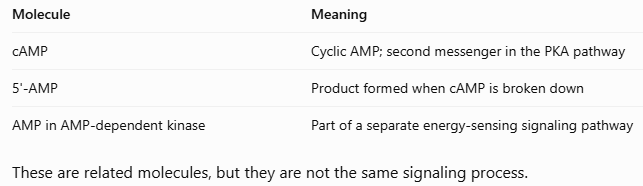
<p><span>The</span> <span>lecture</span> <span>briefly</span> <span>introduced</span> <span>another</span> <span>signaling</span> <span>process</span> <span>involving</span> <span>AMP-dependent</span> <span>protein</span> <span>kinase.</span></p><p class="x1ghz6dp x1717udv x1wty727 x1pd3egz x4gjf66 x1wwbdsi x86bw1a x1lziwak x15472z4 x1qlqyl8 x15bjb6t x1lliihq x14rh7hd"><span>Important</span> <span>points:</span></p><ul><li><p class="x1ghz6dp x1717udv x1wty727 x1pd3egz x4gjf66 x15eiy0f x1lziwak x15472z4 x1qlqyl8 x15bjb6t x1lliihq x14rh7hd xg07evd"><span>This</span> <span>pathway</span> <span>does</span> <span>not</span> <span>require</span> <span>a</span> <span>cell</span> <span>surface</span> <span>receptor.</span></p></li><li><p class="x1ghz6dp x1717udv x1wty727 x1pd3egz x4gjf66 x15eiy0f x1lziwak x15472z4 x1qlqyl8 x15bjb6t x1lliihq x14rh7hd xg07evd"><span>Other</span> <span>proteins</span> <span>inside</span> <span>the</span> <span>cell</span> <span>activate</span> <span>kinases.</span></p></li><li><p class="x1ghz6dp x1717udv x1wty727 x1pd3egz x4gjf66 x15eiy0f x1lziwak x15472z4 x1qlqyl8 x15bjb6t x1lliihq x14rh7hd xg07evd"><span>These</span> <span>kinases</span> <span>can</span> <span>activate</span> <span>AMP-dependent</span> <span>protein</span> <span>kinase.</span></p></li><li><p class="x1ghz6dp x1717udv x1wty727 x1pd3egz x4gjf66 x15eiy0f x1lziwak x15472z4 x1qlqyl8 x15bjb6t x1lliihq x14rh7hd xg07evd"><span>This</span> <span>produces</span> <span>different</span> <span>physiological</span> <span>responses</span> <span>inside</span> <span>the</span> <span>cell.</span></p></li></ul><p class="x1ghz6dp x1717udv x1wty727 x1pd3egz x4gjf66 x1wwbdsi x86bw1a x1lziwak x15472z4 x1qlqyl8 x15bjb6t x1lliihq x14rh7hd"><span style="line-height: inherit; font-size: inherit;"><strong>Do not confuse the different AMP molecules: (See image)</strong></span></p>
52
New cards

What Are Carbohydrates?

Carbohydrates are organic molecules made primarily of:

  • Carbon (C)

  • Hydrogen (H)

  • Oxygen (O)

The name carbohydrate comes from the idea that these molecules have an empirical formula resembling carbon plus water. A common general empirical formula is: (C2H2O)n. Glucose has the molecular formula: C6H1206. It’s empirical formula is: CH2O. Remember the difference:

  • Molecular formula = actual number of each atom in a molecule.

  • Empirical formula = simplest whole-number ratio of the atoms.


53
New cards

How Are Carbohydrates Classified?

There are several ways to classify carbohydrates.

A. Based on the number of sugar units:

  • Monosaccharide: 1 sugar unit

  • Disaccharide: 2 sugar units

  • Trisaccharide: 3 sugar units

  • Oligosaccharide: a small number of sugar units

  • Polysaccharide: Many sugar units.

A monosaccharide is a single sugar molecule. When two monosaccharides are covalently joined, they form a disaccharide.

B. Based on the number of carbon atoms

The suffix -ose is added to the carbon-number prefix.

  • 3 Carbons: Triose

  • 4 Carbons: Tetrose

  • 5 Carbons: Pentose

  • 6 Carbons: Hexose

Examples:

  • Glucose = hexose (6 carbons).

  • Ribose = pentose (5 carbons).

C. Based on the most oxidized carbon

Carbohydrates can also be classified according to whether they contain an aldehyde or ketone functional group (see image).

You can combine the two classification systems.

Examples:

  • Ketohexose = ketone + 6 carbons.

  • Aldopentose = aldehyde + 5 carbons.

  • Aldohexose = aldehyde + 6 carbons.

Example: Glucose is an aldohexose because it has an aldehyde group and six carbons. Fructose is a ketohexose because it has a ketone group and six carbons.

<p><span>There are several ways to classify carbohydrates.</span></p><p><span><strong>A. Based on the number of sugar units:</strong></span></p><ul><li><p>Monosaccharide: 1 sugar unit</p></li><li><p>Disaccharide: 2 sugar units</p></li><li><p>Trisaccharide: 3 sugar units</p></li><li><p>Oligosaccharide: a small number of sugar units</p></li><li><p>Polysaccharide: Many sugar units.</p></li></ul><p><span>A</span> <span>monosaccharide</span> <span>is</span> <span>a</span> <span>single</span> <span>sugar</span> <span>molecule. When</span> <span>two</span> <span>monosaccharides</span> <span>are</span> <span>covalently</span> <span>joined,</span> <span>they</span> <span>form</span> <span>a</span> <span>disaccharide.</span></p><p><span><strong>B.</strong></span><strong> </strong><span><strong>Based</strong></span><strong> </strong><span><strong>on</strong></span><strong> </strong><span><strong>the</strong></span><strong> </strong><span><strong>number</strong></span><strong> </strong><span><strong>of</strong></span><strong> </strong><span><strong>carbon</strong></span><strong> </strong><span><strong>atoms</strong></span></p><p class="x1ghz6dp x1717udv x1wty727 x1pd3egz x4gjf66 x1kw3rcc x104jwf5 x1lziwak x15472z4 x1qlqyl8 x15bjb6t x1lliihq x14rh7hd"><span>The</span> <span>suffix</span> <span>-ose</span> <span>is</span> <span>added</span> <span>to</span> <span>the</span> <span>carbon-number</span> <span>prefix.</span></p><ul><li><p class="x1ghz6dp x1717udv x1wty727 x1pd3egz x4gjf66 x1kw3rcc x104jwf5 x1lziwak x15472z4 x1qlqyl8 x15bjb6t x1lliihq x14rh7hd">3 Carbons: Triose</p></li><li><p class="x1ghz6dp x1717udv x1wty727 x1pd3egz x4gjf66 x1kw3rcc x104jwf5 x1lziwak x15472z4 x1qlqyl8 x15bjb6t x1lliihq x14rh7hd">4 Carbons: Tetrose</p></li><li><p class="x1ghz6dp x1717udv x1wty727 x1pd3egz x4gjf66 x1kw3rcc x104jwf5 x1lziwak x15472z4 x1qlqyl8 x15bjb6t x1lliihq x14rh7hd">5 Carbons: Pentose</p></li><li><p class="x1ghz6dp x1717udv x1wty727 x1pd3egz x4gjf66 x1kw3rcc x104jwf5 x1lziwak x15472z4 x1qlqyl8 x15bjb6t x1lliihq x14rh7hd">6 Carbons: Hexose</p></li></ul><p><span>Examples:</span></p><ul><li><p class="x1ghz6dp x1717udv x1wty727 x1pd3egz x4gjf66 x15eiy0f x1lziwak x15472z4 x1qlqyl8 x15bjb6t x1lliihq x14rh7hd xg07evd"><span>Glucose</span> <span>=</span> <span>hexose</span> <span>(6</span> <span>carbons).</span></p></li><li><p class="x1ghz6dp x1717udv x1wty727 x1pd3egz x4gjf66 x15eiy0f x1lziwak x15472z4 x1qlqyl8 x15bjb6t x1lliihq x14rh7hd xg07evd"><span>Ribose</span> <span>=</span> <span>pentose</span> <span>(5</span> <span>carbons).</span></p></li></ul><p><span><strong>C.</strong></span><strong> </strong><span><strong>Based</strong></span><strong> </strong><span><strong>on</strong></span><strong> </strong><span><strong>the</strong></span><strong> </strong><span><strong>most</strong></span><strong> </strong><span><strong>oxidized</strong></span><strong> </strong><span><strong>carbon</strong></span></p><p class="x1ghz6dp x1717udv x1wty727 x1pd3egz x4gjf66 x1kw3rcc x104jwf5 x1lziwak x15472z4 x1qlqyl8 x15bjb6t x1lliihq x14rh7hd"><span>Carbohydrates</span> <span>can</span> <span>also</span> <span>be</span> <span>classified</span> <span>according</span> <span>to</span> <span>whether</span> <span>they</span> <span>contain</span> <span>an</span> <span>aldehyde</span> <span>or</span> <span>ketone</span> <span>functional</span> <span>group (see image).</span></p><p class="x1ghz6dp x1717udv x1wty727 x1pd3egz x4gjf66 x1kw3rcc x104jwf5 x1lziwak x15472z4 x1qlqyl8 x15bjb6t x1lliihq x14rh7hd"><span><u>You</u></span><u> </u><span><u>can</u></span><u> </u><span><u>combine</u></span><u> </u><span><u>the</u></span><u> </u><span><u>two</u></span><u> </u><span><u>classification</u></span><u> </u><span><u>systems.</u></span></p><p class="x1ghz6dp x1717udv x1wty727 x1pd3egz x4gjf66 x1wwbdsi x86bw1a x1lziwak x15472z4 x1qlqyl8 x15bjb6t x1lliihq x14rh7hd"><span>Examples:</span></p><ul><li><p class="x1ghz6dp x1717udv x1wty727 x1pd3egz x4gjf66 x15eiy0f x1lziwak x15472z4 x1qlqyl8 x15bjb6t x1lliihq x14rh7hd xg07evd"><span>Ketohexose</span> <span>=</span> <span>ketone</span> <span>+</span> <span>6</span> <span>carbons.</span></p></li><li><p class="x1ghz6dp x1717udv x1wty727 x1pd3egz x4gjf66 x15eiy0f x1lziwak x15472z4 x1qlqyl8 x15bjb6t x1lliihq x14rh7hd xg07evd"><span>Aldopentose</span> <span>=</span> <span>aldehyde</span> <span>+</span> <span>5</span> <span>carbons.</span></p></li><li><p class="x1ghz6dp x1717udv x1wty727 x1pd3egz x4gjf66 x15eiy0f x1lziwak x15472z4 x1qlqyl8 x15bjb6t x1lliihq x14rh7hd xg07evd"><span>Aldohexose</span> <span>=</span> <span>aldehyde</span> <span>+</span> <span>6</span> <span>carbons.</span></p></li></ul><p class="x1ghz6dp x1717udv x1wty727 x1pd3egz x4gjf66 x1wwbdsi x86bw1a x1lziwak x15472z4 x1qlqyl8 x15bjb6t x1lliihq x14rh7hd"><span style="line-height: inherit; font-size: inherit;"><strong>Example:</strong></span> <span>Glucose</span> <span>is</span> <span>an</span> <span>aldohexose</span> <span>because</span> <span>it</span> <span>has</span> <span>an</span> <span>aldehyde</span> <span>group</span> <span>and</span> <span>six</span> <span>carbons. Fructose</span> <span>is</span> <span>a</span> <span>ketohexose</span> <span>because</span> <span>it</span> <span>has</span> <span>a</span> <span>ketone</span> <span>group</span> <span>and</span> <span>six</span> <span>carbons.</span></p>
54
New cards

D and L Sugar Configurations

Carbohydrates can also be classified according to their stereochemistry. Their D or L configuration is based on comparison with glyceraldehyde.

How to determine whether a sugar is D or L

Look at the chiral carbon farthest from the carbonyl group.

This is usually the second-to-last carbon in the Fischer projection.

  • OH on the right = D-sugar.

  • OH on the left = L-sugar.

Important: D and L do not tell you whether a molecule rotates light clockwise or counterclockwise. They describe its stereochemical configuration. You should know how to identify D and L sugars from their Fischer projections.

<p><span>Carbohydrates</span> <span>can</span> <span>also</span> <span>be</span> <span>classified</span> <span>according</span> <span>to</span> <span>their</span> <span>stereochemistry. Their</span> <span>D</span> <span>or</span> <span>L</span> <span>configuration</span> <span>is</span> <span>based</span> <span>on</span> <span>comparison</span> <span>with</span> <span>glyceraldehyde.</span></p><p><span><strong><u>How</u></strong></span><strong><u> </u></strong><span><strong><u>to</u></strong></span><strong><u> </u></strong><span><strong><u>determine</u></strong></span><strong><u> </u></strong><span><strong><u>whether</u></strong></span><strong><u> </u></strong><span><strong><u>a</u></strong></span><strong><u> </u></strong><span><strong><u>sugar</u></strong></span><strong><u> </u></strong><span><strong><u>is</u></strong></span><strong><u> </u></strong><span><strong><u>D</u></strong></span><strong><u> </u></strong><span><strong><u>or</u></strong></span><strong><u> </u></strong><span><strong><u>L</u></strong></span></p><p class="x1ghz6dp x1717udv x1wty727 x1pd3egz x4gjf66 x1kw3rcc x104jwf5 x1lziwak x15472z4 x1qlqyl8 x15bjb6t x1lliihq x14rh7hd"><span>Look</span> <span>at</span> <span>the</span> <span>chiral</span> <span>carbon</span> <span>farthest</span> <span>from</span> <span>the</span> <span>carbonyl</span> <span>group.</span></p><p class="x1ghz6dp x1717udv x1wty727 x1pd3egz x4gjf66 x1wwbdsi x86bw1a x1lziwak x15472z4 x1qlqyl8 x15bjb6t x1lliihq x14rh7hd"><span><strong>This</strong></span><strong> </strong><span><strong>is</strong></span><strong> </strong><span><strong>usually</strong></span><strong> </strong><span><strong>the</strong></span><strong> </strong><span><strong>second-to-last</strong></span><strong> </strong><span><strong>carbon</strong></span><strong> </strong><span><strong>in</strong></span><strong> </strong><span><strong>the</strong></span><strong> </strong><span><strong>Fischer</strong></span><strong> </strong><span><strong>projection.</strong></span></p><ul><li><p class="x1ghz6dp x1717udv x1wty727 x1pd3egz x4gjf66 x15eiy0f x1lziwak x15472z4 x1qlqyl8 x15bjb6t x1lliihq x14rh7hd xg07evd"><span>OH</span> <span>on</span> <span>the</span> <span>right</span> <span>=</span> <span>D-sugar.</span></p></li><li><p class="x1ghz6dp x1717udv x1wty727 x1pd3egz x4gjf66 x15eiy0f x1lziwak x15472z4 x1qlqyl8 x15bjb6t x1lliihq x14rh7hd xg07evd"><span>OH</span> <span>on</span> <span>the</span> <span>left</span> <span>=</span> <span>L-sugar.</span></p></li></ul><p class="x1ghz6dp x1717udv x1wty727 x1pd3egz x4gjf66 x1wwbdsi x86bw1a x1lziwak x15472z4 x1qlqyl8 x15bjb6t x1lliihq x14rh7hd"><span style="line-height: inherit; font-size: inherit;"><strong>Important:</strong></span> <span>D</span> <span>and</span> <span>L</span> <span>do</span> <span>not</span> <span>tell</span> <span>you</span> <span>whether</span> <span>a</span> <span>molecule</span> <span>rotates</span> <span>light</span> <span>clockwise</span> <span>or</span> <span>counterclockwise. They</span> <span>describe</span> <span>its</span> <span>stereochemical</span> <span>configuration. You should know how to identify D and L sugars from their Fischer projections.</span></p>
55
New cards

How Do Sugars Form Ring Structures?

In solution, many monosaccharides exist mainly in ring forms rather than straight-chain forms. This happens through an intramolecular reaction. Intramolecular means a reaction occurs within the same molecule.

A. Hemiacetal formation

An aldehyde reacts with an alcohol to form a hemiacetal. In glucose:

  1. Glucose begins in its straight-chain form.

  2. The hydroxyl group on carbon 5 attacks the aldehyde carbon on carbon 1

  3. A ring forms.

  4. The carbonyl oxygen becomes a hydroxyl group after protonation.

The result is a cyclic hemiacetal.

B. Hemiketal formation

A ketone reacts with an alcohol to form a hemiketal.

For example, fructose can form a ring through an intramolecular reaction between a hydroxyl group and its ketone.

Remember:

Aldehyde + alcohol = Hemiacetal

Ketone + alcohol = Hemiketal

<p><span>In</span> <span>solution,</span> <span>many</span> <span>monosaccharides</span> <span>exist</span> <span>mainly</span> <span>in</span> <span>ring</span> <span>forms</span> <span>rather</span> <span>than</span> <span>straight-chain</span> <span>forms. This</span> <span>happens</span> <span>through</span> <span>an</span> <span>intramolecular</span> <span>reaction. </span><span style="line-height: inherit; font-size: inherit;"><strong>Intramolecular means a reaction occurs within the same molecule.</strong></span></p><p><span><strong>A. Hemiacetal formation</strong></span></p><p class="x1ghz6dp x1717udv x1wty727 x1pd3egz x4gjf66 x1kw3rcc x104jwf5 x1lziwak x15472z4 x1qlqyl8 x15bjb6t x1lliihq x14rh7hd"><span>An</span> <span>aldehyde</span> <span>reacts</span> <span>with</span> <span>an</span> <span>alcohol</span> <span>to</span> <span>form</span> <span>a</span> <span>hemiacetal. In</span> <span>glucose:</span></p><ol><li><p class="x1ghz6dp x1717udv x1wty727 x1pd3egz x4gjf66 x15eiy0f x1lziwak x15472z4 x1qlqyl8 x15bjb6t x1lliihq x14rh7hd xg07evd"><span>Glucose</span> <span>begins</span> <span>in</span> <span>its</span> <span>straight-chain</span> <span>form.</span></p></li><li><p class="x1ghz6dp x1717udv x1wty727 x1pd3egz x4gjf66 x15eiy0f x1lziwak x15472z4 x1qlqyl8 x15bjb6t x1lliihq x14rh7hd xg07evd"><span>The</span> <span>hydroxyl</span> <span>group</span> <span>on</span> <span>carbon</span> <span>5</span> <span>attacks</span> <span>the</span> <span>aldehyde</span> <span>carbon</span> <span>on</span> <span>carbon</span> <span>1</span></p></li><li><p class="x1ghz6dp x1717udv x1wty727 x1pd3egz x4gjf66 x15eiy0f x1lziwak x15472z4 x1qlqyl8 x15bjb6t x1lliihq x14rh7hd xg07evd"><span>A</span> <span>ring</span> <span>forms.</span></p></li><li><p class="x1ghz6dp x1717udv x1wty727 x1pd3egz x4gjf66 x15eiy0f x1lziwak x15472z4 x1qlqyl8 x15bjb6t x1lliihq x14rh7hd xg07evd"><span>The</span> <span>carbonyl</span> <span>oxygen</span> <span>becomes</span> <span>a</span> <span>hydroxyl</span> <span>group</span> <span>after</span> <span>protonation.</span></p></li></ol><p class="x1ghz6dp x1717udv x1wty727 x1pd3egz x4gjf66 x1wwbdsi x86bw1a x1lziwak x15472z4 x1qlqyl8 x15bjb6t x1lliihq x14rh7hd"><span>The</span> <span>result</span> <span>is</span> <span>a</span> <span>cyclic</span> <span>hemiacetal.</span></p><p class="x1ghz6dp x1717udv x1wty727 x1pd3egz x4gjf66 x1wwbdsi x86bw1a x1lziwak x15472z4 x1qlqyl8 x15bjb6t x1lliihq x14rh7hd"><span><strong>B.</strong></span><strong> </strong><span><strong>Hemiketal</strong></span><strong> </strong><span><strong>formation</strong></span></p><p class="x1ghz6dp x1717udv x1wty727 x1pd3egz x4gjf66 x1kw3rcc x104jwf5 x1lziwak x15472z4 x1qlqyl8 x15bjb6t x1lliihq x14rh7hd"><span>A</span> <span>ketone</span> <span>reacts</span> <span>with</span> <span>an</span> <span>alcohol</span> <span>to</span> <span>form</span> <span>a</span> <span>hemiketal.</span></p><p class="x1ghz6dp x1717udv x1wty727 x1pd3egz x4gjf66 x1wwbdsi x86bw1a x1lziwak x15472z4 x1qlqyl8 x15bjb6t x1lliihq x14rh7hd"><span>For</span> <span>example,</span> <span>fructose</span> <span>can</span> <span>form</span> <span>a</span> <span>ring</span> <span>through</span> <span>an</span> <span>intramolecular</span> <span>reaction</span> <span>between</span> <span>a</span> <span>hydroxyl</span> <span>group</span> <span>and</span> <span>its</span> <span>ketone.</span></p><p class="x1ghz6dp x1717udv x1wty727 x1pd3egz x4gjf66 x1wwbdsi x86bw1a x1lziwak x15472z4 x1qlqyl8 x15bjb6t x1lliihq x14rh7hd"><span>Remember:</span></p><p class="x1ghz6dp x1717udv x1wty727 x1pd3egz x4gjf66 x1wwbdsi x86bw1a x1lziwak x15472z4 x1qlqyl8 x15bjb6t x1lliihq x14rh7hd"><span>Aldehyde + alcohol = Hemiacetal</span></p><p class="x1ghz6dp x1717udv x1wty727 x1pd3egz x4gjf66 x1wwbdsi x86bw1a x1lziwak x15472z4 x1qlqyl8 x15bjb6t x1lliihq x14rh7hd"><span>Ketone + alcohol = Hemiketal</span></p>
56
New cards

Alpha and Beta Sugar Configurations

When a sugar forms a ring, the carbonyl carbon becomes a new chiral center. This carbon is called the anomeric carbon. For glucose, the anomeric carbon is carbon 1.

How do you identify alpha and beta?

Look at the OH group attached to the anomeric carbon. For the D-sugars discussed in the lecture:

  • Alpha (α): OH points down.

  • Beta (β): OH points up.

The two forms are called anomers because they differ only in configuration at the anomeric carbon.

What is the anomeric carbon?

It is the carbon that was originally the carbonyl carbon in the straight-chain sugar. In the ring structure, it is bonded to:

  • The oxygen in the ring.

  • An OH group (or another substituent if it has formed a glycosidic bond).

Exam tip: To find the anomeric carbon, look for the carbon bonded to both the ring oxygen and an oxygen-containing substituent.

<p><span>When</span> <span>a</span> <span>sugar</span> <span>forms</span> <span>a</span> <span>ring,</span> <span>the</span> <span>carbonyl</span> <span>carbon</span> <span>becomes</span> <span>a</span> <span>new</span> <span>chiral</span> <span>center. This</span> <span>carbon</span> <span>is</span> <span>called</span> <span>the</span> <span style="line-height: inherit; font-size: inherit;"><strong>anomeric carbon</strong></span><span>. For</span> <span>glucose,</span> <span>the</span> <span>anomeric</span> <span>carbon</span> <span>is</span> <span>carbon</span> <span>1.</span></p><p><span><strong>How</strong></span><strong> </strong><span><strong>do</strong></span><strong> </strong><span><strong>you</strong></span><strong> </strong><span><strong>identify</strong></span><strong> </strong><span><strong>alpha</strong></span><strong> </strong><span><strong>and</strong></span><strong> </strong><span><strong>beta?</strong></span></p><p class="x1ghz6dp x1717udv x1wty727 x1pd3egz x4gjf66 x1kw3rcc x104jwf5 x1lziwak x15472z4 x1qlqyl8 x15bjb6t x1lliihq x14rh7hd"><span>Look</span> <span>at</span> <span>the</span> <span>OH</span> <span>group</span> <span>attached</span> <span>to</span> <span>the</span> <span>anomeric</span> <span>carbon. <strong>For</strong></span><strong> </strong><span><strong>the</strong></span><strong> </strong><span><strong>D-sugars</strong></span><strong> </strong><span>discussed</span> <span>in</span> <span>the</span> <span>lecture:</span></p><ul><li><p class="x1ghz6dp x1717udv x1wty727 x1pd3egz x4gjf66 x15eiy0f x1lziwak x15472z4 x1qlqyl8 x15bjb6t x1lliihq x14rh7hd xg07evd"><span><strong>Alpha</strong></span><strong> </strong><span><strong>(α):</strong></span><strong> </strong><span><strong>OH</strong></span><strong> </strong><span><strong>points</strong></span><strong> </strong><span><strong>down.</strong></span></p></li><li><p class="x1ghz6dp x1717udv x1wty727 x1pd3egz x4gjf66 x15eiy0f x1lziwak x15472z4 x1qlqyl8 x15bjb6t x1lliihq x14rh7hd xg07evd"><span><strong>Beta</strong></span><strong> </strong><span><strong>(β):</strong></span><strong> </strong><span><strong>OH</strong></span><strong> </strong><span><strong>points</strong></span><strong> </strong><span><strong>up.</strong></span></p></li></ul><p class="x1ghz6dp x1717udv x1wty727 x1pd3egz x4gjf66 x1wwbdsi x86bw1a x1lziwak x15472z4 x1qlqyl8 x15bjb6t x1lliihq x14rh7hd"><span>The</span> <span>two</span> <span>forms</span> <span>are</span> <span>called</span> <span style="line-height: inherit; font-size: inherit;"><strong>anomers</strong></span> <span>because</span> <span>they</span> <span>differ</span> <span>only</span> <span>in</span> <span>configuration</span> <span>at</span> <span>the</span> <span>anomeric</span> <span>carbon.</span></p><p class="x1ghz6dp x1717udv x1wty727 x1pd3egz x4gjf66 x1wwbdsi x86bw1a x1lziwak x15472z4 x1qlqyl8 x15bjb6t x1lliihq x14rh7hd"><span><strong>What</strong></span><strong> </strong><span><strong>is</strong></span><strong> </strong><span><strong>the</strong></span><strong> </strong><span><strong>anomeric</strong></span><strong> </strong><span><strong>carbon?</strong></span></p><p class="x1ghz6dp x1717udv x1wty727 x1pd3egz x4gjf66 x1kw3rcc x104jwf5 x1lziwak x15472z4 x1qlqyl8 x15bjb6t x1lliihq x14rh7hd"><span>It</span> <span>is</span> <span>the</span> <span>carbon</span> <span>that</span> <span>was</span> <span>originally</span> <span>the</span> <span>carbonyl</span> <span>carbon</span> <span>in</span> <span>the</span> <span>straight-chain</span> <span>sugar. In</span> <span>the</span> <span>ring</span> <span>structure,</span> <span>it</span> <span>is</span> <span>bonded</span> <span>to:</span></p><ul><li><p class="x1ghz6dp x1717udv x1wty727 x1pd3egz x4gjf66 x15eiy0f x1lziwak x15472z4 x1qlqyl8 x15bjb6t x1lliihq x14rh7hd xg07evd"><span>The</span> <span>oxygen</span> <span>in</span> <span>the</span> <span>ring.</span></p></li><li><p class="x1ghz6dp x1717udv x1wty727 x1pd3egz x4gjf66 x15eiy0f x1lziwak x15472z4 x1qlqyl8 x15bjb6t x1lliihq x14rh7hd xg07evd"><span>An</span> <span>OH</span> <span>group</span> <span>(or</span> <span>another</span> <span>substituent</span> <span>if</span> <span>it</span> <span>has</span> <span>formed</span> <span>a</span> <span>glycosidic</span> <span>bond).</span></p></li></ul><p class="x1ghz6dp x1717udv x1wty727 x1pd3egz x4gjf66 x1wwbdsi x86bw1a x1lziwak x15472z4 x1qlqyl8 x15bjb6t x1lliihq x14rh7hd"><span style="line-height: inherit; font-size: inherit;"><strong>Exam tip:</strong></span> <span>To</span> <span>find</span> <span>the</span> <span>anomeric</span> <span>carbon,</span> <span>look</span> <span>for</span> <span>the</span> <span>carbon</span> <span>bonded</span> <span>to</span> <span>both</span> <span>the</span> <span>ring</span> <span>oxygen</span> <span>and</span> <span>an</span> <span>oxygen-containing</span> <span>substituent.</span></p>
57
New cards

Fischer Projections vs. Haworth Projections

These are two different ways to draw carbohydrates.

Converting Fischer to Haworth projections

For the D-sugar examples discussed:

  • Groups on the right in Fischer projections point down in Haworth projections.

  • Groups on the left in Fischer projections point up in Haworth projections.

For glucose, the professor gave an easy way to remember the OH pattern:

Glucose = Down, Up, Down (D-U-D). When drawing the Haworth projection of glucose, remember the OH pattern on carbons 2, 3, and 4:

  • Carbon 2: Down

  • Carbon 3: Up

  • Carbon 4: Down

Pyranose and furanose

Sugars can also be classified according to ring size:

  • Pyranose = six-membered ring.

  • Furanose = five-membered ring.

(this specific classification will not be tested).

<p><span>These are two different ways to draw carbohydrates. </span></p><p><span><strong>Converting</strong></span><strong> </strong><span><strong>Fischer</strong></span><strong> </strong><span><strong>to</strong></span><strong> </strong><span><strong>Haworth</strong></span><strong> </strong><span><strong>projections</strong></span></p><p class="x1ghz6dp x1717udv x1wty727 x1pd3egz x4gjf66 x1kw3rcc x104jwf5 x1lziwak x15472z4 x1qlqyl8 x15bjb6t x1lliihq x14rh7hd"><span>For</span> <span>the</span> <span>D-sugar</span> <span>examples</span> <span>discussed:</span></p><ul><li><p class="x1ghz6dp x1717udv x1wty727 x1pd3egz x4gjf66 x15eiy0f x1lziwak x15472z4 x1qlqyl8 x15bjb6t x1lliihq x14rh7hd xg07evd"><span>Groups</span> <span>on</span> <span>the</span> <span>right</span> <span>in</span> <span>Fischer</span> <span>projections</span> <span>point</span> <span>down</span> <span>in</span> <span>Haworth</span> <span>projections.</span></p></li><li><p class="x1ghz6dp x1717udv x1wty727 x1pd3egz x4gjf66 x15eiy0f x1lziwak x15472z4 x1qlqyl8 x15bjb6t x1lliihq x14rh7hd xg07evd"><span>Groups</span> <span>on</span> <span>the</span> <span>left</span> <span>in</span> <span>Fischer</span> <span>projections</span> <span>point</span> <span>up</span> <span>in</span> <span>Haworth</span> <span>projections.</span></p></li></ul><p class="x1ghz6dp x1717udv x1wty727 x1pd3egz x4gjf66 x1wwbdsi x86bw1a x1lziwak x15472z4 x1qlqyl8 x15bjb6t x1lliihq x14rh7hd"><span>For</span> <span>glucose,</span> <span>the</span> <span>professor</span> <span>gave</span> <span>an</span> <span>easy</span> <span>way</span> <span>to</span> <span>remember</span> <span>the</span> <span>OH</span> <span>pattern:</span></p><p class="x1ghz6dp x1717udv x1wty727 x1pd3egz x4gjf66 x1wwbdsi x86bw1a x1lziwak x15472z4 x1qlqyl8 x15bjb6t x1lliihq x14rh7hd"><span style="line-height: inherit; font-size: inherit;"><strong>Glucose = Down, Up, Down (D-U-D). </strong></span><span>When</span> <span>drawing</span> <span>the</span> <span>Haworth</span> <span>projection</span> <span>of</span> <span>glucose,</span> <span>remember</span> <span>the</span> <span>OH</span> <span>pattern</span> <span>on</span> <span>carbons</span> <span>2,</span> <span>3,</span> <span>and</span> <span>4:</span></p><ul><li><p class="x1ghz6dp x1717udv x1wty727 x1pd3egz x4gjf66 x15eiy0f x1lziwak x15472z4 x1qlqyl8 x15bjb6t x1lliihq x14rh7hd xg07evd"><span>Carbon</span> <span>2:</span> <span>Down</span></p></li><li><p class="x1ghz6dp x1717udv x1wty727 x1pd3egz x4gjf66 x15eiy0f x1lziwak x15472z4 x1qlqyl8 x15bjb6t x1lliihq x14rh7hd xg07evd"><span>Carbon</span> <span>3:</span> <span>Up</span></p></li><li><p class="x1ghz6dp x1717udv x1wty727 x1pd3egz x4gjf66 x15eiy0f x1lziwak x15472z4 x1qlqyl8 x15bjb6t x1lliihq x14rh7hd xg07evd"><span>Carbon</span> <span>4:</span> <span>Down</span></p></li></ul><p><span><strong>Pyranose</strong></span><strong> </strong><span><strong>and</strong></span><strong> </strong><span><strong>furanose</strong></span></p><p><span>Sugars</span> <span>can</span> <span>also</span> <span>be</span> <span>classified</span> <span>according</span> <span>to</span> <span>ring</span> <span>size:</span></p><ul><li><p class="x1ghz6dp x1717udv x1wty727 x1pd3egz x4gjf66 x15eiy0f x1lziwak x15472z4 x1qlqyl8 x15bjb6t x1lliihq x14rh7hd xg07evd"><span>Pyranose</span> <span>=</span> <span>six-membered</span> <span>ring.</span></p></li><li><p class="x1ghz6dp x1717udv x1wty727 x1pd3egz x4gjf66 x15eiy0f x1lziwak x15472z4 x1qlqyl8 x15bjb6t x1lliihq x14rh7hd xg07evd"><span>Furanose</span> <span>=</span> <span>five-membered</span> <span>ring.</span></p></li></ul><p>(this specific classification will not be tested).</p>
58
New cards

Modified Sugars

Sugars can undergo chemical modifications, just like amino acids and lipids. Several important examples were discussed:

1. Glucosamine: A hydroxyl group is replaced with an amino group. This produces an amino sugar.

2. N-acetylglucosamine: An acetyl group is attached to the nitrogen of glucosamine. The N indicates that the acetyl group is attached to nitrogen.

3. Xylitol: A sugar alcohol formed by reducing an aldehyde group to an alcohol. Commonly used as an artificial sweetener in gum.

4. Sugar acids: An alcohol group can be oxidized to a carboxylic acid group.

5. Deoxyribose: A hydroxyl group is replaced by hydrogen. Found in DNA.

6. Methyl glucose: A methyl group is attached through a glycosidic bond.

A glycosidic bond is a covalent bond formed through the anomeric carbon of a sugar and another group. It can connect:

  • A sugar to another sugar.

  • A sugar to a non-sugar molecule.

When two sugars are joined, a glycosidic linkage forms between them.

<p><span>Sugars</span> <span>can</span> <span>undergo</span> <span>chemical</span> <span>modifications,</span> <span>just</span> <span>like</span> <span>amino</span> <span>acids</span> <span>and</span> <span>lipids. Several</span> <span>important</span> <span>examples</span> <span>were</span> <span>discussed: </span></p><p><strong>1. Glucosamine:</strong> A hydroxyl group is replaced with an amino group. This produces an amino sugar.</p><p><strong>2. N-acetylglucosamine: </strong>An acetyl group is attached to the nitrogen of glucosamine. The N indicates that the acetyl group is attached to nitrogen.</p><p><strong>3. Xylitol: </strong>A sugar alcohol formed by reducing an aldehyde group to an alcohol. Commonly used as an artificial sweetener in gum.</p><p><strong>4. Sugar acids: </strong>An alcohol group can be oxidized to a carboxylic acid group.</p><p><strong>5. Deoxyribose: </strong>A hydroxyl group is replaced by hydrogen. Found in DNA.</p><p><strong>6. Methyl glucose: </strong>A methyl group is attached through a glycosidic bond.</p><p><span>A</span> <span style="line-height: inherit; font-size: inherit;"><strong>glycosidic bond</strong></span> <span>is</span> <span>a</span> <span>covalent</span> <span>bond</span> <span>formed</span> <span>through</span> <span>the</span> <span>anomeric</span> <span>carbon</span> <span>of</span> <span>a</span> <span>sugar</span> <span>and</span> <span>another</span> <span>group. It</span> <span>can</span> <span>connect:</span></p><ul><li><p class="x1ghz6dp x1717udv x1wty727 x1pd3egz x4gjf66 x15eiy0f x1lziwak x15472z4 x1qlqyl8 x15bjb6t x1lliihq x14rh7hd xg07evd"><span>A</span> <span>sugar</span> <span>to</span> <span>another</span> <span>sugar.</span></p></li><li><p class="x1ghz6dp x1717udv x1wty727 x1pd3egz x4gjf66 x15eiy0f x1lziwak x15472z4 x1qlqyl8 x15bjb6t x1lliihq x14rh7hd xg07evd"><span>A</span> <span>sugar</span> <span>to</span> <span>a</span> <span>non-sugar</span> <span>molecule.</span></p></li></ul><p class="x1ghz6dp x1717udv x1wty727 x1pd3egz x4gjf66 x1wwbdsi x86bw1a x1lziwak x15472z4 x1qlqyl8 x15bjb6t x1lliihq x14rh7hd"><span>When</span> <span>two</span> <span>sugars</span> <span>are</span> <span>joined,</span> <span>a</span> <span>glycosidic</span> <span>linkage</span> <span>forms</span> <span>between</span> <span>them.</span></p>
59
New cards

Reducing vs. Non-Reducing Sugars

A reducing sugar is a sugar that can undergo oxidation and reduce another substance.

How does this work?

Glucose contains an aldehyde group in its open-chain form. The aldehyde can be oxidized into a carboxylic acid. Aldehyde → Carboxylic acid. This is an oxidation reaction. When glucose is oxidized, another substance must be reduced.

Copper test

The lecture discussed a laboratory test that uses copper ions to detect reducing sugars. During the reaction:

  • Glucose's aldehyde group is oxidized into a carboxylic acid.

  • Cu²⁺ is reduced to Cu⁺.

  • Cu₂O (copper(I) oxide) is produced.

Remember:

  • Oxidation = loss of electrons.

  • Reduction = gain of electrons.

The glucose is oxidized, while the copper is reduced.

Reducing end vs. non-reducing end

  • Reducing end: The end with a free anomeric carbon that can open into the straight-chain form.

  • Non-reducing end: The end where the anomeric carbon is already involved in a glycosidic bond and cannot open.

Important rule: If a sugar has a free anomeric carbon, it can open into its straight-chain form and is capable of acting as a reducing sugar.

<p><span>A</span> <span>reducing</span> <span>sugar</span> <span>is</span> <span>a</span> <span>sugar</span> <span>that</span> <span>can</span> <span>undergo</span> <span>oxidation</span> <span>and</span> <span>reduce</span> <span>another</span> <span>substance.</span></p><p><span><strong>How does this work?</strong></span></p><p class="x1ghz6dp x1717udv x1wty727 x1pd3egz x4gjf66 x1kw3rcc x104jwf5 x1lziwak x15472z4 x1qlqyl8 x15bjb6t x1lliihq x14rh7hd"><span>Glucose</span> <span>contains</span> <span>an</span> <span>aldehyde</span> <span>group</span> <span>in</span> <span>its</span> <span>open-chain</span> <span>form. The</span> <span>aldehyde</span> <span>can</span> <span>be</span> <span>oxidized</span> <span>into</span> <span>a</span> <span>carboxylic</span> <span>acid. Aldehyde</span> <span>→</span> <span>Carboxylic</span> <span>acid. This</span> <span>is</span> <span>an</span> <span>oxidation</span> <span>reaction. When</span> <span>glucose</span> <span>is</span> <span>oxidized,</span> <span>another</span> <span>substance</span> <span>must</span> <span>be</span> <span>reduced.</span></p><p class="x1ghz6dp x1717udv x1wty727 x1pd3egz x4gjf66 x1kw3rcc x104jwf5 x1lziwak x15472z4 x1qlqyl8 x15bjb6t x1lliihq x14rh7hd"><span><strong>Copper</strong></span><strong> </strong><span><strong>test</strong></span></p><p class="x1ghz6dp x1717udv x1wty727 x1pd3egz x4gjf66 x1kw3rcc x104jwf5 x1lziwak x15472z4 x1qlqyl8 x15bjb6t x1lliihq x14rh7hd"><span>The</span> <span>lecture</span> <span>discussed</span> <span>a</span> <span>laboratory</span> <span>test</span> <span>that</span> <span>uses</span> <span>copper</span> <span>ions</span> <span>to</span> <span>detect</span> <span>reducing</span> <span>sugars. During</span> <span>the</span> <span>reaction:</span></p><ul><li><p class="x1ghz6dp x1717udv x1wty727 x1pd3egz x4gjf66 x15eiy0f x1lziwak x15472z4 x1qlqyl8 x15bjb6t x1lliihq x14rh7hd xg07evd"><span>Glucose's</span> <span>aldehyde</span> <span>group</span> <span>is</span> <span>oxidized</span> <span>into</span> <span>a</span> <span>carboxylic</span> <span>acid.</span></p></li><li><p class="x1ghz6dp x1717udv x1wty727 x1pd3egz x4gjf66 x15eiy0f x1lziwak x15472z4 x1qlqyl8 x15bjb6t x1lliihq x14rh7hd xg07evd"><span>Cu²⁺</span> <span>is</span> <span>reduced</span> <span>to</span> <span>Cu⁺.</span></p></li><li><p class="x1ghz6dp x1717udv x1wty727 x1pd3egz x4gjf66 x15eiy0f x1lziwak x15472z4 x1qlqyl8 x15bjb6t x1lliihq x14rh7hd xg07evd"><span>Cu₂O</span> <span>(copper(I)</span> <span>oxide)</span> <span>is</span> <span>produced.</span></p></li></ul><p><span style="line-height: inherit; font-size: inherit;"><strong>Remember:</strong></span></p><ul><li><p class="x1ghz6dp x1717udv x1wty727 x1pd3egz x4gjf66 x15eiy0f x1lziwak x15472z4 x1qlqyl8 x15bjb6t x1lliihq x14rh7hd xg07evd"><span>Oxidation</span> <span>=</span> <span>loss</span> <span>of</span> <span>electrons.</span></p></li><li><p class="x1ghz6dp x1717udv x1wty727 x1pd3egz x4gjf66 x15eiy0f x1lziwak x15472z4 x1qlqyl8 x15bjb6t x1lliihq x14rh7hd xg07evd"><span>Reduction</span> <span>=</span> <span>gain</span> <span>of</span> <span>electrons.</span></p></li></ul><p class="x1ghz6dp x1717udv x1wty727 x1pd3egz x4gjf66 x1wwbdsi x86bw1a x1lziwak x15472z4 x1qlqyl8 x15bjb6t x1lliihq x14rh7hd"><span>The</span> <span>glucose</span> <span>is</span> <span>oxidized,</span> <span>while</span> <span>the</span> <span>copper</span> <span>is</span> <span>reduced.</span></p><p><span><strong>Reducing end vs. non-reducing end</strong></span></p><ul><li><p class="x1ghz6dp x1717udv x1wty727 x1pd3egz x4gjf66 x15eiy0f x1lziwak x15472z4 x1qlqyl8 x15bjb6t x1lliihq x14rh7hd xg07evd"><span>Reducing</span> <span>end:</span> <span>The</span> <span>end</span> <span>with</span> <span>a</span> <span>free</span> <span>anomeric</span> <span>carbon</span> <span>that</span> <span>can</span> <span>open</span> <span>into</span> <span>the</span> <span>straight-chain</span> <span>form.</span></p></li><li><p class="x1ghz6dp x1717udv x1wty727 x1pd3egz x4gjf66 x15eiy0f x1lziwak x15472z4 x1qlqyl8 x15bjb6t x1lliihq x14rh7hd xg07evd"><span>Non-reducing</span> <span>end:</span> <span>The</span> <span>end</span> <span>where</span> <span>the</span> <span>anomeric</span> <span>carbon</span> <span>is</span> <span>already</span> <span>involved</span> <span>in</span> <span>a</span> <span>glycosidic</span> <span>bond</span> <span>and</span> <span>cannot</span> <span>open.</span></p></li></ul><p><span style="line-height: inherit; font-size: inherit;"><strong>Important rule: </strong></span><span>If</span> <span>a</span> <span>sugar</span> <span>has</span> <span>a</span> <span>free</span> <span>anomeric</span> <span>carbon,</span> <span>it</span> <span>can</span> <span>open</span> <span>into</span> <span>its</span> <span>straight-chain</span> <span>form</span> <span>and</span> <span>is</span> <span>capable</span> <span>of</span> <span>acting</span> <span>as</span> <span>a</span> <span>reducing</span> <span>sugar.</span></p>
60
New cards

Important Disaccharides

A disaccharide consists of two monosaccharides joined by a glycosidic bond. The professor emphasized three important disaccharides:

A. Maltose

Maltose consists of:

  • Glucose + Glucose

  • Alpha-1,4 glycosidic linkage.

The first glucose uses its anomeric carbon to form the bond. The second glucose still has a free anomeric carbon. Therefore: Reducing sugar. Maltose is a reducing sugar because one of its glucose units can still open into its straight-chain form.

B. Lactose

Lactose consists of:

  • Galactose + Glucose

  • Beta-1,4 glycosidic linkage.

The glucose unit has a free anomeric carbon. Therefore: Reducing sugar Lactose is a reducing sugar. Lactose is the sugar commonly found in milk.

C. Sucrose

Sucrose consists of:

  • Glucose + Fructose

  • An alpha-1,2-beta glycosidic linkage.

Both anomeric carbons are involved in the glycosidic bond. Therefore, neither sugar unit can open into its normal straight-chain form while part of sucrose. Non-reducing sugar. Sucrose is NOT a reducing sugar. Sucrose is ordinary table sugar.
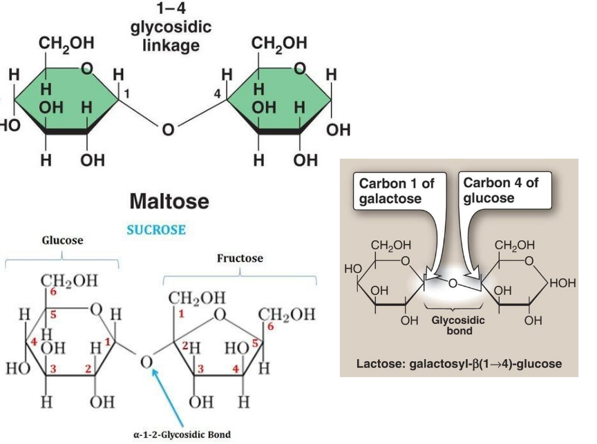
<p><span>A</span> <span>disaccharide</span> <span>consists</span> <span>of</span> <span>two</span> <span>monosaccharides</span> <span>joined</span> <span>by</span> <span>a</span> <span>glycosidic</span> <span>bond. The</span> <span>professor</span> <span>emphasized</span> <span>three</span> <span>important</span> <span>disaccharides:</span></p><p><span><strong>A.</strong></span><strong> </strong><span><strong>Maltose</strong></span></p><p><span>Maltose</span> <span>consists</span> <span>of:</span></p><ul><li><p class="x1ghz6dp x1717udv x1wty727 x1pd3egz x4gjf66 x15eiy0f x1lziwak x15472z4 x1qlqyl8 x15bjb6t x1lliihq x14rh7hd xg07evd"><span>Glucose</span> <span>+</span> <span>Glucose</span></p></li><li><p class="x1ghz6dp x1717udv x1wty727 x1pd3egz x4gjf66 x15eiy0f x1lziwak x15472z4 x1qlqyl8 x15bjb6t x1lliihq x14rh7hd xg07evd"><span>Alpha-1,4</span> <span>glycosidic</span> <span>linkage.</span></p></li></ul><p class="x1ghz6dp x1717udv x1wty727 x1pd3egz x4gjf66 x1wwbdsi x86bw1a x1lziwak x15472z4 x1qlqyl8 x15bjb6t x1lliihq x14rh7hd"><span>The</span> <span>first</span> <span>glucose</span> <span>uses</span> <span>its</span> <span>anomeric</span> <span>carbon</span> <span>to</span> <span>form</span> <span>the</span> <span>bond. The</span> <span>second</span> <span>glucose</span> <span>still</span> <span>has</span> <span>a</span> <span>free</span> <span>anomeric</span> <span>carbon. Therefore: <strong>Reducing sugar. </strong>Maltose</span> <span>is</span> <span>a</span> <span>reducing</span> <span>sugar</span> <span>because</span> <span>one</span> <span>of</span> <span>its</span> <span>glucose</span> <span>units</span> <span>can</span> <span>still</span> <span>open</span> <span>into</span> <span>its</span> <span>straight-chain</span> <span>form.</span></p><p class="x1ghz6dp x1717udv x1wty727 x1pd3egz x4gjf66 x1wwbdsi x86bw1a x1lziwak x15472z4 x1qlqyl8 x15bjb6t x1lliihq x14rh7hd"><span><strong>B.</strong></span><strong> </strong><span><strong>Lactose</strong></span></p><p class="x1ghz6dp x1717udv x1wty727 x1pd3egz x4gjf66 x1wwbdsi x86bw1a x1lziwak x15472z4 x1qlqyl8 x15bjb6t x1lliihq x14rh7hd"><span>Lactose</span> <span>consists</span> <span>of:</span></p><ul><li><p class="x1ghz6dp x1717udv x1wty727 x1pd3egz x4gjf66 x15eiy0f x1lziwak x15472z4 x1qlqyl8 x15bjb6t x1lliihq x14rh7hd xg07evd"><span>Galactose</span> <span>+</span> <span>Glucose</span></p></li><li><p class="x1ghz6dp x1717udv x1wty727 x1pd3egz x4gjf66 x15eiy0f x1lziwak x15472z4 x1qlqyl8 x15bjb6t x1lliihq x14rh7hd xg07evd"><span>Beta-1,4</span> <span>glycosidic</span> <span>linkage.</span></p></li></ul><p class="x1ghz6dp x1717udv x1wty727 x1pd3egz x4gjf66 x1wwbdsi x86bw1a x1lziwak x15472z4 x1qlqyl8 x15bjb6t x1lliihq x14rh7hd"><span>The</span> <span>glucose</span> <span>unit</span> <span>has</span> <span>a</span> <span>free</span> <span>anomeric</span> <span>carbon. Therefore: <strong>Reducing sugar </strong>Lactose</span> <span>is</span> <span>a</span> <span>reducing</span> <span>sugar. Lactose</span> <span>is</span> <span>the</span> <span>sugar</span> <span>commonly</span> <span>found</span> <span>in</span> <span>milk.</span></p><p class="x1ghz6dp x1717udv x1wty727 x1pd3egz x4gjf66 x1wwbdsi x86bw1a x1lziwak x15472z4 x1qlqyl8 x15bjb6t x1lliihq x14rh7hd"><span><strong>C.</strong></span><strong> </strong><span><strong>Sucrose</strong></span></p><p class="x1ghz6dp x1717udv x1wty727 x1pd3egz x4gjf66 x1wwbdsi x86bw1a x1lziwak x15472z4 x1qlqyl8 x15bjb6t x1lliihq x14rh7hd"><span>Sucrose</span> <span>consists</span> <span>of:</span></p><ul><li><p class="x1ghz6dp x1717udv x1wty727 x1pd3egz x4gjf66 x15eiy0f x1lziwak x15472z4 x1qlqyl8 x15bjb6t x1lliihq x14rh7hd xg07evd"><span>Glucose</span> <span>+</span> <span>Fructose</span></p></li><li><p class="x1ghz6dp x1717udv x1wty727 x1pd3egz x4gjf66 x15eiy0f x1lziwak x15472z4 x1qlqyl8 x15bjb6t x1lliihq x14rh7hd xg07evd"><span>An</span> <span>alpha-1,2-beta</span> <span>glycosidic</span> <span>linkage.</span></p></li></ul><p class="x1ghz6dp x1717udv x1wty727 x1pd3egz x4gjf66 x1wwbdsi x86bw1a x1lziwak x15472z4 x1qlqyl8 x15bjb6t x1lliihq x14rh7hd"><span>Both</span> <span>anomeric</span> <span>carbons</span> <span>are</span> <span>involved</span> <span>in</span> <span>the</span> <span>glycosidic</span> <span>bond. Therefore,</span> <span>neither</span> <span>sugar</span> <span>unit</span> <span>can</span> <span>open</span> <span>into</span> <span>its</span> <span>normal</span> <span>straight-chain</span> <span>form</span> <span>while</span> <span>part</span> <span>of</span> <span>sucrose. <strong>Non-reducing sugar. </strong>Sucrose</span> <span>is</span> <span>NOT</span> <span>a</span> <span>reducing</span> <span>sugar. Sucrose</span> <span>is</span> <span>ordinary</span> <span>table</span> <span>sugar.</span></p>
61
New cards

Understanding Glycosidic Linkage Numbers

The numbers in a glycosidic linkage tell you which carbon atoms are connected. For example:

Alpha-1,4 glycosidic linkage

  • Alpha = configuration of the anomeric carbon forming the bond.

  • 1 = carbon 1 of the first sugar.

  • 4 = carbon 4 of the second sugar.

Beta-1,4 glycosidic linkage

  • Beta = configuration of the anomeric carbon forming the bond.

  • 1 = carbon 1 of the first sugar.

  • 4 = carbon 4 of the second sugar.

Comparison of the three disaccharides: (see image)

Exam tip: The most important thing when determining whether a disaccharide is reducing is checking whether at least one anomeric carbon remains free.

<p><span>The</span> <span>numbers</span> <span>in</span> <span>a</span> <span>glycosidic</span> <span>linkage</span> <span>tell</span> <span>you</span> <span>which</span> <span>carbon</span> <span>atoms</span> <span>are</span> <span>connected. For</span> <span>example:</span></p><p class="x1ghz6dp x1717udv x1wty727 x1pd3egz x4gjf66 x1wwbdsi x86bw1a x1lziwak x15472z4 x1qlqyl8 x15bjb6t x1lliihq x14rh7hd"><span style="line-height: inherit; font-size: inherit;"><strong>Alpha-1,4 glycosidic linkage</strong></span></p><ul><li><p class="x1ghz6dp x1717udv x1wty727 x1pd3egz x4gjf66 x15eiy0f x1lziwak x15472z4 x1qlqyl8 x15bjb6t x1lliihq x14rh7hd xg07evd"><span>Alpha</span> <span>=</span> <span>configuration</span> <span>of</span> <span>the</span> <span>anomeric</span> <span>carbon</span> <span>forming</span> <span>the</span> <span>bond.</span></p></li><li><p class="x1ghz6dp x1717udv x1wty727 x1pd3egz x4gjf66 x15eiy0f x1lziwak x15472z4 x1qlqyl8 x15bjb6t x1lliihq x14rh7hd xg07evd"><span>1</span> <span>=</span> <span>carbon</span> <span>1</span> <span>of</span> <span>the</span> <span>first</span> <span>sugar.</span></p></li><li><p class="x1ghz6dp x1717udv x1wty727 x1pd3egz x4gjf66 x15eiy0f x1lziwak x15472z4 x1qlqyl8 x15bjb6t x1lliihq x14rh7hd xg07evd"><span>4</span> <span>=</span> <span>carbon</span> <span>4</span> <span>of</span> <span>the</span> <span>second</span> <span>sugar.</span></p></li></ul><p class="x1ghz6dp x1717udv x1wty727 x1pd3egz x4gjf66 x1wwbdsi x86bw1a x1lziwak x15472z4 x1qlqyl8 x15bjb6t x1lliihq x14rh7hd"><span style="line-height: inherit; font-size: inherit;"><strong>Beta-1,4 glycosidic linkage</strong></span></p><ul><li><p class="x1ghz6dp x1717udv x1wty727 x1pd3egz x4gjf66 x15eiy0f x1lziwak x15472z4 x1qlqyl8 x15bjb6t x1lliihq x14rh7hd xg07evd"><span>Beta</span> <span>=</span> <span>configuration</span> <span>of</span> <span>the</span> <span>anomeric</span> <span>carbon</span> <span>forming</span> <span>the</span> <span>bond.</span></p></li><li><p class="x1ghz6dp x1717udv x1wty727 x1pd3egz x4gjf66 x15eiy0f x1lziwak x15472z4 x1qlqyl8 x15bjb6t x1lliihq x14rh7hd xg07evd"><span>1</span> <span>=</span> <span>carbon</span> <span>1</span> <span>of</span> <span>the</span> <span>first</span> <span>sugar.</span></p></li><li><p class="x1ghz6dp x1717udv x1wty727 x1pd3egz x4gjf66 x15eiy0f x1lziwak x15472z4 x1qlqyl8 x15bjb6t x1lliihq x14rh7hd xg07evd"><span>4</span> <span>=</span> <span>carbon</span> <span>4</span> <span>of</span> <span>the</span> <span>second</span> <span>sugar.</span></p></li></ul><p><span>Comparison</span> <span>of</span> <span>the</span> <span>three</span> <span>disaccharides: (see image)</span></p><p><span style="line-height: inherit;"><strong>Exam tip:</strong></span><span> The most important thing when determining whether a disaccharide is reducing is checking whether at least one anomeric carbon remains free.</span></p>



