Human Anatomy and Physiology Chapter 12 Nervous Tissue
 Call Kai
 Learn
Learn Practice Test
 Spaced Repetition
 Match
Match Flashcards
 Knowt Play
1/79

There's no tags or description
Looks like no tags are added yet.
Name | Mastery | Learn | Test | Matching | Spaced | Call with Kai | Chat |
|---|
No analytics yet
Send a link to your students to track their progress
80 Terms
Primary role of the nervous system
The bodys main control and communication system
Gathers information from internal and external environments, processes it, and transmits signals to trigger responses.
Three essetnial functions of the nervous sytem
Sensory input: sensory receptors detect internal and external stimuli
Integration: Processing and interpreting sensory input to determine the appropriate response
Motor output: Activating effector organs (muscles or glands) to produce a response

Anatomical components of the central nervous system (CNS)
Brain (occupies cranium) and spinal cord (occupies vertebral canal).
Acts as the integrative and control center
Anatomical components of the peripheral nervous system (PNS)
Cranial nerves, spinal nerves, and ganglia (clusters of neuron cell bodies outside the CNS)
Communication lines linking all body regions to the CNS

Functional divisions of the PNS
Sensory (Afferent) Division: somatic and visceral sensory nerve fibers carrying impulses from receptors to the CNS
Motor (Efferent) Division: Motor nerve fibers carrying impulses from the CNS to effectors (muscles, glands, adipose tissue)
Subdivisions of the Motor (Efferent) Division
Somatic Nervous System (SNS): Somatic motor fibers conducting impulses to skeletal muscle (voluntary; also controls involuntary somatic reflexes)
Autonomic Nervous system (ANS): Visceral motor fibers regulating smooth muscle, cardiac muscle and glands (involuntary).
Functional branches of the Autonomic Nervous System (ANS)
Sympathetic divisions: Mobilizes body during activity (“fight or flight”)
Parasympathetic divisions: conserves energy and promotes housekeeping functions during rest (“rest and digest”)
Typically exert antagonistic (opposite) effects
Structure and autonomy of the Enteric Nervous System (ENS)
Third division of the ANS consisting of neuron plexuses (myenteric and submucosal) within the digestive tract walls.
Influenced by sympathetic and parasympathetic inputs, but can initiate and coordinate digestive reflexes independently without CNS input
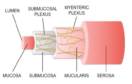
Physiological roles of the enteric nervous system
Motility: coordinate peristalsis to move substances through the digestive tract
Secretion: regulates gastric acid, digestive enzymes, and gut hormones
Blood flow: alters local mucosal/wall blood flow to match digestive demands
Immune interaction: modulates local mucosal barriers and immune responses

Peripheral sensory receptors: Somatic vs visceral vs special
Somatic: Monitor skeletal muscles, joints and skin surface
Visceral: Monitor internal organs and cardiovascular/digestive status
Special: Specialized organs monitoring vision, hearing, balance, taste, and smell
Structural regions of a typical neuron
Soma
Dendrites
Axon
Telodentria (terminal axonal arborizations)
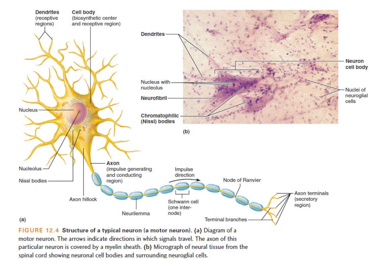
Cytoplasm and cytoskeleton of the neuronal cell body (soma)
Cytoplasm surrounding the nucleus is called perikaryon.
Cytoskeleton contains neurofilaments (intermediate filaments) and neurotubules (microtubules)
Bundles called neurofibrils extend into processes, resistig tensile (pulling) forces.
Structure and microscopic significance of Nissl bodies
Dense clusters of rough endoplasmic reticulum (RER) and free ribosomes in the perikaryon
Stan darkly with cresyl violet and give gross gray matter its dusky gray hue

Centriole deficiency and neurogenesis limitations in mature neurons
Most mature neurons lack centrioles and cannot undergo mitotic division
Neural stem cells remail largely inactive in adults, except in the olfactory epithelium (smell receptor renewal) and the hippocampus
Metabolic characteristics and longevity of neurons
Extreme longevity (can function over 100 years).
Exceptionally high metabolic rate requiring constant glucose and oxygen; vulnerable to irreversible damage within minutes of hypoxia
Receptive morphology and specializations of dendrites
Short, tapering, highly branched processes serving as input regions
Contain dendritic spines (bulbous projections for synaptic contact) and house most organelles found in the cell body
Axon hillock and initial segment
The axon hillock is the cone-shaped junction where the axon originates from the soma
The initial segment lie immediately distal to the hillock and functions as the trigger zone containing high densities of voltage-gated ion channels
Organelle composition of the axon vs. the cell body
Contains mitochondria, neurotubules, and neurofilaments, but completely lacks rough endoplasmic reticulum and golgi apparatus
Entirely dependent on axonal transport from the cell body for protein synthesis and membrane renewal
Anterograde axonal transport mechanism and cargo
Movement away from the soma toward axon terminals
Powered by the ATP-dependent motor protein kinesin along microtubules (speeds up to 40cm/day)
Transports mitochondria, cytoskeletal components, membrane lipids, and neurotransmitter synthesis enzymes.
Retrograde axonal transport mechanism and cargo
Movement from axon terminals back toward the cell body
Powered by the ATP-dependent motor protein dynein along microtubules.
Returns degraded organelles for recycling and delivers intracellular communication molecules (signaling vesicles, neurotrophic factors).
Anaxonic neuron: Structure and location
Small with numerous dendritic processes but no distinguishable, visible axon
Found in the brain and special sense organs; functions remain poorly understood.

Bipolar neuron: Structure and location
Two distinct processes: one fused dendrite branching distally and one axon, with the cell body positioned between them.
Rare; specialized sensory neurons for vision (retina), olfaction (nasal mucosa), and hearing/inner ear.

Unipolar (Pseudounipolar) neuron: structure and location
Single short process emerging from soma that branches T-shaped into a peripheral process (axon with distal receptive endings) and a central process entering the CNS.
Primary sensory neurons with cell bodies clustered in dorsal root ganglia of the spinal cord and sensory cranial ganglia

Multipolar neuron: structure and location
Three or more processes: multiple dendrites and a single long axon
Most common structural class
Examples include: cerebellar Purkinje cells and cerebral pyramidal cells.

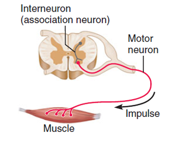
Functional definition of sensory (afferent) neurons
Transmit sensory impulses from internal or external receptors toward the CNS
Most are structurally unipolar with cell bodies in peripheral sensory ganglia
Functional definition of motor (efferent) neurons
Carry impulses away from the CNS to effector organs (muscles, glands).
Structurally multipolar; cell bodies reside within the CNS gray matter (except for postganglionic autonomic neurons)
Functional definition and distribution of interneurons
Lie between sensory and motor neurons; integrate inputs and coordinate outputs.
Confined entirely to the CNS; make up >99% of all multipolar
General characteristics of neuroglial cells vs neurons
Smaller, darker-staining nuclei, far outnumber neurons, and make up 50% of total nervous tissue volume
Retain the ability to undergo cell division throughout life
Do not conduct action potentials; act as a supportive, protective, and homeostatic framework
Pathogenesis and clinical features of gliomas
Primary brain tumors originating from abnormally proliferating glial cells (which retain division capacity, unlike neurons).
Account for 60% of primary brain tumors and 2% of all cancers; highly malignant with 1-year survival rates <50%
Astrocyte morphology and structural connections
Star-shaped, largest and most numerous CNS glial cells with radiating cytoplasmic processes ending in expanded perivascular feet
Packed with intermediate microfilaments; interconnected to one another via gap junctions to transmit calcium waves
Metabolic and chemical homeostatic roles of astrocytes
Regulate extracellular microenvironment- by absorbing leaked potassium ions (K+) and recapturing released neurotransmitter.
Guide the migration of developing neurons and synapse formation during neurogenesis.
Astrocytes and the Blood-Brain Barrier (BBB)
Astrocytes perivascular feet envelop brain capillary endothelial cells.
Secrete regulatory chemicals that induce endothelial cells to form tight junctions, restricting capillary permeability and shielding the CNS from circulating fluctuations
Microglia structure, origin and defense functions
Smallest and least abundant CNS neuroglia; possess thorny, branching processes that monitor neuronal health.
Differentiate into mobile, macrophage- like phagocytes upon pathogen detection or tissue damage to remove dead neurons and debris.
Primary defense since circulating leukocytes have restricted CNS access.

Ependymal cells: Histological structure and ventricular lining
Simple cuboidal to columnar epithelial layer lining the cerebral ventricles and the spinal cord central canal
Apical surfaces possess microvilli and monitoring CSF

Oligodendrocytes: morphology and myelination mechanism
Possess fewer, sheet-like cytoplasmic processes extending from a small soma
Each process wraps around an axon segment to produce a multilayered myelin sheath; a single cell myelinates segments of multiple adjacent axons

Structural distinction between CNS white matter and gray matter
White matter: Regions dominated by lipid-rich, glossy-white myelinated axons (tracts).
Gray matter: regional containing unmyelinated axons, cell bodies, dendrites, and terminal synaptic arborizations.
Satellite cells in the PNS
Flattened glial cells that surround and cushion neuronal cell bodies in peripheral sensory and autonomic ganglia.
Functionally analogous to astrocytes: regulate local nutrients, O2, CO2, and the neurotransmitter levels in ganglia.

Schwann cells (Neurolemmocytes): PNS myelination and regeneration
Envelop all peripheral nerve fibers
Form myelin sheaths around thick peripheral axons (each Schwann cell wraps a single internode segment of one axon, unlike oligodendrocytes)
Play a vital role in axonal repair and regeneration following peripheral nerve trauma.

Resting membrane potential (RMP) polarity and standard value
Electrical charge difference across the resting neuronal plasma membrane, typically -70mV
Inner cytoplasmic face is net negative relative to the extracellular fluid

Chemical gradient differences across the resting plasma membrane
Extracellular fluid (ECF): High concentration of sodium (Na+) and chloride (Cl-)
Intracellular cytosol (ICF): High concentration of potassium (K+) and impermeable negative proteins/phosphates

Resting membrane permeability to K+ vs Na+
The resting membrane is roughly 25 times more permeable to K+ than to Na+ due to abundant open potassium leak channels
Large intracellular protein anions cannot pass through the membrane
Primary mechanism established the resting negative potential
Unopposed outward diffusion of K+ down its concentration gradient through open leak channels leaves behind impermeable organic anions
This loss of positive charge makes the interior increasingly negative until electrical attraction pulls K+ back, reaching equilibrium.