Honors Anatomy Unit 2
 Call Kai
 Learn
Learn Practice Test
 Spaced Repetition
 Match
Match Flashcards
 Knowt Play
1/137

There's no tags or description
Looks like no tags are added yet.
Name | Mastery | Learn | Test | Matching | Spaced | Call with Kai | Chat |
|---|
No analytics yet
Send a link to your students to track their progress
138 Terms

simple squamous epithelial

simple cuboidal epithelial

simple columnar epithelial

nonkeratinized stratified squamous epithelial

keratinized stratified squamous epithelial

pseudostratified columnar (with cillia)
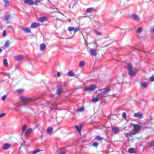
loose connective (areolar)

adipose

dense regular connective

dense irregular connective

hyaline cartilage

osseus (bone)
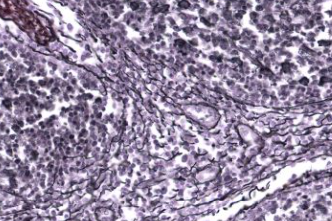
hematopoietic tissue (lymphatic)

hematopoietic tissue (blood)

striated (skeletal) muscle

smooth (visceral) muscle

cardiac muscle

nervous tissue
levels of cellular organization
cell
tissue
organ
organ system
organism
histology
study of tissues
4 types of tissues
epithelial, muscle, connective, nervous
epithelial tissue
-upper (apical) surface of epithelium is free space
-lower (basal) surface rests on connective tissue
-basement membrane
-densely packed
-avascular, nutrients diffuse from neighboring connective tissue
-high rate of mitosis
epithelial tissue function
protection, filtration, secretion, absorption, excretion
simple squamous
located in alveoli and lining of blood vessels, nucleus is centrally located
Simple Cuboidal
located in kidney tubules
Ciliated Simple Columnar
located in oviducts/upper respiratory tract
Nonciliated Simple Columnar
located in the Ileum, Gallbladder, and Urinary tract; may contain goblet cells (secrete mucus)
Simple Columnar with microvilli brush border
located in duodenum; microvilli found at apical surface to increase surface area/ absorption
Keratinized Stratified Squamous
located in superficial skin layer; contains filaments of protein keratin → cells accumulate keratin as they age, become tough and water resistant
Non-keratinized Stratified Squamous
located in esophagus, Oral cavity, Vagina, Anal canal; no keratin, must be kept moist
Stratified Cuboidal
found in ducts of mammary, sweat, and salivary glands
Stratified Columnar
located in lining of developing ovarian follicles; very rare
Ciliated Pseudostratified Columnar
found in respiratory passages; cilia = movement of materials; single layer, but nuclei are at different levels and not all cells reach the free surface
Nonciliated Pseudostratified Columnar
located in prostate and vas deferens; no mucus or cilia
Transitional epithelium
located in urinary bladder'; Cells change shape (from squamous to columnar) as body parts stretch and move
Glandular Epithelium
- makes up the secreting portions of exocrine and endocrine glands
- epithelial tissue specialized for secretion
Endocrine Glands
-secretions enter the extracellular fluid and diffuse into the bloodstream
-no ducts
-secretions are called hormones
-Glands include adrenal, thyroid, pituitary
Exocrine Gland
-secrete products into a duct that leads to the surface of the skin or into the lumen (interior space) of a hollow organ
-Glands include sweat, salivary
- hollow
-lined by glandular epithelium.
Merocrine glands
-type of exocrine gland
-Cells secrete their substances by exocytosis
-Salivary and Sudoriferous (sweat) Glands
-think of “emerge” (exocytosis to open spaces)
Apocrine gland
-type of exocrine gland
-portion of the cell membrane that contains the secretion buds off
-Mammary and Ceruminous (earwax) Glands
-think “a-part”/”apical surface” breaks off
Holocrine gland
-type of exocrine gland
-entire cell disintegrates to excrete its substance
-Sebaceous (oil) Glands
-think “whole” cell
Connective tissue
- Most abundant tissue
- Major transport tissue (blood)
- Stored energy (adipose)
- Doesn’t occur on free surfaces
- rich nerve and blood supply (except cartilage)
connective tissue function
- Supports/strengthens/protects/insulates
Mesenchymal Connective Tissue
-first connective tissue to develop in the embryo
-all connective tissues are later derived from
- clusters are scattered throughout adult tissue and supply the cells needed for replacement and repair after connective tissue injury
all connective tissues derive from
mesodermal layer of the embryo
Extracellular Matrix
-material found in space between tissue cells
-contains ground substance and fibers
-produced by the connective tissue cells that are embedded within it
- composed of ground substance + protein fibers
Ground Substance
part of extracellular matrix; can be fluid/semi-fluid/gelatinous/calcified
Protein Fibers
part of extracellular matrix; composed of one or more types: collagen, elastic, reticular
“-blast” cells
immature; secrete the matrix and produce new cells; all major tissues contain
Chondroblasts
-Cartilage
-secrete gelatinous matrix
Osteoblasts
-bone formation
-secretion of organic fibers and inorganic salts to create crystalline matrix
Mature Fibroblasts
secrete fibers and ground substance of matrix in loose and dense connective tissue, lots of collagen
Red Blood Cells/Erythrocytes
mature resident cell, transport oxygen
White Blood cells/Leukocytes
mature resident cell, immune response; i.e. plasma cells – secrete antibodies/immune system
Platelets/Thrombocytes
blood clotting
Mast cells
produce histamine (chemical that triggers inflammatory response which increases vascular swelling) and attracts more phagocytic cells to the site.
Adipocytes
- fat cells/store triglycerides
- found below skin and around organs
Macrophages
-develop from WBC’s
-move about in connective tissue as scavengers clearing foreign particles
subclasses of connective tissue proper
loose (areolar, reticular, adipose)
dense (regular, irregular)
cells of loose connective tissue proper
fibroblasts
adipocytes
mast cells
macrophages
cells in dense connective tissue proper
fibroblasts scattered scarcely
extracellular matrix in loose connective tissue proper
reticular includes fibers; areolar is gel-like; adipose has very little
extracellular matrix in dense connective tissue proper
mainly fibers; little else
extracellular fibers in loose connective tissue proper
all three
- collagen
- reticular
- elastic
general features of loose connective tissue proper
-wide variety of locations
-wide variety of functions → support, strength, and elasticity
-store triglycerides
general features of dense connective tissue proper
-dense fibers provide tensile strength
-resist stretching
-found in tendons/ligaments or dermis of skin
subclasses of cartilage connective tissue
hyaline
elastic
fibrocartilage
cells of cartilage connective tissue
chondroblasts
chondrocytes
extracellular matrix of cartilage connective tissue
secreted by chondroblasts
extracellular fibers of cartilage connective tissue
collagen in all; elastic in some
general features of cartilage connective tissue
-provides support and flexibility
-minimizes friction
-forms features and found in joints
subclasses of bone connective tissue
-compact
-spongy (trabecular)
cells of bone connective tissue
osteoblasts
osteocytes
extracellular matrix of bone connective tissue
secreted by osteoblasts
extracellular fibers of bone connective tissue
collagen
general features of bone connective tissue
provides framework; protects organs; supports body
subclasses of liquid connective tissue
-hemopoietic lymph
-hemopoietic blood
cells of hemopoietic lymph connective tissue
macrophages lymphocytes
cells of hemopoietic blood connective tissue
-Thrombocytes (platelets)
-Erythrocytes (RBCs)
-Leukocytes (WBCs)
extracellular matrix of liquid connective tissue (blood + lymph)
plasma
extracellular fibers of hemopoietic blood connective tissue
none
extracellular fibers of hemopoietic lymph connective tissue
reticular
general features of hemopoietic blood connective tissue
-provides transportation, regulation, and protection
- carries oxygen and nutrients to cells
-carries away wastes and carbon dioxide
general features of hemopoietic lymph connective tissue
-plays role in immune defenses
-found in spleen and lymph nodes
skeletal muscle tissue
-striated
-voluntary
-Attached to bones of the skeleton
-function: movements and locomotion
cardiac muscle tissue
-Striated/Intercalated Discs
-involuntary
-walls of heart
-function: pump blood
smooth muscle tissue
-non-striated
-involuntary
-in walls of hollow organs, blood vessels, intestines, urinary tract
-function: change diameter of blood vessels, eliminate waste, move materials along digestive tract
nervous tissue
initiates and transmits nerve impulses to coordinate activities of
the body
muscle tissue
contract to produce voluntary or involuntary movements
two major cells of nervous tissue
neurons + neuroglia
neurons
-specialized to transmit electrical (nerve) impulses
- Sense stimuli → convert stimuli into nerve impulses → signal other nerves, muscles, or glands respond
neuroglia
- do not generate or conduct nerve impulses
- smaller than neurons, many times more numerous
Astrocytes (CNS)
-neuroglia
-Help provide proper environment for generation of nerve impulses
-Provide nutrients to neurons
-Take up excess neurotransmitters
-Maintain proper ion balance (Ca 2+ and K+)
Microglial cells (CNS)
-neuroglia
-Engulf invading microbes
-Clear debris of dead cells
Oligodendrocytes (CNS)
-neuroglia
-provide support for surrounding neurons
-produce myelin sheaths around adjacent neuron axons
Ependymal (CNS)
-neuroglia
-Line cavities of the brain filled with CSF
-Forms CSF and aids with its circulation
CSF
cerebral spinal fluid; clear, colorless liquid that cushions, nourishes, and protects the brain and spinal cord
Schwann Cell (PNS)
Produce myelin sheath around axons of PNS neurons
Satellite Cell (PNS)
Form a protective layer around neuron cell bodies in PNS, providing nutritional and metabolic support
organ
two or more tissues, work together to perform a specific function