histo self quiz- non-glandular and glandular stomach
 Call Kai
 Learn
 Practice Test
 Spaced Repetition
 Match
 Flashcards
 Knowt Play
1/25

There's no tags or description
Looks like no tags are added yet.
26 Terms

which type of epithelium lines the ruminant forestomachs?
stratified squamous, keratinized/cornified (unique in appearance because it must provide protection and be permeable enough to absorb VFAs)

based on your knowledge of the lamina muscularis in the ruminant forestomachs, which forestomach is this?
omasum

there are 3 layers of smooth muscle in the laminae of the omasum.
which tunic does the smooth muscle in the yellow brackets belong to?
which tunic does the smooth muscle in the green bracket belong to?
yellow brackets- tunica mucosa (lamina muscularis)
green bracket- tunica muscularis

based upon your knowledge of the lamina muscularis in the ruminant forestomachs, which forestomach is this?
rumen

based upon your knowledge of the lamina muscularis in the ruminant forestomachs, which forestomach is this?
reticulum

based upon your knowledge of the lamina muscularis in the ruminant forestomachs, which forestomach is this?
omasum

ID the ruminant forestomach
rumen

ID the ruminant forestomach
omasum

ID the ruminant forestomach
omasum

ID the ruminant forestomach
reticulum

based upon your knowledge of the lamina muscularis in the ruminant forestomachs, which foretomach is this?
reticulum

nerve plexuses of the GI tube
encircled in yellow highlighter is an enteric autonomic ganglion between the 2 layers of muscle in the tunica muscularis of the GI tube.
what do we call the nerve plexus that innervates the smooth muscle of the tunica musclularis in the GI tube?
myenteric nerve plexus (auerbachs plexus)

nerve plexuses of the GI tube
these enteric autonomic ganglia outlines in yellow highlighter are near the submucosal glands
what do we call the nerve plexus that innervates the smooth muscle and glands of the tunica mucosa and the tunica submucosa?
the submucosal plexus (meissners plexus)

ID the 2 types of epithelia seen at this gastroesophageal junction
right- stratified squamous epithelium of the esophagus, non cornified
left- simple columnar surface epithelium of the glandular stomach

these gastric glands are approximately the same depth as their pits, secrete mucus, and are located near the gastroesophageal junction.
which type of the three gastric glands are these?
cardiac glands

these gastric glands are shorter than the depth of their deep pits, secrete mucus, and are locted near the gastoduodenal junction.
which of the three types of gastric glands are these?
pyloric glands

which of the 3 types of gastric glands are these?
pyloric glands (coiled tubular, deep pits)

pyloric gastric glands
which image shows gastric pits? gastric glands?
what do the glands produce?
pits on left, glands on right
mucus

the cardiac and pyloric gastric glands produce mucus.
what is another source of the protective mucus seen in this image (marked by yellow asterisks) in addition to these gastric glands?
the epithelium lining both the surface of the stomach and the gastric pits

which type of epithelium lines both the luminal surface and the pits in the glandular stomach?
are the nuclei of this epithelium positioned at the apical or basal surface of the cells?
simple columnar
basal surface (adjacent to basement membrane)

are gastric glands mucosal or submucosal glands? aka are they located in the tunica mucosa or tunica submucosa?
mucosal, located in tunica mucosa
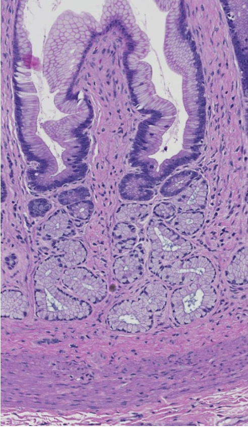
which lamina of the tunica mucosa are the gastric glands invaginated down into?
which lamina of the tunica mucosa do the fundic portions of the gastric glands approach but not penetrate?
lamina propria
lamina muscularis

are these cardiac, proper gastric, or pyloric glands?
what do these gastric glands produce?
proper gastric glands
digestive enzymes, hydrochloric acid, and mucus

ID the encircled cells in this proper gastric gland
what do these cells produce?
parietal cells
hydrogen ions, which join with chloride ions pumped into the canaliculi, to form HCl
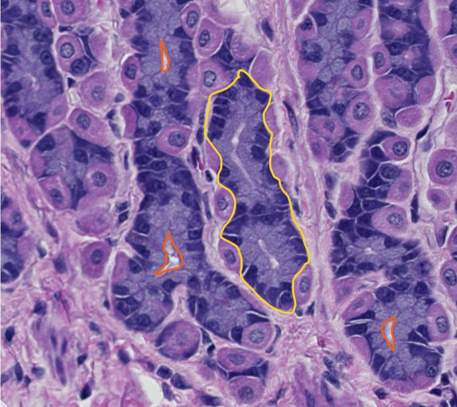
ID the cells encircled in yellow within this proper gastric gland
which inactive enzyme do these cells produce?
how is the enzyme activated?
what are the small spaces encircld with orange lines?
chief cells
pepsinogen
hydrochloric acid converts pepsinogen to pepsin
gastric gland lumens
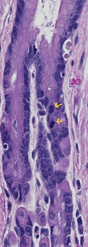
the yellow arrowed lines are indicating 2 mitotic figures, indicating cell division.
where are the stem cells of the gastric glands located?
what do the stem cells produce?
in the neck of the gland
stem cells migrate bidirectionally to replace surface and pit mucous cells or any of the cells of the gastric glands (mucus neck cells, chief cells, parietal cells, and enteroendocrine cells)