SOL & Raised ICP
 Call Kai
 Learn
 Practice Test
 Spaced Repetition
 Match
 Flashcards
 Knowt Play
1/98

There's no tags or description
Looks like no tags are added yet.
99 Terms
Match: childhood/adult brain tumours to supra/infra-tentorial
Adult brain tumours = supra-tentorial
Childhood brain tumours = infra-tentorial
How commonly do malignant CNS tumours metastasise outside the CNS?
It is rare for malignant CNS tumours to metastasise outside the CNS
(but other tumours can metastasise into the CNS)
What are the major groups of primary CNS tumours
Gliomas
Glioneuronal and neuronal tumours
Ependymal tumours
Meningioma
Pituitary adenoma
Vestibular schwannoma/acoustic neuroma
Medulloblastoma
Haemangioblastoma
Primary CNS lymphoma
Name 3 adult-type diffuse gliomas
Astrocytoma, IDH-mutant
Oligodendroglioma, IDH-mutant and 1p/19q-co-deleted
Glioblastoma, IDH-wildtype
Normal role of astrocytes
maintain blood–brain barrier
Normal role of oligodendrocytes
Produce myelin in CNS
(called Schwann cells in PNS)
Normal role of glia
General support cells of the nervous system
Match:
IDH-wild-type
IDH mutation + 1p/19q deletion
IDH mutation without 1p/19q deletion
astrocytoma
oligodendroglioma
glioblastoma
IDH mutation without 1p/19q deletion = astrocytoma.
IDH mutation + 1p/19q deletion = oligodendroglioma.
IDH-wild-type + high-grade features = glioblastoma
Which tumour type is the most frequent malignant primary brain tumour in adults
Glioblastoma, IDH-wildtype
Name a circumscribed astrocytic glioma
Pilocytic astrocytoma
Name a glioneuronal/neuronal tumour
Ganglioneuroma
Name a ependymal tumour
Ependymoma
What is an astrocytoma, IDH-mutant?
A diffusely infiltrating glioma with a mutation in either IDH1 or IDH2
(IDH = isocitrate dehydrogenase)
What is the biological range (rage of grades) of IDH-mutant astrocytomas
They range from well-differentiated, slow-growing CNS grade 2 tumours to highly anaplastic, hypercellular and rapidly progressive grade 4 tumours
Where can an IDH-mutant astrocytoma occur
It can occur in any region of the CNS, but most commonly develops supratentorially, usually centred near or within the frontal lobes
3 main presenting features of astrocytoma, IDH-mutant?
Seizures
Headaches
Focal neurological deficits
How can an IDH mutation be identified (2 methods)
Methylation profiling can identify the IDH mutation.
Immunohistochemistry for IDH1 p.R132H can be used as an alternative.
What molecular feature distinguishes astrocytoma, IDH-mutant from oligodendroglioma?
Astrocytoma, IDH-mutant lacks the 1p/19q co-deletion
Is it possible to completely resect an infiltrative astrocytoma
It is often not possible to resect infiltrative astrocytomas completely without compromising neurologic function
What molecular abnormalities define oligodendroglioma
IDH mutation & 1p/19q co-deletion
Where do oligodendrogliomas commonly occur?
In the cerebral hemispheres, often in the anterior fossa
What WHO grades can oligodendrogliomas be
2 or 3
Name the 2 classic histological features of oligodendroglioma
Delicate capillary network → "chicken-wire vessels"
Cells with a perinuclear halo → "fried egg" appearance
How is a oligodendroglioma diagnosed
Demonstration of IDH mutation by IDH1 p.R132H immunohistochemistry and/or sequencing of the IDH1 or IDH2 gene
&
Demonstration of 1p/19q co-deletion
Which has better prognosis: oligodendroglioma / astrocytoma
Oligodendroglioma
Median survival time of Oligodendroglioma
>10 yrs
What % of all intracranial neoplasms are glioblastomas?
15%
What % of all primary malignant brain tumours are glioblastomas?
45–50%
Which age group is most affected by glioblastoma
Older adults - peak incidence 55–85 years
What’s a risk factor for glioblastomas
ionising radiation
(that’s radiation from like a bomb - not a phone)
Name 3 histological features usually present in glioblastoma
Microvascular proliferation
Necrosis
Sometimes nuclear palisading
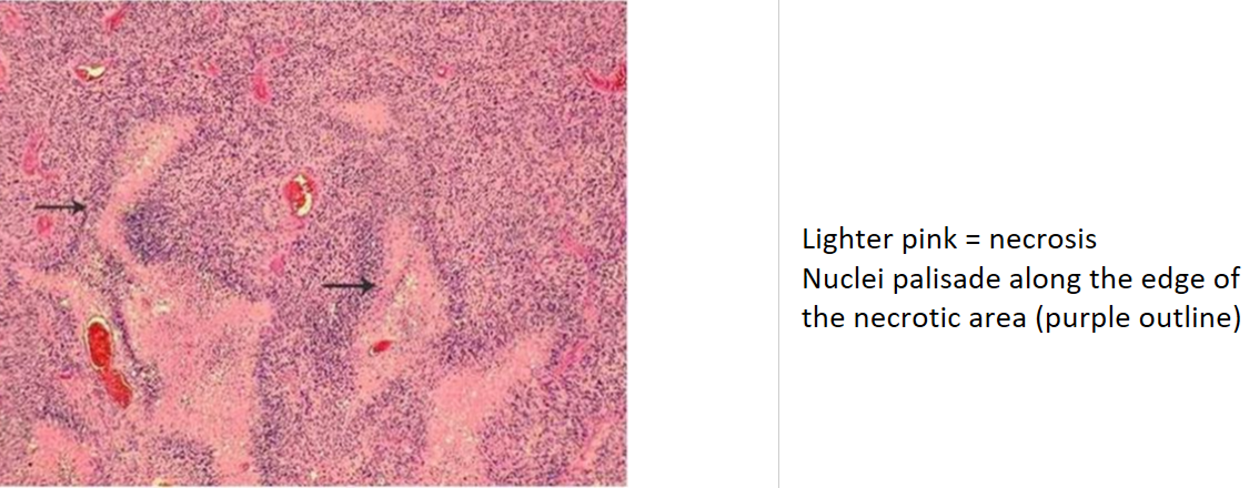
What WHO grade is glioblastoma
4 - so all v. aggressive
Is the prognosis for glioblastomas good? What is it?
No! Very poor prognosis; most glioblastoma patients die within 15–18 months after therapy with chemoradiation
Differentiate a Pilocytic astrocytoma from an astrocytoma, IDH-mutant
Pilocytic astrocytoma = non-infiltrating astrocytoma
What grade is pilocytic astrocytoma
1 - relatively benign behaviour
Who is most commonly affected by Pilocytic astrocytomas
Children and young adults
Where is pilocytic astrocytoma most commonly located?
cerebellum
What treatment is used for pilocytic astrocytomas
May be cured by resection
Where do ependymomas arise?
Next to the ependyma-lined ventricular system
Are glial / non-glial tumours more common
Glial are much more common
What is the most common neuronal (so non-glial) tumour
Ganglioglioma
Which patients are neuronal tumours particularly associated with
Children and young adults with epilepsy
Meningiomas are usually fast/slow growing & aggressive/benign
Slow growing, benign (usually sit on surface of brain without infiltrating)
Meningioma treatment
May be cured by resection
In which age group are meningiomas most commonly seen
Adults
Location of meningiomas
Attached to dura
What cells do meningiomas arise from?
Meningoendothelial cells of the arachnoid
What is the classic histological pattern of a meningioma
A whorled pattern of meningothelial cells with Psammomatous (in rings) calcification
How are pituitary adenomas classified
According to the hormone they produce (e.g. ACTH, TSH, LH, FSH, GH, Prolactin [oxytocin, ADH - but post. pituitary tumours far less common])
Which presents first - functional/non-functional pituitary tumours
Functional tumours present earlier than non-functional tumours
What syndrome is classically associated with pituitary adenomas
Multiple endocrine neoplasia syndrome (MEN)
(You get tumours in the 3 Ps - pituitary, pancreas, parathyroid)
What visual field defect is classically produced by a pituitary adenoma?
Bitemporal hemianopia
What is another name for acoustic neuroma
Vestibular schwannoma
From which cells does a vestibular schwannoma arise
Schwann cells of cranial nerve VIII (CN VIII)
How dangerous are acoustic neuromas
Not the most dangerous - they are benign
What are the 3 classic presenting symptoms of acoustic neuroma
Hearing loss
Dizziness
Tinnitus
Are most vestibular schwannomas sporadic or inherited
>90% are sporadic
Which inherited condition is associated with some vestibular schwannomas with bilateral acoustic neuroma
Neurofibromatosis type 2 (NF2).
What type of tumour is medulloblastoma
A type of primitive neuroectodermal tumour (PNET)
What age is mostly affected by medulloblastoma
Children
Where does medulloblastoma occur
cerebellum
What does medulloblastoma look like histologically
small round blue cell tumour
Which medulloblastoma subtype has the best survival rate
WNT-activated medulloblastoma, associated with nearly 100% survival at 5 years
What is the 5-year survival for other subtypes of medulloblastoma
20-30% survival
Where is haemangioblastoma typically located?
cerebellum
How common is haemangioblastoma
Rare
What is a major pathological characteristic of haemangioblastoma
It is a highly vascular tumour
Treatment & prognosis of haemangioblastoma
Excellent prognosis after complete excision
What genetic syndrome is associated with haemangioblastoma
Von Hippel–Lindau (VHL) syndrome
What is the most common CNS tumour in immunocompromised individuals
Primary CNS lymphoma
Histology of Primary CNS lymphoma
Usually large B cell lymphoma (primary CNS-LBCL)
What is the biological behaviour of primary CNS lymphoma? (benign/aggressive)
Aggressive
Which has a better response to chemotherapy - Primary CNS lymphoma vs peripheral lymphomas
Peripheral lymphomas has a better response
What is the most common brain tumour seen in hospitals
Metastatic tumours
What type of tumour most commonly metastasises to the brain
Metastatic carcinoma
What 6 primary cancers are common sources of brain metastases
Lung
Breast
Melanoma
Kidney
Gastrointestinal tract
Choriocarcinoma

Where are brain metastases typically located
At the grey-white matter junction
Name 2 main causes of raised ICP
Cerebral oedema
Hydrocephalus
Name 2 types of Cerebral oedema
Vasogenic & cytotoxic
What is vasogenic cerebral oedema
Oedema caused by disruption of the blood-brain barrier and increased vascular permeability, allowing fluid to move from the intravascular compartment into the intercellular spaces of the brain
What is cytotoxic cerebral oedema
An increase in intracellular fluid caused by injury to neuronal, glial or endothelial cell membranes
Give 2 examples of causes of cytotoxic oedema
Generalised hypoxic/ischaemic insult
Metabolic damage
What is a very common cause of cerebral oedema
Cerebral oedema extremely common after a substantial head injury, esp. in children
What 2 brain injuries may cerebral oedema be related to?
Diffuse axonal injury (DAI) and concussion.
By how much may brain weight increase following substantial head injury
Brain weight may increase by approximately 100 g.
What part of the brain is most of this increase in brain weight found after a head injury
Mainly in the white matter.
Is it ever the case that cerebral oedema is the only intracranial abnormality found at autopsy after a substantial head injury has occurred
Yes - it may be
(Swelling blocks blood vessels -> cuts off blood supply → death)
What is hydrocephalus
Accumulation of excessive CSF within the ventricular system
What happens if hydrocephalus develops during infancy before closure of the cranial sutures
There is enlargement of the head
Name 2 causes of raised ICP and possible subsequent brain herniation
Generalised cerebral oedema
or
A focal space-occupying lesion, such as:
Haemorrhage
Tumour
Abscess
What are the three main types of brain herniation
Subfalcine (cingulate)
Transtentorial
Tonsillar
What is the location of each type of hernia
Subfalcine (cingulate)
Transtentorial
Tonsillar
Subfalcine (cingulate): the cingulate gyrus gets displaced under the falx cerebri
Transtentorial: the medial aspect of the temporal lobe is compressed against the free margin of the tentorium
Tonsillar: displacement of the cerebellar tonsils through the foramen magnum

Which type of hernia is known as coning
Tonsillar
(Coning is worst case scenario - basically guarantees death)
What is a physical sign of Transtentorial herniation
Uncal grooves
What are the brainstem haemorrhages secondary to herniation called
Duret haemorrhages
What type of brain herniation do Duret haemorrhages often accompany
Progressive transtentorial herniation
How does transtentorial herniation cause duret haemorrhages
Herniation → distortion or tearing of penetrating veins and arteries supplying the upper brainstem
What are four main causes of cerebral oedema/increased ICP
Brain haemorrhage
Brain tumours
Diffuse axonal injury
Hypoxic injury
Name 4 examples of hypoxic injury
Cardiac arrest
Stroke
Asphyxial deaths
Carbon monoxide (CO) poisoning