Ear
 Call Kai
 Learn
Learn Practice Test
 Spaced Repetition
 Match
Match Flashcards
 Knowt Play
1/299

There's no tags or description
Looks like no tags are added yet.
Name | Mastery | Learn | Test | Matching | Spaced | Call with Kai | Chat |
|---|
No analytics yet
Send a link to your students to track their progress
300 Terms
Swelling can block sound from entering the external acoustic meatus
Why can an auricular hematoma cause hearing loss?
Varicella zoster (shingles); dormant in the geniculate ganglion (CN VII)
What virus causes Ramsay Hunt syndrome, and where does it lie dormant?
CN VII makes a small sensory contribution to the external ear, so the shingles rash appears in that dermatome
Why does Ramsay Hunt syndrome affect the external ear?
Touching the external acoustic meatus/concha, which is supplied by a small CN X branch
What triggers the Arnold cough reflex?
Pars tensa: the taut majority; pars flaccida: the small, lax superior portion
Which part of the tympanic membrane is taut, and which is lax?
Anterior and inferior
Where does the cone of light point on a normal tympanic membrane?
The tip of the handle of the malleus, at the center (apex) of the cone-shaped tympanic membrane
What is the umbo?
Perforation of the tympanic membrane
Main risk of inserting objects (e.g., cotton swabs) into the ear canal
The tympanic portion of the temporal bone
What forms most of the bony external acoustic meatus?
Cochlea anterior, semicircular canals posterior, middle ear cavity lateral to both, beneath the tegmen tympani
Location of the ear structures on radiologic images
CN VII, in the facial canal
Which nerve runs along the medial wall of the middle ear?
The stapedius muscle
What does the pyramidal eminence enclose?
Posteriorly from the middle ear to the mastoid air cells (mastoid antrum)
Where does the aditus lead?
Stylomastoid foramen
Where does CN VII exit the skull after the facial canal?
Petrotympanic fissure
Where does the chorda tympani exit the skull?
Hiatus for the greater petrosal nerve
Where does the greater petrosal nerve emerge onto the middle cranial fossa floor?
Tympanic canaliculus
Through what opening does the tympanic nerve (Jacobson's n.) enter the middle ear?
The malleus (head = scoop) and incus (body = cone) seen together on CT
What is the "ice cream cone" on radiologic imaging?
In the oval window
Where does the base (footplate) of the stapes sit?
Sound -> tympanic membrane -> malleus -> incus -> stapes -> oval window -> cochlear fluid vibrations -> organ of Corti -> CN VIII action potentials
Full sound conduction pathway to CN VIII
Whether air conduction is stronger than bone conduction
What does the Rinne test check?
Sounds from chewing and loud noises
What kinds of loud sounds does the tensor tympani dampen?
Tensor and levator veli palatini open the pharyngotympanic tube, equalizing middle ear pressure
Why do your ears "pop" when you yawn or swallow on a plane?
Their pharyngotympanic tube is more horizontal, so the middle ear drains poorly
Why is otitis media more common in children?
Vestibular labyrinth (semicircular ducts, utricle, saccule) and cochlear labyrinth (cochlear duct)
2 parts of the membranous labyrinth
Utriculosaccular duct
What connects the utricle and saccule?
Ductus reuniens
What connects the cochlear duct to the saccule?
Suspended by delicate filaments within the bony labyrinth
How is the membranous labyrinth held in place?
Gravity and linear acceleration (utricle: horizontal plane; saccule: vertical plane)
What do the maculae of the utricle and saccule detect?
Rotational movement of the head
What do the semicircular ducts detect?
Helicotrema (at the apex of the cochlea)
Where do the scala vestibuli and scala tympani communicate?
Scala vestibuli begins at the oval window; scala tympani ends at the round window
Where does the scala vestibuli begin, and where does the scala tympani end?
Scala media
What is another name for the cochlear duct?
Vestibular membrane
What separates the cochlear duct from the scala vestibuli?
Basilar membrane
What separates the cochlear duct from the scala tympani?
On the basilar membrane, under the tectorial membrane
Where do the hair cells of the organ of Corti sit?
Spiral ligament
What lies on the lateral wall of the cochlear duct?
Runs in the vestibular aqueduct to end in the endolymphatic sac
Path of the endolymphatic duct
The perilymphatic space to the subarachnoid space (CSF)
What does the cochlear aqueduct connect?
Both the cochlear (tinnitus, hearing loss) and vestibular (vertigo) systems
Which systems does Ménière's disease affect?
Greater petrosal n., nerve to stapedius, chorda tympani
Branches of CN VII in/near the facial canal
Motor to the stapedius muscle
What does the nerve to stapedius supply?
Exits via the petrotympanic fissure, joins the lingual nerve (V3), and continues to the submandibular ganglion
Path of the chorda tympani after crossing the middle ear
Superior salivatory nucleus -> greater petrosal n. -> joins deep petrosal n. -> Vidian n. (pterygoid canal) -> pterygopalatine ganglion -> lacrimal gland and nasal/palatal mucosa
Greater petrosal parasympathetic pathway
The deep petrosal nerve (postganglionic sympathetic, from the internal carotid plexus)
What joins the greater petrosal nerve to form the Vidian nerve?
Superior salivatory nucleus -> chorda tympani -> lingual n. (V3) -> submandibular ganglion -> submandibular and sublingual glands
Chorda tympani parasympathetic pathway
Hyperacusis (stapedius paralysis)
CN VII lesion proximal to the nerve to stapedius causes:
Loss of taste (anterior 2/3 tongue) and reduced submandibular/sublingual salivation
CN VII lesion proximal to the chorda tympani causes:
Reduced lacrimation (dry eye), plus the chorda tympani and stapedius deficits
CN VII lesion proximal to the greater petrosal nerve causes:
Proximal to the greater petrosal nerve (at or before the geniculate ganglion)
Bell's palsy with hyperacusis, loss of taste, and dry eye: where is the lesion?
To avoid the chorda tympani (which crosses superiorly) and to allow better drainage
2 reasons myringotomy tubes are placed inferiorly
It involves loss of cochlear hair cells, which don't regrow
Why doesn't sensorineural hearing loss recover?
Canal blockage, middle ear fluid, tympanic membrane or ossicle problems, impaired oval/round window movement
Examples of conductive hearing loss causes
Sensorineural: cochlea or cochlear nerve; conductive: external or middle ear (sound never reaches the cochlea properly)
Sensorineural vs. conductive hearing loss: site of the problem
The external ear, middle ear, and organ of Corti; it stimulates the cochlear branch of CN VIII directly
What does a cochlear implant bypass?
External
Middle
Inner
The ear has three parts:
Equilibrium (balance)
Hearing
2 main functions of the ear
cochlear system
system within the ear in charge of hearing:
vestibular system
system within the ear in charge of balance:
Auricle
Identify the structure

External acoustic meatus
Identify the structure

external ear
the ear auricle is part of the:
external ear
the external acoustic meatus is part of the:
Auricle (pinna)
What is the irregular shaped cartilage plate covered by thin skin?
lobule
what part of the ear auricle does not have cartilage?
Helix
Identify the structure at #1
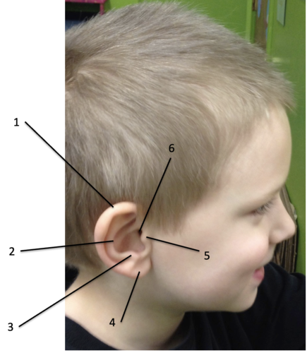
Antihelix
Identify the structure at #2

Antitragus
Identify the structure at #3

Lobule
Identify the structure at #4

Tragus
Identify the structure at #5

External acoustic meatus
Identify the structure at #6

auricular hematoma
patient is a wrestler and presents as such. What is the pathology?

Cauliflower ear
If a patient has a persistent auricular hematoma and it is not drained, it can lead to...
auriculotemporal n. (V3)
sensory innervation for the external ear over the anterior helix and tragus:
lesser occipital n. (C2)
great auricular n. (C2-3)
sensory innervation for the external ear over the posterior helix and anti-tragus:
Small sensory contributions from CN VII (blue circles) and X (red triangles)
What do the triangles and circles represent?

CN X
What cranial nerve is involved with the arnold cough reflex?
CN VII
What cranial nerve is involved with Ramsay-Hunt Syndrome?
CN VII
somatic motor innervation to the ear is done by:
Auriculotemporal n. (V3)
Identify the structure at #1

Lesser occipital n. (cervical plexus)
Identify the structure at #2

Greater auricular n. (cervical plexus)
Identify the structure at #3

Motor branches of CN VII
Auricular muscles are innervated by what nerve?
Posterior auricular branch of CN VII
Identify the structure at #1

Posterior auricular m.
Identify the structure at #2

Superior auricular m.
Identify the structure at #3

Anterior auricular m.
Identify the structure at #4

Temporal branch of CN VII
Identify the structure at #5

Cartilaginous portion of the external acoustic meatus
Identify the structure at #1

Bony portion of the external acoustic meatus
Identify the structure at #2

- About 1 inch long: Slightly sigmoid (S-shaped)
- Lined with skin containing sebaceous and ceruminous glands which produce ear wax
Characteristics of the external acoustic meatus (canal)
perforation of the tympanic membrane
identify the trauma:

perforation of the tympanic membrane
identify the trauma:

tympanic membrane
what is the medial boundary of the external ear?
tympanic membrane
what structure separates external and middle ear?
A sulcus of the temporal bone
What is the tympanic membrane set in?
Oriented laterally, anteriorly and inferiorly
How is the tympanic membrane oriented?
Tympanic membrane
What is a cone shaped with apex attached to the handle of the malleus at the umbo?
capture changes in air pressure (sound) and transfer to the bones of the middle ear
What is the function of the tympanic membrane?
malleus
the tympanic membrane is attached to which ossicle?