Anat and Phys Ch. 6
 Call Kai
 Learn
Learn Practice Test
 Spaced Repetition
 Match
Match Flashcards
 Knowt Play
1/26

There's no tags or description
Looks like no tags are added yet.
Name | Mastery | Learn | Test | Matching | Spaced | Call with Kai | Chat |
|---|
No analytics yet
Send a link to your students to track their progress
27 Terms
skeletal cartilage
highly resilient, molded cartilage tissue that consists primarily of water
contains no blood vessels or nerves
perichondrium
layer of dense connective tissue surrounding cartilage
contains blood vessels for nutrient delivery to cartilage
helps cartilage resist outward expansion
cartilage is made up of (blank), cells encased in small cavities called (blank), within a jelly-like (blank)
chondrocytes, lacunae, extracellular matrix
cartilage grows in two ways:
appositional growth
interstitial growth
appositional growth
cartilage forming cells in the perichondrium secrete matrix against external face of existing cartilage
interstitial growth
chondrocytes within lacunae divide and secrete new matrix, expanding cartilage from within
seven important functions of bones
support
protection
movement
mineral and growth factor storage
blood cell formation (hematopoiesis)
triglyceride (fat) storage
hormone production (osteocalcin)
cartilage in the external ear is (blank)
cartilage in the intervertebral discs is (blank)
cartilage connecting the ribs to the sternum is (blank)
elastic
fibrocartilage
hyaline
your humerus is a (blank) bone
your sternum is a (blank) bone
your vertebra are (blank) bones
your talus is a (blank) bone
long
flat
irregular
short

are bones organs?
bones are organs because they contain different types of tissue
bone (osseous) tissue dominates, but bones also have nervous tissue, cartilage, fibrous connective tissue, muscle cells and epithelial cells
3 levels of bone structure
gross
microscopic
chemical
describe compact and spongy bone
compact bone is smooth and solid
spongy bone has a mesh of boney spines called trabeculae
true/false: flat bones only contain compact bone
FALSE: flat bones have a layer of spongy bone sandwiched between compact bone
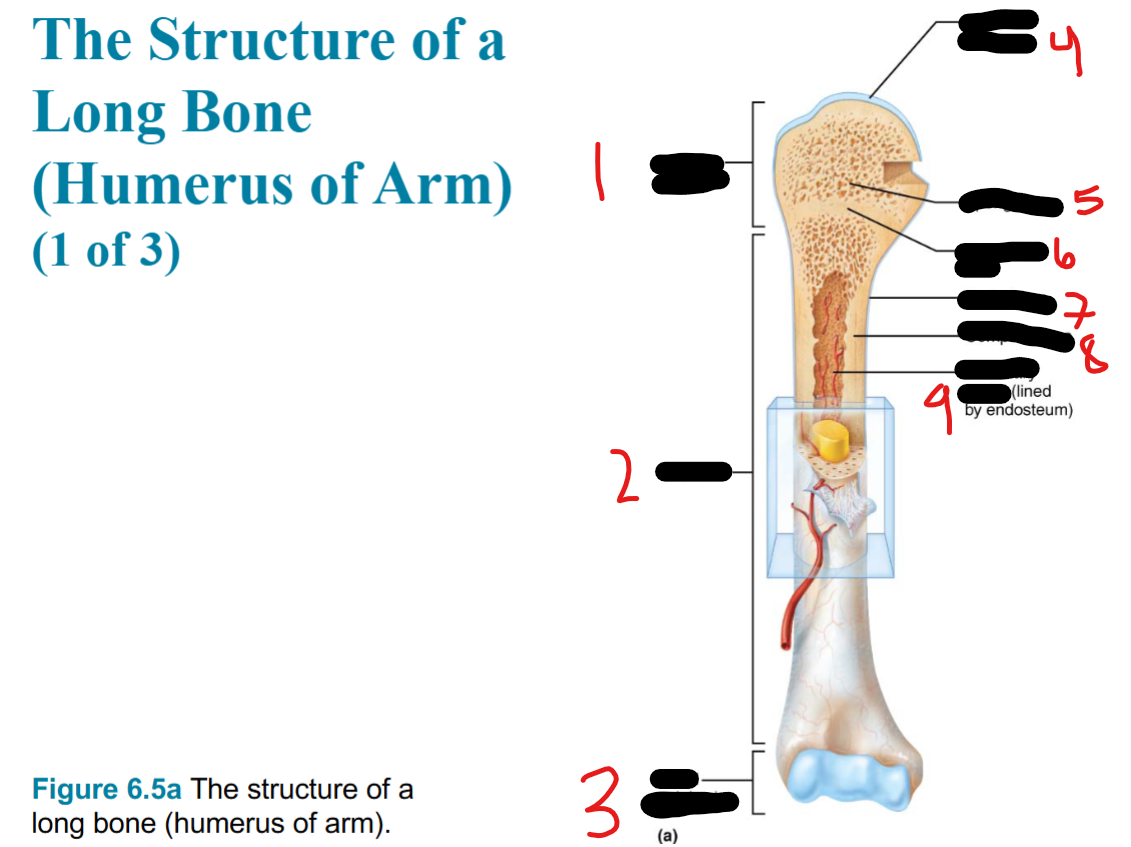
proximal epiphysis
diaphysis
distal epiphysis
articular cartilage
spongy bone
epiphyseal plate
periosteum
compact bone
medullary cavity (lined by endosteum)


Endosteum
yellow bone marrow
compact bone
nutrient foramen
perforating fibers
nutrient artery
Bone markings
sites of (blank), (blank) and (blank) attachment on external surfaces
areas involved in (blank) formation or conduits for (blank) vessels and (blank)
muscle, tendon, ligament
joint, blood, nerves
3 types of bone markings
projection
depression
opening

spinous process
5 major cell cell types of bone tissue
osteogenic cells
osteoblasts
osteocytes
bone-lining cells
osteoclasts
osteoprogenitor cells are (blank) cells
osteoprogenitor cells become (blank), which regulate (blank)
stem
osteoblasts
bone growth and matrix synthesis

osteoblasts (responsible for bone growth and matrix synthesis) become (blank), which are (blank) bone cells and function in what
osteocytes
mature
monitoring and maintaining the mineralized bone matrix

osteoclasts arise from the (blank) lineage and function in what
white blood cell, reabsorbing bones
NOTE: osteoclasts arise separate from the bone cell lineage and instead arise from the white blood cell lineage

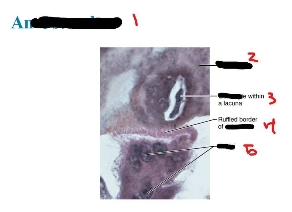
osteoclast
bone matrix
osteocyte
osteoclast
nuclei
compact bone is also called (blank) bone
what does compact bone consist of (OCI)
lamellar
osteon (haversian system), canals and canaliculi, interstitial and circumferential lamellae
two methods of bone formation
endochondral
intramembranous
Endochondrial Ossification (6 steps) (Big Cats Pounce, Dogs Sleep Everywhere)
bone collar forms around diaphysis of hyaline cartilage model
cartilage calcifies in center of diaphysis and forms cavities
periosteal bud invades internal cavity and spongy bone forms
diaphysis elongates and medullary cavity forms
secondary ossification centers appear in the epiphyses
epiphyses ossify, hyaline cartilage remains only in epiphyseal plates and articular cartilage when ossification is complete
Intramembranous ossification (MOOO VC)
Mesenchymal cells cluster and differentiate into osteoblasts, forming ossification center
osteoblasts secrete osteoid, which calcifies. trapped osteoblasts become osteocytes
osteoid is laid down between embryonic blood vessels, forming spongy bone
vascularized mesenchyme on the outside of the bone condenses and becomes periosteum
compact bone replaces immature spongy bone just deep to the periosteum and red bone marrow develops