Anatomy 2300 Unit 3: The Axial Skeleton and Deep Muscles of the Back
 Call Kai
 Learn
Learn Practice Test
 Spaced Repetition
 Match
Match Flashcards
 Knowt Play
1/58

There's no tags or description
Looks like no tags are added yet.
Name | Mastery | Learn | Test | Matching | Spaced | Call with Kai | Chat |
|---|
No analytics yet
Send a link to your students to track their progress
59 Terms
Primary Spinal Curvatures
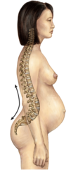
The curvatures present at birth (fetal position), where the spine curves in one continuous C-shape.
They are concave anteriorly. Found in the thoracic and sacral regions, and they persist into adulthood.

Secondary Spinal Curvatures
Curvatures that develop after birth as an infant gains motor control (lifting head, then walking).
They are concave posteriorly (curve the opposite direction of primary curves). Found in the cervical and lumbar regions.

Kyphosis
An exaggerated posterior deviation of the normal thoracic curvature (i.e., an over-accentuated primary curve), producing a rounded, hunchback appearance of the upper back.

Lordosis
An exaggerated anterior deviation of the normal lumbar curvature (i.e., an over-accentuated secondary curve), producing a pronounced inward "swayback" appearance of the low back.
Scoliosis
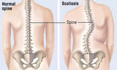
An abnormal lateral (side-to-side) curvature of the spine, often with rotation of the vertebrae, that can occur in the thoracic and/or lumbar region.
Occurs in the coronal plane and deviates right or left
Vertebral Body
The thick, weight-bearing, anterior portion of a vertebra; roughly disc/cylinder-shaped and stacked on top of adjacent vertebral bodies (separated by intervertebral discs).
Vertebral Arch
The posterior, bony arch attached to the vertebral body; formed by the pedicles + laminae. It surrounds and protects the vertebral foramen (spinal cord).

Pedicles
Short, thick bony pillars that project posteriorly from the vertebral body and connect it to the laminae.
They form the sides of the vertebral arch.

Laminae
Flat plates of bone that extend from the pedicles and fuse posteriorly at the midline to form the spinous process.
Completes the vertebral arch posteriorly.

Transverse processes
Bony projections that extend laterally (one on each side) from where the pedicle meets the lamina.
Serve as attachment sites for muscles and ligaments (and articulate with ribs in the thoracic region).

Spinous process
The single bony projection extending posteriorly (and often inferiorly) from the junction of the two laminae.
Palpable as the bumps down your back; site of muscle/ligament attachment.

Superior articular process/facet
Bony projections pointing upward to articulate with the vertebra above.

Inferior articular process/facet
Bony projections pointing downward to articulate with the vertebra below.

Intervertebral disc
A fibrocartilage pad located between adjacent vertebral bodies that absorbs shock and allows slight movement. (Functional Discs: C2-C3 → L5-S1)

Anulus fibrosus
The tough, ring-shaped outer layer of the disc, made of concentric layers of fibrocartilage; contains and protects the nucleus pulposus. (A)

Nucleus Pulposus
The soft, gel-like core in the center of the disc; acts as a cushion/shock absorber. (B)

Vertebral foramen and vertebral canal
The vertebral foramen is the opening in each vertebra bounded by the body (anteriorly) and the vertebral arch (posteriorly).
When stacked, all the vertebral foramina line up to form the vertebral canal, which houses and protects the spinal cord.

Intervertebral notch
A concave notch found on the superior and inferior borders of each vertebra's pedicle. Every vertebra has one on top and one on the bottom.

Intervertebral foramen
The opening formed when the inferior notch of one vertebra lines up with the superior notch of the vertebra just below it. This opening is the passageway through which spinal nerve roots exit the vertebral canal.
Cervical region (of spine), identifying traits?
Definition: The most superior portion of the vertebral column, located in the neck, consisting of 7 vertebrae (C1-C7).
It supports the head while allowing the greatest range of motion of any spinal region (flexion, extension, lateral flexion, and rotation).
Identifying traits:
- Vertebral bodies are small and wide side-to-side
- Vertebral foramen is large and triangular
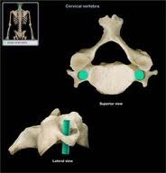
- Bifid spinous processes — Spinous processes (C3-C6) that are split/forked at the tip.
- Contains transverse foramen

Transverse foramina
Openings within the transverse processes of cervical vertebrae, formed by the anterior and posterior roots of the transverse process, that allow the vertebral blood vessels to pass through.
Bifid spinous processes
Spinous processes that are split into two branches at their tips, a characteristic feature of cervical vertebrae C2-C6.

C1
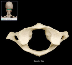
(atlas )The first cervical vertebra; has no vertebral body and no spinous process.
Ring-shaped, with lateral masses that articulate with the occipital condyles of the skull (allows "yes" nodding motion).
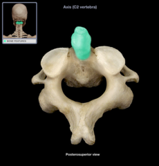
C2
(axis) The second cervical vertebra; has a bony peg called the dens projecting superiorly. Allows the atlas (and skull) to pivot/rotate around it (allows "no" head-shaking motion).

Dens (C2)
Tooth-like projection rising from the body of the axis.
It sits within the ring of the atlas, held in place by the transverse ligament; acts as the pivot point for head rotation
Thoracic region (of spine), identifying traits?
Definition: The thoracic region is located in the mid-back, inferior to the cervical region and superior to the lumbar region.
It consists of 12 vertebrae (T1-T12) that uniquely articulate with the ribs to form the posterior attachment of the rib cage.
Identifying traits:
- Vertebral bodies are heart-shaped and larger than cervical vertebral bodies
- Vertebral foramen is circular and intermediate sized
- Contains Costal facets
- Long and pointy spinous process

Costal facets
Small, smooth cartilage-covered surfaces on the vertebral body (and transverse processes) of thoracic vertebrae where the ribs articulate ("costal" = rib).

Where do the thoracic vertebrae and ribs articulate?
(1) The head of the rib articulates with the body of the vertebra (at costal facets).
(2) The tubercle of the rib articulates with the transverse process of the vertebra.
(Mnemonic: "TNT" — Tubercle/Transverse process.)

Lumbar region (of spine), identifying traits?
Definition: The lumbar region is located in the lower back, inferior to the thoracic region and superior to the sacrum.
It consists of 5 vertebrae (L1-L5) built to bear the greatest amount of body weight, allowing flexion, extension, and some lateral flexion, but preventing rotation.
Identifying traits:
- Vertebral bodies are massive and kidney-shaped (largest of all regions, built for weight-bearing)
- Vertebral foramen is small and triangular
- Spinous processes are short, blunt, and erect straight posteriorly

Sacral region (of spine)
The sacrum is a single, triangular bone formed by the fusion of 5 sacral vertebrae (S1-S5).
It connects the vertebral column to the pelvis (articulates with the pelvic bones) and has anterior/posterior sacral foramina for nerve passage.

Coccygeal region (of spine)
The coccyx ("tailbone") is formed by the fusion of 3-5 small, rudimentary coccygeal vertebrae at the very inferior end of the vertebral column.

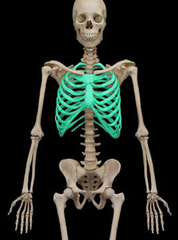
ribs/rib cage (how many total?)
Bones that form the protective framework around the thoracic cavity, protecting the heart and lungs. (12 pairs, 24 total)
Rib Head (articulates w/?)
The rounded, posterior end of a rib; articulates with the body (and costal facets) of the thoracic vertebra/vertebrae.

Rib Tubercle (articulates w/?)
A bony bump located near the rib's neck/angle; articulates with the transverse process of the thoracic vertebra.

Costal Cartilage (on ribs)
Hyaline cartilage that caps the anterior end of each rib and connects it to the sternum (or to the cartilage of another rib); allows some flexibility in the rib cage during breathing.

"costal"
An anatomical term meaning pertaining or relating to the ribs.

TRUE Ribs (pair #, how they attach)
(pairs 1-7) — Attach directly to the sternum, each via its own costal cartilage.

FALSE Ribs (pair #, how they attach)
(pairs 8-10) — Do not attach directly to the sternum; instead, their costal cartilage attaches to the costal cartilage of the rib just above (superior to) it.

FLOATING Ribs (pair #, how they attach)
(pairs 11-12) — Have no connection to the sternum at all; they only attach posteriorly to the vertebrae.

Sternum
The breastbone, a flat bone located in the center of the chest.

Manubrium
The broad, superior-most portion of the sternum; articulates with the clavicles and the first two ribs.

Sterum Body
The long, middle/largest portion of the sternum; articulates with costal cartilages of ribs 2-7 (mostly).

Xyphoid process
The small, cartilaginous (later ossified) inferior tip of the sternum; a landmark for CPR hand placement.

Jugular notch
The concave notch at the superior border of the manubrium, between the two clavicular notches.

Clavicular notches
The two depressions on either side of the jugular notch, at the top of the manubrium, where the clavicles articulate.

Sternal angle
The palpable ridge/joint where the manubrium meets the sternum body; corresponds to the level of rib 2 — an important surface landmark for counting ribs.

Erector Spinae Muscles (3)
iliocostalis, longissimus, spinalis
Iliocostalis m.
The most lateral column of the erector spinae group; runs vertically along the back closest to the angles of the ribs
(its name reflects its attachments between the ilium and the ribs/costals).
Longissimus m. (action & innervation)
The middle (intermediate) column of the erector spinae group, sandwiched between the iliocostalis (lateral) and spinalis (medial); it's the longest and largest of the three, running from the lumbar region up toward the skull.
Spinalis m. (action & innervation)
Definition: The most medial column of the erector spinae group, running directly alongside the spinous processes (its name reflects this close midline position).
Spinalis m. action (Bilaterally)
Extend head and
vertebral column
Spinalis m. action (unilaterally)
Lateral flexion of
head and vertebral
Spinalis m. Innervation
Dorsal rami of spinal nerves
Longissimus m. action (bilaterally)
Extend head and vertebral column
Longissimus m. action (unilaterally)
Lateral flexion of head and vertebral column
Longissimus m. innervation
Dorsal rami of spinal nerves
Iliocostalis m. action (bilaterally)
Extend head and vertebral column
Illiocostalis m. action (unilaterally)
Lateral flexion of head and vertebral column
Illiocostalis m. action
Dorsal rami of spinal nerves