A&P Lecture - Week 2 - Tissue & Membranes

0.0(0)
Studied by 4 people
call kaiCall Kai
learnLearn
examPractice Test
spaced repetitionSpaced Repetition
heart puzzleMatch
flashcardsFlashcards
GameKnowt Play
Card Sorting

1/215

encourage image

There's no tags or description

Looks like no tags are added yet.

Last updated 10:27 PM on 9/21/26
Name
Mastery
Learn
Test
Matching
Spaced
Call with Kai
Chat

No analytics yet

Send a link to your students to track their progress

216 Terms

1
New cards

What are the major tissue membranes?

Two broad categories: connective-tissue membranes and epithelial membranes. Connective-tissue membrane example = synovial membrane in joints. Epithelial membranes = mucous, serous, and cutaneous membranes. Cutaneous membrane = skin.

2
New cards

What are the 4 major tissue types?

Epithelial, connective, muscle, and nervous/neuronal tissue. Different structures allow each tissue to perform different functions.

3
New cards

What are the 3 embryonic germ layers and the easiest way to distinguish their derivatives?

Ectoderm = skin/epidermis + nervous system; Mesoderm = muscle, blood, bone/connective tissues; Endoderm = much of the internal respiratory/digestive lining and associated glands.

4
New cards

A neuron, epidermal cell, or pigment cell most likely came from which germ layer?

Ectoderm. Think ECTO = outside + nervous system.

5
New cards

Skeletal, cardiac, or smooth muscle most likely came from which germ layer?

Mesoderm. Mesoderm also contributes to blood and many connective tissues.

6
New cards

Lung epithelial cells or pancreatic cells are associated with which germ layer?

Endoderm. Think ENDO = internal linings and associated glands.

7
New cards

What are the main characteristics of epithelial tissue?

Tightly packed cells with little intercellular material; anchored to a basement membrane; generally avascular; replaced frequently; polarized into apical and basal/basolateral regions; functions include protection, secretion, absorption, excretion, and sensory reception.

8
New cards

What does epithelial polarity mean, and how do tight junctions relate to it?

Epithelial cells have different apical and basal/basolateral regions. Above a tight junction is considered apical; below it is basal/basolateral.

9
New cards

What is the basic job of a tight junction?

It forms a seal between neighboring epithelial cells and helps separate the apical and basal/basolateral compartments.

10
New cards

What is the basic job of a gap junction?

Communication between adjacent cells through electrical and metabolic/chemical coupling. In the heart, gap junctions help cardiac cells contract together as a unit.

11
New cards

What is the difference between desmosomes and hemidesmosomes?

Desmosomes connect cell-to-cell and use cadherins. Hemidesmosomes connect an epithelial cell to the basal lamina/extracellular matrix and use integrins.

12
New cards

Cadherin vs integrin?

Cadherin = cell-to-cell attachment. Integrin = cell-to-extracellular-matrix attachment.

13
New cards

What is important about adherens junctions?

They participate in attachment and connect internally with actin, helping influence cell shape and folding.

14
New cards

Alport syndrome vs Knobloch syndrome?

Alport = sex-linked inherited collagen disorder affecting multiple systems, especially kidneys/ears/eyes. Knobloch = autosomal recessive collagen disorder with severe eye problems and skull abnormalities such as occipital encephalocele.

15
New cards

What are the two things used to classify epithelial tissue?

Number of layers + shape of the cells.

16
New cards

What do simple, stratified, and pseudostratified mean?

Simple = one layer and all cells contact the basement membrane. Stratified = multiple layers and only the basal/deepest layer contacts the basement membrane. Pseudostratified = looks multilayered, but it is actually one layer and all cells contact the basement membrane.

17
New cards

What do squamous, cuboidal, and columnar mean?

Squamous = flat; cuboidal = cube-shaped; columnar = tall/column-shaped.

18
New cards

If every epithelial cell touches the basement membrane but the nuclei appear at different heights, what is the tissue?

Pseudostratified epithelium. It looks stratified, but all cells contact the basement membrane.

19
New cards

How do you name a stratified epithelial tissue?

Use the shape of the cells at the outermost/apical surface, not the shape of the basal cells.

20
New cards

One layer of flat cells, one layer of cube-shaped cells, and one layer of tall cells are called what?

Simple squamous, simple cuboidal, and simple columnar epithelium, respectively.

21
New cards
<p><span>Identify this junction. What does it attach?</span></p>

Identify this junction. What does it attach?

Hemidesmosome — epithelial cell → basal lamina/ECM; integrins. It anchors epithelial cells to the underlying basal lamina and extracellular matrix using integrins.

22
New cards
<p><span>Identify this junction and give its major function</span></p>

Identify this junction and give its major function

Gap junction — cell-to-cell communication; electrical/metabolic coupling.Allow ions and small molecules to pass.

23
New cards
What is a goblet cell and what does it do?
A specialized epithelial cell that secretes mucus. It is commonly found among columnar epithelial cells, including in the intestine. Mucus helps form a protective/lubricating barrier.
24
New cards
What are the structure, function, and major locations of simple squamous epithelium?
One layer of thin, flat cells. Function = diffusion/exchange. Major locations = lungs/alveoli, blood and lymph vessels, capillaries, body cavities, and filtering regions of the kidney.
25
New cards
Why is simple squamous epithelium well suited for diffusion?
It is only one thin layer of flattened cells, so the diffusion distance is very small.
26
New cards
What is endothelium?
Simple squamous epithelium that lines blood vessels.
27
New cards
What are the structure, function, and major locations of simple cuboidal epithelium?
One layer of cube-shaped cells with centrally located nuclei. Function = secretion and absorption. Major locations = kidney tubules and glands.
28
New cards
What visual clue should make me think simple cuboidal epithelium?
A single ring/layer of cube-shaped cells surrounding a lumen, usually with round centrally located nuclei.
29
New cards
If I see kidney tubules lined by one layer of cube-shaped cells, what tissue is it?
Simple cuboidal epithelium.
30
New cards
What are the structure, function, and major locations of simple columnar epithelium?
One layer of tall elongated cells. Functions = absorption and secretion. Major locations = stomach, intestines, and uterus.
31
New cards
What special structures may be associated with simple columnar epithelium?
Microvilli, goblet cells, and sometimes cilia. Microvilli increase surface area for absorption; goblet cells secrete mucus.
32
New cards
Why are microvilli useful in simple columnar epithelium?
They increase surface area, increasing the capacity for absorption.
33
New cards
If a patient has difficulty absorbing nutrients and secreting mucus in the intestine, which epithelial tissue should I think of?
Simple columnar epithelium.
34
New cards
What is the major difference between simple columnar and pseudostratified columnar epithelium?
Simple columnar has nuclei arranged more uniformly in one layer; pseudostratified looks multilayered because nuclei occur at different heights, although it is still one layer.
35
New cards
What defines pseudostratified columnar epithelium?
It looks stratified because nuclei are at different heights, but it is actually a single layer and all cells contact the basement membrane.
36
New cards
Where is ciliated pseudostratified columnar epithelium commonly found and what does it do?
Trachea and upper respiratory tract. Goblet cells produce mucus and cilia move mucus/debris toward the pharynx.
37
New cards
Where is non-ciliated pseudostratified columnar epithelium found in these slides?
Epididymis.
38
New cards
If an exam says all epithelial cells contact the basement membrane but the nuclei are at different levels, what tissue is being described?
Pseudostratified columnar epithelium.
39
New cards
What are the structure, function, and major locations of stratified squamous epithelium?
Multiple layers with flattened cells at the outer surface. Function = protection from abrasion. Locations = skin, mouth, throat, vagina, and anal canal.
40
New cards
How do I name a stratified epithelial tissue?
Look at the shape of the OUTERMOST/apical cells, not the shape of the deepest cells.
41
New cards
If a tissue has many layers, basal cells that are not flat, but flat cells at the outer surface, what is it?
Stratified squamous epithelium.
42
New cards
Why is stratified squamous epithelium suited for protection?
Its multiple layers provide resistance to friction, abrasion, and mechanical stress.
43
New cards
<p><span><em>Identify the epithelial tissue lining this vessel.</em></span></p>

Identify the epithelial tissue lining this vessel.

Simple squamous epithelium — endothelium

44
New cards
<p><span><em>Identify this epithelial tissue</em></span></p>

Identify this epithelial tissue

Simple cuboidal — kidney tubule; secretion/absorption.
45
New cards
<p><span><em>Identify the tissue and give its major function</em></span></p>

Identify the tissue and give its major function

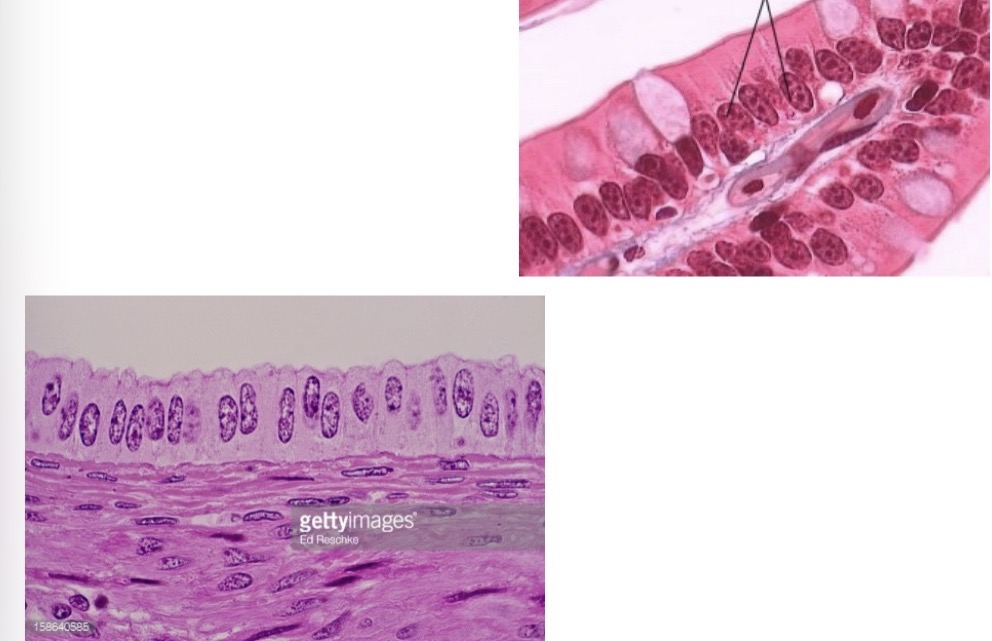
Simple columnar — absorption/secretion; GI tract; may contain goblet cells/microvilliand may be ciliated in the respiratory tract.

46
New cards
<p><span><em>Identify this tissue. What feature proves your answer?</em></span></p>

Identify this tissue. What feature proves your answer?

Pseudostratified columnar — nuclei at different heights, but all cells contact the basement membrane

47
New cards
What is the difference between keratinized and nonkeratinized stratified squamous epithelium?
Keratinized stratified squamous is found in skin and has a tough keratinized surface. Nonkeratinized stratified squamous stays moist and is found in areas such as the throat, vagina, and anal canal.
48
New cards
How do I recognize stratified squamous epithelium?
Multiple cell layers with FLAT cells at the outermost/apical surface. Function = protection from abrasion.
49
New cards
When naming any stratified epithelial tissue, which cells determine the name?
The cells at the OUTERMOST/apical surface, not the deeper basal cells.
50
New cards
What are the structure, function, and major locations of stratified cuboidal epithelium?
Usually 2–3 layers of cuboidal cells. Function = protection. Locations include ducts of sweat glands, mammary glands, salivary glands, and pancreas.
51
New cards
If I see multiple layers of cells surrounding a gland duct and the surface cells are cuboidal, what tissue is it?
Stratified cuboidal epithelium.
52
New cards
What are the structure and major locations of stratified columnar epithelium?
Several layers with columnar cells at the surface. Found in parts of the male urethra, vas deferens, and pharynx.
53
New cards
If a tissue has many layers and the OUTERMOST cells are columnar, what is it?
Stratified columnar epithelium.
54
New cards
What is the main feature of transitional epithelium?
It can stretch/distend and then return toward its original shape, so its appearance changes depending on how stretched it is.
55
New cards
What are the major locations of transitional epithelium?
Urinary bladder, ureters, and renal pelvis; the lecture also includes portions of the urethra and some prostate-associated ducts.
56
New cards
What is the classic organ associated with transitional epithelium?
The urinary bladder.
57
New cards
Why does transitional epithelium look different in an empty bladder versus a full bladder?
The tissue changes shape as it stretches; relaxed cells look thicker/rounder and become flatter when stretched.
58
New cards
If a question describes an epithelial tissue designed to stretch as an organ fills with urine, what is the answer?
Transitional epithelium.
59
New cards
What epithelial types are found along the male urethra from proximal to distal?
Near bladder = transitional; middle = columnar/pseudostratified columnar; near external opening = stratified squamous.
60
New cards
Simple squamous: structure, function, location?
One layer of flat cells; diffusion/filtration; alveoli and vessel lining.
61
New cards
Simple cuboidal: structure, function, location?
One layer of cube-shaped cells with central nuclei; secretion/absorption; kidney tubules and glands.
62
New cards
Simple columnar: structure, function, location?
One layer of tall cells; absorption/secretion; stomach and intestines, often with microvilli and goblet cells.
63
New cards
Pseudostratified columnar: structure, function, location?
Looks multilayered but all cells contact the basement membrane; secretion and movement of mucus; trachea/upper respiratory tract.
64
New cards
Match the protective epithelial tissues with their main clues.
Stratified squamous = many layers + flat surface cells, abrasion protection. Stratified cuboidal = multiple cuboidal layers, gland ducts. Stratified columnar = multiple layers + columnar surface cells, male urethra/gland ducts.
65
New cards
Match the 8 major epithelial tissues to their strongest clue.
Simple squamous = diffusion; simple cuboidal = secretion/absorption in kidney; simple columnar = GI absorption/secretion; pseudostratified = trachea/mucus/cilia; stratified squamous = abrasion protection; stratified cuboidal = gland ducts; stratified columnar = male urethra; transitional = urinary stretch.
66
New cards
<p><span><em>Identify this tissue. What feature proves it?</em></span></p>

Identify this tissue. What feature proves it?

Stratified squamous — many layers, flat outermost (top) cells.

67
New cards
<p><span><em>Identify this tissue.</em></span></p>

Identify this tissue.

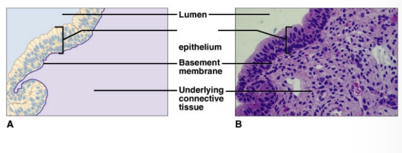
Stratified cuboidal — multiple cuboidal layers around a duct/lumen; protective.
68
New cards
<p><span><em>Identify this tissue.</em></span></p>

Identify this tissue.

Stratified columnar — multiple layers with columnar surface cells

69
New cards
<p><span><em>These images show the same tissue relaxed and stretched. Identify it and name its classic location.</em></span></p>

These images show the same tissue relaxed and stretched. Identify it and name its classic location.

Transitional epithelium — urinary bladder; stretches/distends

70
New cards
What is glandular epithelium, and what is the key difference between exocrine and endocrine glands?
Glandular epithelium produces and secretes substances. Exocrine glands release products through ducts to a surface; endocrine glands release into interstitial fluid and then blood.
71
New cards
How are exocrine glands classified?
By duct pattern and shape of the secretory portion. Simple = unbranched duct; compound = branched ducts. Secretory portions may be tubular, alveolar/acinar, or a combination.
72
New cards
How do I decode an exocrine gland name such as “compound tubular”?
First word = duct pattern; second word = secretory shape. Compound = branching ducts; tubular = tube-shaped secretory portions.
73
New cards
What is the difference between simple and compound exocrine glands?
Simple glands have a single/unbranched duct system. Compound glands have branching duct systems.
74
New cards
What is the difference between tubular and alveolar/acinar exocrine glands?
Tubular glands have tube-shaped secretory portions; alveolar/acinar glands have rounded or sac-like secretory portions.
75
New cards
What are merocrine, apocrine, and holocrine secretion?
Merocrine = secretion by exocytosis and the cell stays intact. Apocrine = the apical portion of the cell buds off with the secretion. Holocrine = the entire cell is destroyed/released and becomes part of the secretion.
76
New cards
Which secretion mode causes the least and greatest gland-cell damage?
Least damage = merocrine because the cell remains intact. Greatest damage = holocrine because the whole cell is lost. Apocrine is intermediate.
77
New cards
What is the easiest memory trick for merocrine, apocrine, and holocrine?
Mero = material exits but cell remains; Apo = a portion comes off; Holo = whole cell is lost.
78
New cards
What are useful examples of each secretion mode from the slides?
Merocrine = salivary glands, pancreatic glands, some sweat glands. Apocrine = mammary glands. Holocrine = sebaceous glands, Meibomian glands, some sweat glands.
79
New cards
If a picture says “whole cells are lost during the secretory process,” which mode is it?
Holocrine secretion.
80
New cards
What does endocrine secretion mean in this lecture?
Products are released into interstitial fluid and then diffuse into the blood; the products are hormones.
81
New cards
What are the major characteristics of connective tissue?
Connective tissue mainly supports, binds, protects, fills spaces, transports, stores, and repairs. Its defining feature is abundant extracellular matrix surrounding relatively separated cells; most connective tissues have a good blood supply, except cartilage.
82
New cards
How should I visualize connective tissue and extracellular matrix?
Professor’s Jell-O analogy: cells are like grapes and the extracellular matrix is like the Jell-O surrounding them.
83
New cards
What are the 3 broad connective-tissue groups shown in the lecture?
Connective tissue proper = loose and dense; supportive connective tissue = cartilage and bone; fluid connective tissue = blood and lymph.
84
New cards
What is adipose tissue and what are its major functions/locations?
Adipose is loose connective tissue specialized for fat storage. It is found under the skin and around organs such as the kidneys, where it provides energy storage, cushioning/protection, and helps conserve heat.
85
New cards
How can adipose tissue be recognized microscopically?
Adipocytes look large and mostly empty because they contain lipid; the fat pushes the nucleus toward the edge of the cell.
86
New cards
What are the key general characteristics of cartilage?
Cartilage is supportive connective tissue. It has poor blood supply, so it heals slowly. Cartilage cells are chondrocytes, which sit in spaces called lacunae within a gel-like extracellular matrix.
87
New cards
Why does cartilage heal slowly?
It has a poor vascular supply.
88
New cards
What is a chondrocyte, and where is it located?
A chondrocyte is a cartilage cell, and it sits inside a small space called a lacuna.
89
New cards
<p><span><em>Identify this exocrine gland from its duct pattern and secretory shape.</em></span></p>

Identify this exocrine gland from its duct pattern and secretory shape.

Name it using: simple vs compound + tubular/alveolar/acinar shape.

90
New cards
<p><span><em>Which secretion mode is shown?</em></span></p>

Which secretion mode is shown?

  • intact cell/exocytosis → merocrine

  • apical part lost → apocrine

  • whole cell lost → holocrine


91
New cards
<p><span><em>Identify this connective tissue and give one recognition clue.</em></span></p>

Identify this connective tissue and give one recognition clue.

Adipose tissue — large lipid-filled cells with nuclei displaced toward the edge.

92
New cards
What are the 3 types of cartilage and the best location clues for each?
Hyaline = trachea, fetal skeleton, epiphyseal/growth plate, ends of bones, nose/ribs. Fibrocartilage = pubic symphysis, intervertebral discs, TMJ. Elastic = epiglottis and external ear/pinna.
93
New cards
Which cartilage is the most common and where is it found?
Hyaline cartilage. It is found at the ends of bones, in the trachea/respiratory passages, fetal skeleton, epiphyseal growth plate, nose, and ribs.
94
New cards
Which cartilage is associated with the pubic symphysis, intervertebral discs, and TMJ?
Fibrocartilage.
95
New cards
What is the main function of fibrocartilage?
It is compressible and absorbs pressure.
96
New cards
Which cartilage is associated with the external ear and epiglottis?
Elastic cartilage.
97
New cards
What is the flexibility ranking of cartilage from most to least flexible?
Elastic > Hyaline > Fibrocartilage.
98
New cards
If an exam asks which cartilage is at the tip of the nose, what should I think of?
Hyaline cartilage.
99
New cards
What is a chondrocyte and where is it found?
A chondrocyte is a cartilage cell, and it sits in a space called a lacuna within the extracellular matrix.
100
New cards
Why does cartilage heal slowly?
Cartilage has a poor blood supply.



