Exam 2 Study Guide
 Call Kai
 Learn
 Practice Test
 Spaced Repetition
 Match
 Flashcards
 Knowt Play
1/110

There's no tags or description
Looks like no tags are added yet.
111 Terms
transduction
the process of converting one form of energy into another. Refers to changing physical energy into electrical/chemical signals (neural impulses) that can make their way into the brain.
→ ex) your ears receive energy (sound waves) and transduce (or convert) this energy into neural messages that make their way to your braina nd are processed as sounds
→ The process through which environmental energy is transformed into neural impulses
nociception
perception of pain, temperature, and itch.
hapsis
perceptual ability to discriminate objects on the basis of touch.
fine touch, pressure
proprioception
perception of the position and movement of the body, limbs, and head.
body awareness
initiating a motor sequence
1) prefrontal cortex plans movement
2) premotor cortex organizes movement sequences
3) motor cortex produces specific movements
motor sequence
movement modules preprogrammed by the brain and produced as a unit.
homunculus
a distorted map of the human body laid across the surface of the brain
2 elements:
1) topographical organization (neurons that respond to neighboring body parts are next to each other (i.e., toes are next to feet)
2) size of the region in the somatosensory cortex and extent of sensitivity (a large part of the cortex is dedicated to the lips even though the lips are so small because there is a large number of sensory receptors on the lips)

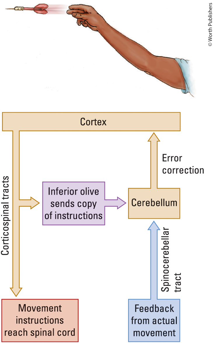
cerebellum (in movement)
error correction in motor movements
motor cortex sends information to the spinal cord; an afferent copy goes to the cerebellum. Cerebellum integrates between intended and actual signals and sends it back to the neocortex to adjust motor signals (for a free throw, making the shot not go short next time)
ensures motor sequences are executed properly and appropriate to the task goals
rods
more numerous than cones, more sensitive to dim light. Mainly used for night vision, low threshold for activation
cones
responsive to bright light, responsible for color vision and our ability to see fine detail
color blindness
Loss of one or more types of cone photoreceptors (one, two, or all types of cone photoreceptors)
the most common color blindness is..
red-green colorblindness; the red or green photoreceptor gene is broken, resulting in individuals who struggle with red and green differentiation
→ 1:100 males have this colorblindness
→ 1:10000 females have this colorblindness
ganglion cells
a cell found in a ganglion (a cluster of neurons in the peripheral nervous system)
(ganglion cell) magnocellular cell (m-cells)
magno-, large
receives input primarily from rods
sensitive to light and moving stimuli
large receptive fields, lower density
contribute to perceptions of space and motion
(ganglion cell) parvocellular cell (p-cell)
parvo-, small
receives input primarily from cones
sensitive to color
ideally suited for the creation of object-detailed vision
high density - smaller receptive fields
primary visual cortex (V1, striate cortex)
Striate, layering
Two visual paths emerge from the striate cortex
→ one route goes to vision-related regions of the parietal lobes
→ one route goes to vision-related regions of the temporal lobes
first location that processes visual information in the occipital lobe
People who cannot see can still react to visual stimuli (orientation)
receptive fields
the specific region of sensory space (such as a patch of skin, a volume of auditory space, or an area of the visual field) in which the presence of a stimulus changes the electrical activity or firing rate (action potentials) of a given sensory neuron
simple cells
located in the primary visual cortex in the occipital lobe of the neocortex
Receptive field where a response is elicited for a neuron
Retinal ganglion cells all send convergent input to a simple cell. Lower-order neurons to higher-order neurons
in lower simple cells, respond to bars of light vs. dots of light
complex cells
smaller receptive fields, respond to a bar of light in specific orientations, and respond best to motion in a particular direction
dorsal stream (of vision)
visual processing pathway from region V1 to the parietal lobe; guides actions relative to objects.
ventral stream (of vision)
visual processing pathway from region V1 to the temporal lobe that mediates perception, as opposed to action mediated by the dorsal stream.
learning
a change in an organism’s behavior as a result of experience
pain pathway
the neural circuit that detects, transmits, and processes noxious (tissue-damaging or potentially damaging) stimuli from the body to the brain, converting them into the conscious experience of pain
binding problem
philosophical question focused on how the brain ties single and varied sensory and motor events together into a unified perception or behavior.
The brain does not process an object like a whole picture in one single location. Instead, different regions of the brain specialize in different attributes
declarative memory
a type of long-term memory that involves the conscious, intentional recollection of facts, events, and general knowledge that can be verbally described or "declared"
Conscious memory, effort to recall, effortful, information you need to apply
(declarative memory) episodic
events that have happened to you, events marked as important
(declarative memory) semantic
facts and general knowledge of the world
non-declarative memory
a type of long-term memory that stores skills, habits, priming effects, and conditioned responses that operate without conscious recall
Implicit memory, memory in your brain that you don’t have to put effort into remembering it, applied but no effort needed to use it
(non-declarative memory) priming
unconscious association in your mind
consolidation
Process of stabilizing a memory trace after learning.
the process by which a memory becomes hippocampal-independent
reconsolidation
restabilizing a memory trace after the memory is revisited
short-term memory
few minutes
information is held in memory only briefly, then discarded; involves the frontal lobes
long-term memory
indefinite duration
information is held in memory indefinitely, perhaps for a lifetime; involves the temporal lobe
HM-henry molaison
Henry suffered from severe, debilitating epileptic seizures starting in his childhood (following a bicycle accident at age 7 or 9). By age 27, medications could no longer control his seizures.
On September 1, 1953, neurosurgeon Dr. William Beecher Scoville performed an experimental bilateral medial temporal lobectomy. Surgeons removed the hippocampus and most of the amygdala complex
had trouble forming new memories, but retained information for moments to moments (anterograde amnesia)
memory each uses specific different brain sections
Semantic memory is different from procedural memory
retained learned skills but couldn’t remember how he learned them
motor skills, language skills, perception, intellect intact
lost explicit memory
place cells
hippocampal neurons whose response properties are where the animal is in space
emotional memory
acquired memory
memory for the affective properties of stimuli or events
can be implicit or explicit
amygdala is critical for emotional memory
damage to the amygdala abolishes memory but has little effect on implicit or explicit memory
long-term potentiation (LTP)
long-lasting increase in synaptic effectiveness after high-frequency stimulation.
spatial memory
the cognitive system that allows you to encode, store, and retrieve information about the physical layout of your environment, object locations, and navigational routes
long-term depression (LTD)
Long-lasting decrease in synaptic effectiveness after low-frequency electrical stimulation.
response properties
the specific, measurable characteristics of how a neuron, a neural circuit, or a brain region reacts to a defined internal or external stimulus
periaqueductal gray matter (PAG)
Nuclei in the midbrain that surround the cerebral aqueduct joining the third and fourth ventricles; PAG neurons contain circuits for species-typical behaviors (e.g., female sexual behavior) and play an important role in the modulation of pain.
leads to descending inhibition of a signal that will inhibit the pain pathway
→ electrical stimulation of the PAG suppresses pain
→ PAG neurons produce their pain-suppressing effects by exciting pathways in the brainstem that project to the spinal cord, where they inhibit neurons that form the ascending pain pathways
Intention, Action, Feedback chart
Karl Lashley (1890 - 1958)
how do rats get the memory to learn a maze
if he lesions the brain of the rat and can't find food previously, it means that's where memory is stored solely
equipotentiality
more wrong than right; when one area is damaged, the area next to it will take over all the functions it had
mass action
more right than wrong; the larger the legion, the more impairment you will experience
two memory tasks for monkeys
with parenthetical lesions monkeys can’t do object discrimination but can do spacial
with hippocampal lesions monkeys can’t do spacial but object discrimination
Inferring spacial memory
Size of the hippocampi in both birds and mammals appears to be related to the cognitive demands of two highly spatial activities: foraging for and storing food.
Posterior region of the hippocampus in London taxi drivers is significantly larger than the same region in the control participants.
effects of unilateral somatosensory system damage
Hemisection cut (hemi = half)
If you completely cut the spinal cord, you’ll lose all somatosensory feelings in all sensation in the legs/motor control in the legs
Spinal cord is a huge connection to the brain
Lose touch sensation from the left side of the body; lose pain sensation from the right side of the body
Pain crosses at the bottom, and touch doesn’t cross till medula
Soft touch stays on the same side during ascension and crosses over the medula/brainstem

receptive field hierarchy

mechanoreceptors
peripheral receptors that respond to mechanical energy, like pressure. When the resting ion channel is closed, when pressure happens, it stretches the filaments that lead to the ion channel opening
touch
kiss, punch in the face, etc
hearing
region of inner ear called cochlea, hair cells. Anytime a sound is produced, it changes the patterns of air pressure.
chemical signaling
chemical doesn’t equal energy
smell
olfactory nerves breathe in chemicals that bind to receptors that activate nerves to the brain. Odor molecules bind to receptors, opening ion channels that lead to action potentials being developed
taste
contact, gustatory system
photoreceptors
Relationship with vision. Electromagnetic energy in the environment that reacts with photoreceptors. Opsins. Light reaches the rhodopsin in the retina
heuristics
general rules that navigate how the world works
haptic neuron of the posterior root ganglion
1) posterior-root ganglion neurons carry fine-touch and pressure information
2) have large, myelinated axons whose receptors are located in the skin, muscles, and tendons
3) the cell body is located in a posterior-root ganglion
4) fine-touch and pressure axons ascend in the ipsilateral spinal cord, forming the posterior spinothalamic tract
crossing contralateral
the anatomical structure where nerve fibers cross the body's midline to connect one side of the brain with the opposite side of the body
→ primary recipient to ascending somatosensory to the thalamus
thalamus
crucial relay status for ascending sensory input before they reach the neocortex
→ signal is progressing from lower-order processing regions to the higher-order processing regions (neocortex)
topographic organization
similar regions on a sensory space are processed by neighboring regions within the sensory space; these regions are processed by regions in the brain
retinal ganglion cell (RGC)
One of a group of retinal neurons with axons that give rise to the optic nerve.
model of convergence output
retinal ganglion cells (large number in the retina, dots of light) → LGN (dots of light) → V1 simple cells (smaller numbers, bars of light) → V1 complex cells (one, bars of light moving)
somatosensation, subjective nature
2 people can experience the same trauma to the same part of the body and experience different levels of pain
feedback pathways
Producing sounds makes somatosensory feedback (how good does this sound compared to the model sound?)
auditory signal
The auditory system is part of the speech pathway
Needs somatosensory feedback to represent which motor system is working
Speech is not trivial
visible light
Light is electromagnetic energy that we see
Range of electromagnetic energy visible to humans
→ about 400 nanometers (violet) to 700 nanometers (red)
nanometer (nm)
one-billionth of a meter
wavelength
the distance between the peak of one wave and the peak of the next
gamma rays
very small, radiation of an atomic bomb, potent genetic mutation potential
radio waves
long, small
x-rays
small, less dangerous than gamma rays but still can cause genetic mutation
ultraviolet rays
can’t see them, genetic mutations, mild
infrared waves - radio waves
longer, harmless
tetrachromatids
4 different kinds of photoreceptors that have different kinds of photo sensitivities, breaks down into smaller pieces
lateral geniculate nucleus (LGN)
6 layers; the first 2 are where m-cells will synapse; p-cells synapse in 3-6. Leads to the extraction of object information and m-cells space and motion
geniculostraite system
visual information → eye → optic nerve → LGN → v1 → other visual cortical areas
tectopulvinar system
visual information → eye → optic nerve → superior colliculus → pulvinar → other visual cortical areas
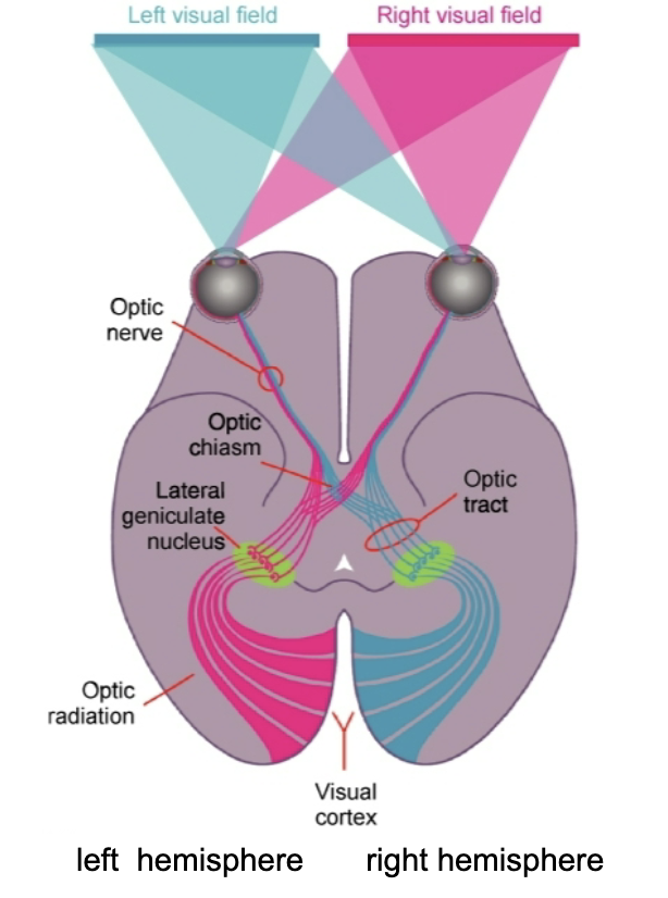
functional organization of the visual system
Visual field is the environment of space that you can see with your eyes at a given moment: 200-220 degrees
Right and left visual field information goes to both eyes
Activates neurons on the temporal part of the eye -> reaches the optic chiasm (crossing)
The retinal ganglion cell axons that have been activated from the opposite eye will project to that side
Left visual field goes to the right side / right visual field goes to the left side
→ mixed in cortex
nasal portion of eye and activates neurons on the temporal side of the other eye
reaches optic chiasm of brain; synapses on other side of LGN
binocular overlap
where both eyes are seeing the same space
because both eyes are separated, where objects fall on one retina it might be a different location in the other retina
Extent of that difference depends on how close/far away from the stimuli you are
Disparity neurons cause us to construct our three dimensional experiences
serial processing in parallel pathways
6 layers organization, same in every mammal
2,3,5,6 → output layers
2,3 → other neocortical regions
5,6 → out of the neocortex
1 → neuropill, axon and dendrites come together
Primary sensory cortex: 4 → largest layer, lots of ascending sensory input
Motor cortex: 4 → tiny, doesn’t receive asending motor stuff
association cortex
convenience, compromise, thought, language, memory, cognitive
retinotopy
radial component
on-off receptivity
Response properties of neurons; what types of light information they contain; the idea of neurons working/not working in the visual system
Defining the response properties of visually sensitive neurons
Two elements
1) where does light need to be in the visual field for a neuron to respond
2) What are the properties of that light
For retinal ganglion cells, they are small
Restricting light in the center of the field has you look for action potentials
Direct light to the outside of the receptive field -> inhibition of action potentials
A response can be excitatory or inhibitory response
Classical response property of a retinal ganglion cell responses to dots of light in a visual field, where light in the center of the receptive field produces excitation, and on the outside it’s inhibition
Code of visual reception

pole of the temporal lobe
responds to clocks, hands, real objects
attention
shifting saline visual stimuli, colorful, large, surprising things, orientation
ataxia
have trouble coordinating movements in visual space (initiating voluntary eye movements) (such as following a physician’s finger)
apraxia
inaccurate arm pointing (a patient can see an object, but is unable to pick it up)
attentional
constriction of the visual field (ask a patient to look at a parking lot, and all she sees is a lamppost or a car)
agnosia
not knowing
visual form agnosia
inability to recognize drawings of objects
color agnosia (achromatopsia)
inability to recognize colors
face agnosia (prosopagnosia)
inability to recognize faces
memory trace
a mental representation of a previous experience, physical change in the brain involving synapses
emotional memory-amygdala
assigns emotions to memory - amygdala
when recalling, you can feel similar to the way you felt when the event happened
episodic memory
autobiographical memory for events pegged to specific place and time contexts
episodic amnesia
inability to recall any personal experience or think about future experiences
associated with frontal lobe injury or reduced blood flow to the frontal lobes
frontal lobes may allow us to mentally travel through our past
retrograde amnesia
cannot remember events prior to brain damage
anterograde amnesia
cannot later remember events that occur after brain damage
distributed reinstatement theory
a learning episode rapidly produces a stored memory representation that is strong in the hippocampus
the memory is replayed on the time scale of hours or days after learning, learning to enhance representations outside the hippocampus