Anatomy & Physiology: Skeletal, Muscular, Nervous & Thoracic Systems
 Call Kai
 Learn
Learn Practice Test
 Spaced Repetition
 Match
Match Flashcards
 Knowt Play
1/342

There's no tags or description
Looks like no tags are added yet.
Name | Mastery | Learn | Test | Matching | Spaced | Call with Kai | Chat |
|---|
No analytics yet
Send a link to your students to track their progress
343 Terms
Frontal Bone
Cranial bone forming the forehead and roof of the eye sockets.

Parietal Bones (2)
Paired cranial bones forming the sides and roof of the cranium.

Temporal Bones (2)
Paired cranial bones situated at the sides and base of the skull.

Occipital Bone
Cranial bone forming the rear and base of the skull.

Sphenoid Bone
Cranial bone situated at the base of the skull behind the eye.

Ethmoid Bone
Cranial bone separating the nasal cavity from the brain.

Maxillae (2)
Paired upper jaw bones forming the hard palate and upper teeth sockets.

Mandible
Lower jaw bone, the largest and strongest bone of the face.

Zygomatic Bones (2)
Paired cheekbones articulating with the maxilla and temporal bones.

Nasal Bones (2)
Paired small bones forming the bridge of the nose.

Lacrimal Bones (2)
Paired small, fragile facial bones forming part of the front inner wall of each eye socket.

Palatine Bones (2)
Paired facial bones forming the back part of the hard palate.

Vomer
Unpaired facial bone forming the lower posterior part of the nasal septum.

Inferior Nasal Conchae (2)
Paired scroll-like facial bones extending horizontally inside the nasal cavity.

Coronal Suture
Skull suture connecting the frontal bone to the parietal bones.

Sagittal Suture
Skull suture connecting the two parietal bones along the midline.

Lambdoid Suture
Skull suture connecting the parietal bones to the occipital bone.

Squamous Suture
Skull suture connecting the temporal bone to the parietal bone.

Hyoid Bone
U-shaped bone in the neck that supports the tongue without direct bone articulation.

Malleus
(1) Auditory ossicle (hammer) attached to the tympanic membrane.
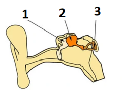
Incus
(2) Auditory ossicle (anvil) located between the malleus and stapes.

Stapes
(3) Auditory ossicle (stirrup) transmitting vibrations to the oval window.

Clavicle
S-shaped collarbone connecting the sternum to the scapula.

Scapula
Triangular shoulder blade connecting the humerus and clavicle.

Humerus
Upper arm bone extending from the shoulder to the elbow.

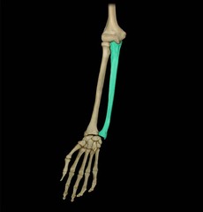
Radius
Lateral forearm bone on the thumb side.

Ulna
Medial forearm bone on the pinky side.
Metacarpals (I-V)
Five bones forming the palm of the hand.

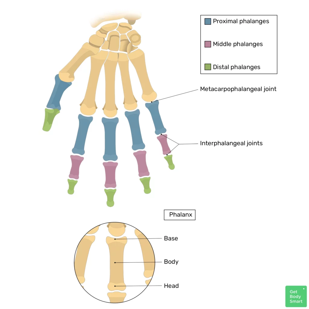
Phalanges Hand Bones
I - Proximal, distal (aka, pollux)
II - proximal, middle, distal
III - proximal, middle, distal
IV - proximal, middle, distal
V - proximal, middle, distal
Ilium
Part of the hip bone and articulates with the sacrum.

Ischium
The bottom part of the hip bone posterior to pubis.

Pubis
The bottom part of the hip bone anterior to ischium.

Femur
Proximal End
Shaft
Distal End

Patella
Known as the kneecap

Tibia
paired with a smaller fibula

Calcaneus
The heel bone of the foot.

Talus
The saddle-shaped bone in the ankle that connects the lower leg to the foot.

Metatarsals (I-V) Foot
Located in the midfoot region and the big toe starts as I while the pinky ends as V.

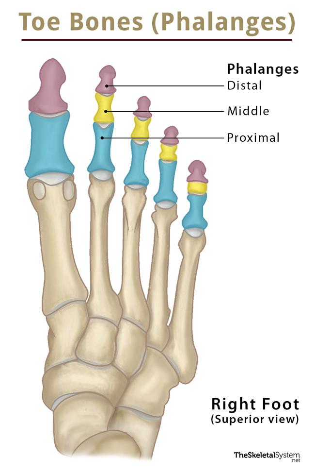
Phalanges Foot
The bones that make up the toes. As always proximal to middle to distal. The big toe only has proximal and distal.
Sternoclavicular (SC) Joint
Articulation between the sternum and clavicle.

Acromioclavicular (AC) Joint
Articulation between the acromion of the scapula and the clavicle.

Glenohumeral Joint
Ball-and-socket shoulder joint between the glenoid fossa and humerus head.

Scapulothoracic Articulation
Functional physiological articulation between the scapula and the thoracic cage.

Elbow Joint
Compound joint comprising the humeroulnar and humeroradial articulations.
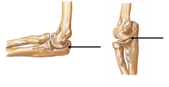
Radioulnar Joints
Proximal and distal joints allowing supination and pronation of the forearm.

Radiocarpal (Wrist) Joint
Condyloid articulation between the radius/articular disc and proximal carpals.

Carpometacarpal (CMC) Joints
Articulations between the distal carpals and metacarpals, notably the thumb saddle joint.

Synovial Joint Structure
Features include a joint capsule, synovial membrane, and articular cartilage.

Cervical Vertebrae (C1-C7)
Seven vertebrae comprising the neck region of the spinal column.

Thoracic Vertebrae (T1-T12)
Twelve vertebrae in the chest region articulating with ribs.

Lumbar Vertebrae (L1-L5)
Five large vertebrae in the lower back supporting major weight.

Sacrum
Triangular bone formed by five fused vertebrae at the base of the spine.

Coccyx
The tailbone, formed by four fused small vertebrae.

Vertebral Column Curvatures
Primary curvatures (thoracic and sacral) and secondary curvatures (cervical and lumbar).

Intervertebral Discs
Fibrocartilaginous pads composed of an annulus fibrosis and nucleus pulposus.

Herniated Disc
protrusion of a fragmented intervertebral disc in the intervertebral foramen with potential compression of a nerve

Trapezius
Superficial back muscle elevating, depressing, and retracting the scapula.

Latissimus Dorsi
Superficial back muscle extending, adducting, and medially rotating the arm.

Conus Medullaris
Tapered terminal end of the spinal cord.

Cauda Equina
Bundle of spinal nerves and nerve roots occupying the vertebral canal below the conus medullaris.

Brachial Plexus
Network of nerve fibers formed by spinal roots supplying the upper limb.

Subclavian Artery
Major blood vessel supplying blood to the head and upper limbs.

Median Cubital Vein
Superficial forearm vein commonly used for venipuncture.

True Ribs (1-7)
Vertebrosternal ribs attaching directly to the sternum via costal cartilage.

False Ribs (8-12)
Vertebrochondral ribs attaching indirectly or not at all to the sternum.

Floating Ribs (11-12)
Ribs with no anterior attachment to the sternum.

Mediastinum
Central compartment of the thoracic cavity between the pleural sacs.
Superior border: T1 to jugular notch of manubrium
Inferior border: T4 to diaphragm

Tricuspid Valve (Right AV)
Atrioventricular valve located between the right atrium and right ventricle.

Mitral Valve (Bicuspid/Left AV)
Atrioventricular valve located between the left atrium and left ventricle.

Chordae Tendineae
Fibrous cords anchoring AV valve cusps to papillary muscles.

Coronary Sinus
Large venous channel collecting deoxygenated blood from the heart wall into the right atrium.

Hemothorax
blood accumulation

Pleural Effusion
Fluid (non-blood) accumulatesin the pleural cavity

Vertebral body
Thick, oval-shaped anterior portion of the vertebra that bears weight.

Vertebral arch
Posterior structure formed by pedicles and laminae that encloses the vertebral foramen.

Vertebral foramen
Opening formed by the vertebral body and arch that contains the spinal cord.

Spinous process
Posterior bony projection pointing backward from the vertebral arch.

Transverse process
Lateral bony projection extending from the junction of the pedicle and lamina.

Superior articular processes
Projections upward from a vertebra to articulate with the vertebra above.

Inferior articular processes
Projections downward from a vertebra to articulate with the vertebra below.

Intervertebral foramen
Opening formed by adjacent vertebrae for the passage of a spinal nerve.

Transverse foramina
Openings found in cervical vertebrae for vertebral vessels.

Costal facets
Articulation sites for ribs found on thoracic vertebrae.

Lordosis
An inward curve of the lumbar spine, commonly known as swayback.

Kyphosis
An excessive outward curve of the thoracic spine, commonly known as hunchback.

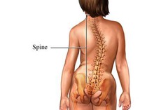
Scoliosis
A sideways curvature of the spine often diagnosed during adolescence.
Levator Scapulae
Muscle of the back and neck that elevates the scapula.

Rhomboid Minor
Superficial back muscle that retracts and rotates the scapula downward.

Rhomboid Major
Superficial back muscle situated below the rhomboid minor that retracts and stabilizes the scapula.

Serratus Posterior Superior
Superficial back muscle that elevates the upper ribs during inspiration.

Serratus Posterior Inferior
Superficial back muscle that depresses the lower ribs during expiration.

Erector Spinae
Large muscle group of the back that extends and laterally flexes the vertebral column.

Iliocostalis
Lateral column of the erector spinae muscle group.

Longissimus
Intermediate and largest column of the erector spinae muscle group.

Spinalis
Medial column of the erector spinae muscle group closest to the spine.

Transversospinalis Group
Deep muscle group of the back involved in stabilization, extension, and rotation of the spine.
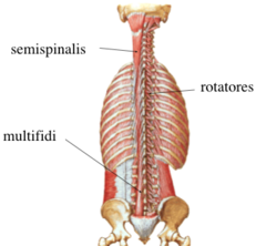
Semispinalis
Superficial muscle of the transversospinalis group spanning multiple vertebrae.

Multifidus
Intermediate muscle of the transversospinalis group spanning 2 to 4 vertebrae to stabilize the spine.

Rotatores
Deepest muscles of the transversospinalis group spanning adjacent vertebrae for rotation and proprioception.

Origin
The attachment site of a muscle's tendon to the more stationary bone.