Sono Sect Anatomy/Abdom Physiology (Urinary, Adrenal , & Retro)
 Call Kai
 Learn
 Practice Test
 Spaced Repetition
 Match
 Flashcards
 Knowt Play
1/64

There's no tags or description
Looks like no tags are added yet.
65 Terms
What is the serous membrane that forms the lining of the abdominal cavity and covers most of the abdominal organs. It consists of two layers.
Peritoneum
What are the 2 layers of the peritoneum and what is between them?
Parietal peritoneum (outer layer): lines the abdominal wall
Visceral peritoneum (inner layer): covers the abdominal organs
The space between the 2 layers is the peritoneal cavity
What are the 2 peritoneal cavity compartments called?
Lesser and greater sac
What is the retroperitoneum and what organs are within it?
The posterior area of the abdominopelvic cavity, located posterior to the peritoneal lining.
ORGANS:
Kidneys
Adrenal glands
Ureters
Pancreas
Aorta
IVC
Urinary bladder
Prostate gland
Ascending/Descending colon
Most of the duodenum
What is the shape of the aorta? Where does it originate from? What is the physiologic function?
Candy cane
Left ventricle of the heart
Transport oxygenated blood from the heart to the body
For the aorta, which direction does the vessel and blood flow? How many sections and their names?
Superior to inferior
4 sections (thoracic, proximal, mid, distal)
For the aorta, What are the 3 types of AAA discussed? Can atherosclerosis plaque develop within stents?
Fusiform, saccular, and dissecting
Yes
What is the sonographic appearance of the aorta?
Anechoic lumen with bright, ehcogenic walls that remains the same size and circular shape, may appear pulsatile, on the left side of the spine
What does the crus of the diaphragm do during inspiration?
Descends to increase thoracic volume and draw in air
What is the physiologic function of the IVC? Which direction does the vessel and blood flow?
Transports deoxygenated blood from he body to the heart
Inferior to superior
For the IVC, how many sections are there and what are the names? What causes dilation?
4 sections (hepatic, prerenal, renal, postrenal
Cardiac failure and fluid overload from CHF
What are the reasons for displacement for the IVC?
Liver masses, right renal artery aneurysm, lymphadenopathy, tortuous aorta, right renal or adrenal mass
What is the sonographic appearance of the IVC?
Anechoic lumen with this bright, echogenic walls that changes shape and size (due to blood flow changes with respiration), on the right dies of the spine
Are the aorta and IVC peritoneal or retroperitoneal? What is the anatomic position of each in relation to the spine?
Both are retroperitoneal, aorta is left of the spine, the IVC to the right
Are the kidneys and ureters located in the peritoneum or retroperitoneum?
Retroperitoneum
What 3 structures are at the kidney hilum and how do they lay?
Anterior to posterior
The renal vein, renal artery, and ureter
What is Morison’s pouch?
Peritoneal space between liver and right kidney, potentially can be fluid filled
Describe the location of the adrenal glands in relationship to the kidneys.
Superior and medial to the respective kidney
The ureters pass _________ to the psoas muscle.
Anterior
Where do the ureters begin?
At the renal pelvis
What is the length of the ureters? Diameter?
25-30cm long, 4-7mm diameter
What is the trigone?
The portion of the bladder that corresponds with the urethral orifices (where the ureter attaches) and the urethral insertion, forming a triangle
What is the mechanism called that transports urine from the kidneys to the bladder?
Peristalsis
Where does the bladder sit in respect to the reproductive organs? Male? Female?
Male: anterior to the seminal vesicles, rectum, and superior to the prostate gland
Female: anterior to the vagina, posterior cul-de-sac, and rectum
How thick should the bladder wall be?
3-6mm
What is the purpose of the urethra?
Membranous canal that allows urine to leave the bladder
What is the length of the urethra? Male? Female?
Male: 20 cm
Female: 3.5 cm
What measurements are considered normal size for the kidney? When may it decrease and increase in size?
9-12 cm length, 2.5-4 cm depth, 4-6 cm diameter
Decrease in size: age, decreased blood flow, disease
Increase in size: one is not functioning
How many poles does the kidney have and what are they called?
3 poles (upper, mid, lower)
What covers the kidneys?
4 layers: a fibrous capsule, perirenal fat, Gerota fascia, Pararenal fat (for cushion)
What are the 2 main parts of the kidney?
Renal parenchyma and renal sinus
What is the information about the renal parenchyma?
The cortex is the outer portion which contains the nephrons.
The medulla is the inner portion that contains the loops of Henle, where filtration/reabsorption occurs
What is the information given on the renal sinus?
Central portion of kidney, contains the collecting system and renal hilum
What structures are located anteriorly to right kidney?
Rt adrenal gland, rt liver lobe, 2nd part of duodenum, hepatic fixture, jejunum
What structures are located anteriorly to left kidney?
Tail of pancreas, left adrenal gland, spleen, jejunum, stomach, splenic flexure
What is posterior to the kidenys?
Diaphragm, psoas muscle, transverse muscle, quadratus lumborum muscle
What is the arterial branching for the kidneys?
Aorta - renal artery - interlobar artery - arcuate artery - interlobular artery - afferent artery
What is resistive index used to evaluate? (3 things)
Renal transplant rejection, medical renal disease, and low resistance flow
Normally, should the kidneys have high-resistive flow or low-resistive flow?
Low-resistive flow
Renal dysfunction results in a loss of _________ flow that results in a ____________________ waveform.
Diastolic, higher resistance
What is the formula for figuring RI?
Peak systolic frequency - end diastolic frequency / peak systolic frequency
Normal resistive index is typically _______.
<7
What are the symptoms for renal artery stenosis?
Sudden onset hypertension, uncontrollable hypertension
Significant renal artery stenosis may cause what change in the kidney?
Decreased renal size less than 9cm in length
How do you distinguish between direct and indirect evaluation of the renal arteries?
Direct: take measurement 3 locations (prox, mid, distal), renal art/Ao (RAR), greater than 3.5 peak systolic velocity (PSV) greater than 200m/s (60% RAS)
Indirect: intrarenal wave form evaluation
What are the formulas for determining renal artery stenosis as well as what number is considered abnormal?
Renal artery/aorta
It is abnormal when greater than 3.5 and when PSV is greater than 200 m/s
What is considered the functional unit of kidney?
Nephron
What is the functional process of the nephron?
Osmosis
What body system do the kidneys belong to?
Excretory system
How do the kidneys help to maintain homeostasis?
Excrete metabolic waste, maintain blood volume
What are the lab values done to evaluate kidneys?
Blood urea nitrogen (BUN)
Creatinine (Cr)
Glomerular filtration rate (GFR)
What is the renal echogenicity of the different parts of the kidneys , ureters, and urinary bladder?
Renal Capsule: highly reflective thin line along the periphery
Renal Cortex: homogeneous, hypoechoic or isoechoic to liver/spleen
Renal Medulla: anechoic when urine filled, triangular shaped
Renal Sinus: echogenic ovoid center with irregular borders
Ureters: not normally visualized, can identify urine entering the bladder with color Doppler
Urinary Bladder: anechoic when filled with fluid
What are the normal variants and define them?
Dromedary Hump: bulge on the lateral border of kidney (left more common)
Hypertrophied Column of Bertin: extension of the cortical tissue into the renal sinus
Double Collecting System: division of the renal sinus each will have own renal pelvis (double ureter)
Horseshoe Kidneys: connected at lower pole (isthmus) anterior to aorta lower in abdomen then normal
Renal Ectopia: 1 or both kidneys outside of normal fossa (pelvic kidney most common)
Who are the adrenal glands easiest to identify in?
Fetuses and young children
What is the sonographic appearance of adrenal glands in neonate and adults?
Adult: Small, low-gray structures (hypoechoic to adjacent anatomy)
Neonate: To have a thin hyperechoic core surrounded by thick anechoic zone
What is the normal size of the adult adrenal gland?
2 × 1.1 x .4in.
What is the name of the fascia that surrounds the adrenal gland?
Gerota fascia
The adrenal gland is made up of 2 main parts. What are the names of these parts and their function?
Outer cortex: produces steriod hormones aka corticoids
Inner cortex: secretes norepinephrine and epinephrine
Where does the adrenal gland get its vasculature from?
Supplied by the suprarenal arteries
Drained by the suprarenal veins
The right adrenal gland will drain into what vessel?
Suprarenal vein to IVC
The left adrenal gland will drain into what vessel?
Suprarenal vein to left renal vein then to IVC

What is this normal variant of the kidney?
Dromedary Hump

What is this normal variant of the kidney?
Hypertrophied Column of Bertin

What is this normal variant of the kidney?
Double Collecting System
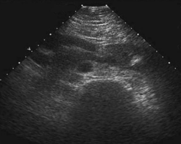
What is this normal variant of the kidney?
Horseshoe Kidney?