0. Post Seg Exam 2 - Know These
 Call Kai
 Learn
Learn Practice Test
 Spaced Repetition
 Match
Match Flashcards
 Knowt Play
1/50

There's no tags or description
Looks like no tags are added yet.
Name | Mastery | Learn | Test | Matching | Spaced | Call with Kai | Chat |
|---|
No analytics yet
Send a link to your students to track their progress
51 Terms
Conditions that can cause MNV (CNV):
Choroidal Rupture,
Histoplasmosis,
Best’s Disease,
Angioid Streaks,
Lacquer Cracks,
AMD

Diseases that can cause Angioid Streaks:
Pseudoxanthoma Elasticum,
Ehler-Danlos Syndrome,
Paget’s Disease of Bone,
Sickle Cell Disease,
Idiopathic

(lacquer cracks/ angioid streaks) are due to high myopia
lacquer cracks

(lacquer cracks/ angioid streaks) are due to disease
angioid streaks

Stargardt Disease think:
excessive lipofuscin in RPE
Best’s Vitelliform Dystrophy think:
Egg Yolk, Abnormal Arden Ratio (abnormal EOG)
Dominant Drusen/ Doyne Honeycomb think:
AMD presentation in young folk
Progressive Cone Dystrophy think:
progressive loss of cones by 30 yo, maculopathy
Rod Monochromatism think:
No Cones (lack of cones) from birth, non-recordable ERG
Retinitis Pigmentosa think:
nyctalopia,
arterial attenuation,
waxy ON pallor,
bony spicules (unless sine pimento),
systemic disease? (Usher’s, Bardet-Biedl, Leber’s),
reduced ERG
Congenital Stationary Night Blindness (CSNB) think:
STABLE/ non-progressive nyctalopia,
collection of genetic diseases,
Fundus Albipunctatus,
Oguchi Disease
Fundus Albipunctatus think:
congenital stationary night blindness (CSNB),
dull, peripheral, white spots,
abnormal → normal scotopic ffERG (after full dark adaptation)
Oguchi Disease think:
congenital stationary night blindness (CSNB),
mizuo phenomenon (unremarkable fundus when dark adapted, yellow/ iridescent fundus after light exposure)
Choroidemia think:
nyctalopia,
progressive VF constriction,
progressive RPE/chorioretinal deterioration + pigment clumping,
X-linked
Gyrate Atrophy think:
nyctalopia,
progressive chorioretinal deterioration,
progressive VF constriction,
scalloped RPE
OAT enzyme deficiency (autosomal recessive)
cRORA indicates:
Geographic Atrophy

iRORA indicates:
pre-GA / AMD warning sign

Wet AMD Neo:
(Type 1/ Type 2) is well-demarcated hyper-fluorescence on FA
Type 2

Wet AMD Neo:
(Type 1/ Type 2) is an undefined source of hyper-fluorescence on FA
Type 1

Wet AMD Neo:
Prevalence of Polypoidal Choroidal Vasculopathy (PCV) is highest in what countries/ populations???
Asian (Japan)
Wet AMD Neo:
Gold standard test for polypoidal choroidal vasculopathy (PCV)?
ICG-A

Wet AMD Neo:
common sign of polypoidal choroidal vasculopathy (PCV)?
two PEDs

Wet AMD Neo:
How does treatment for polypoidal choroidal vasculopathy (PCV) differ from other wet AMD treatments?
anti-VEGF and PDT (verteporfin)
Wet AMD Neo:
“stars in the sky” FA pattern corresponds to what finding?
basal laminar/ cuticular drusen

Wet AMD Neo:
What type of neo is commonly associated with hemorrhagic PEDs?
Polypoidal Choroidal Vasculopathy (PCV)

other causes of MNV:
most common cause of angioid streaks?
pseudoxanthoma elasticum (PXE)
other causes of MNV:
(T/F): MNV due to inflammation can only occur due to a physical break in Bruch’s
False

How common is pathological myopia in myopic patients?
25% (1/4) of myopes will develop pathological myopia

other causes of MNV:
what’s the most common cause of CNV in individuals younger than 50?
myopia

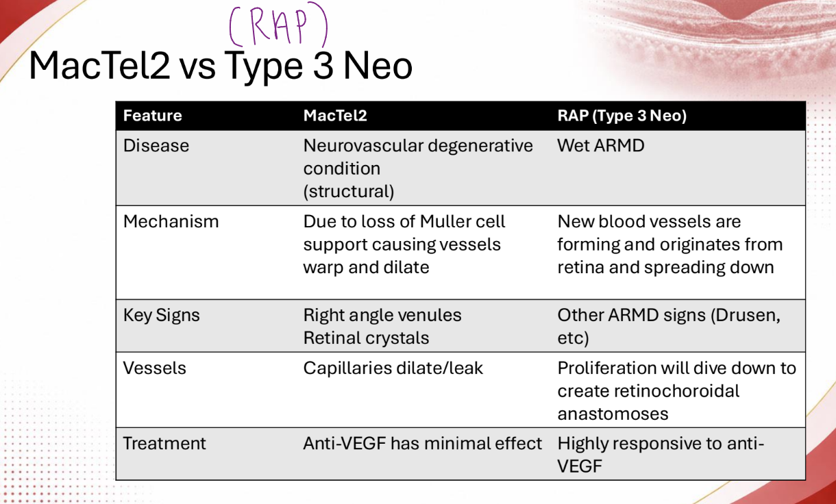
(mactel 1 / mactel 2) is more common
mactel 2

mactel 2, type of disease?
neurovascular degenerative condition (structural)

T/F: mactel 2 leakage is due to new blood vessels forming in the retina
false - likely due to muller cell support causing vessel dilation and warpage
Can CSCR and PED occur simultaneously?
yes

most typical treatment of VMT?
monitoring (spontaneous release)

At what stage of a macular hole will you first see a (+) Watzke-Allen sign?
Macular Hole - Stage 2

if you do an OCT and find a macular hole with an operculum, what stage is the mac hole?
Stage 3
At what stage macular hole should you consider more treatment than observation?
2, 3, 4

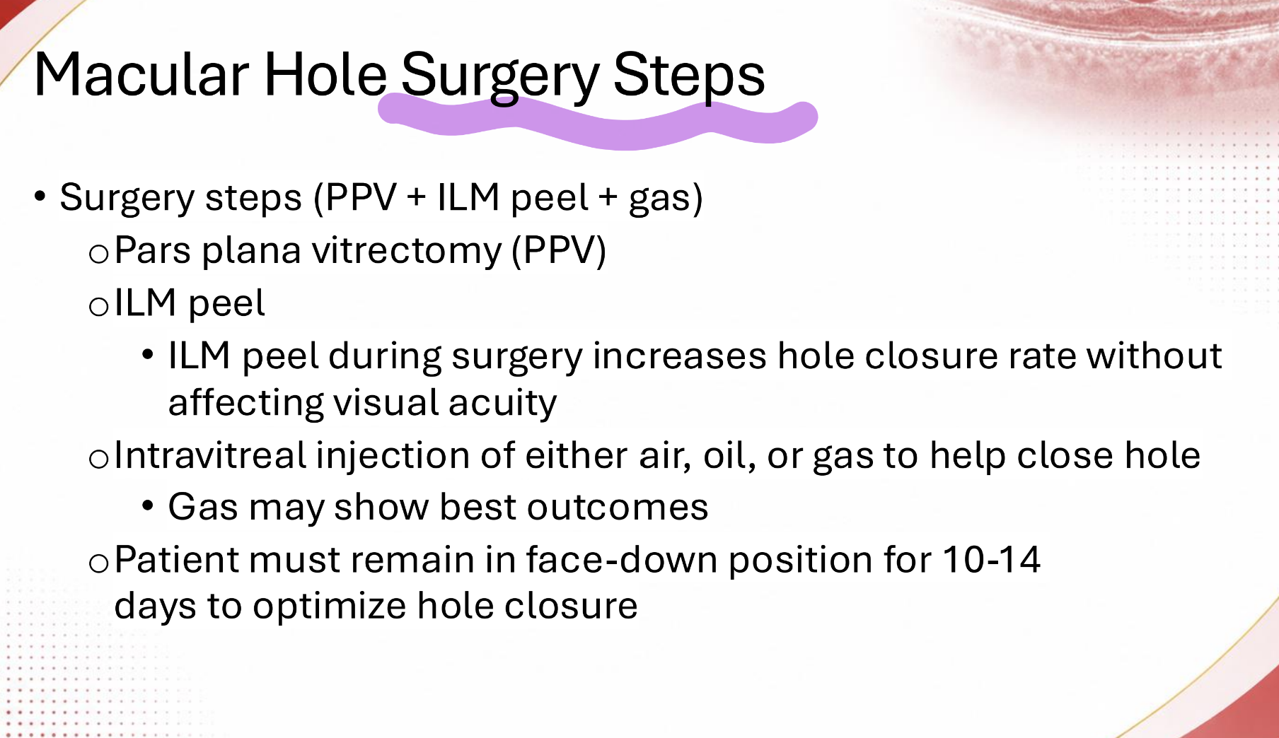
Mac Hole surgery steps?
PPV + ILM peel + Gas
ERM is more commonly caused by (aging / secondary) factors
aging (idiopathic)

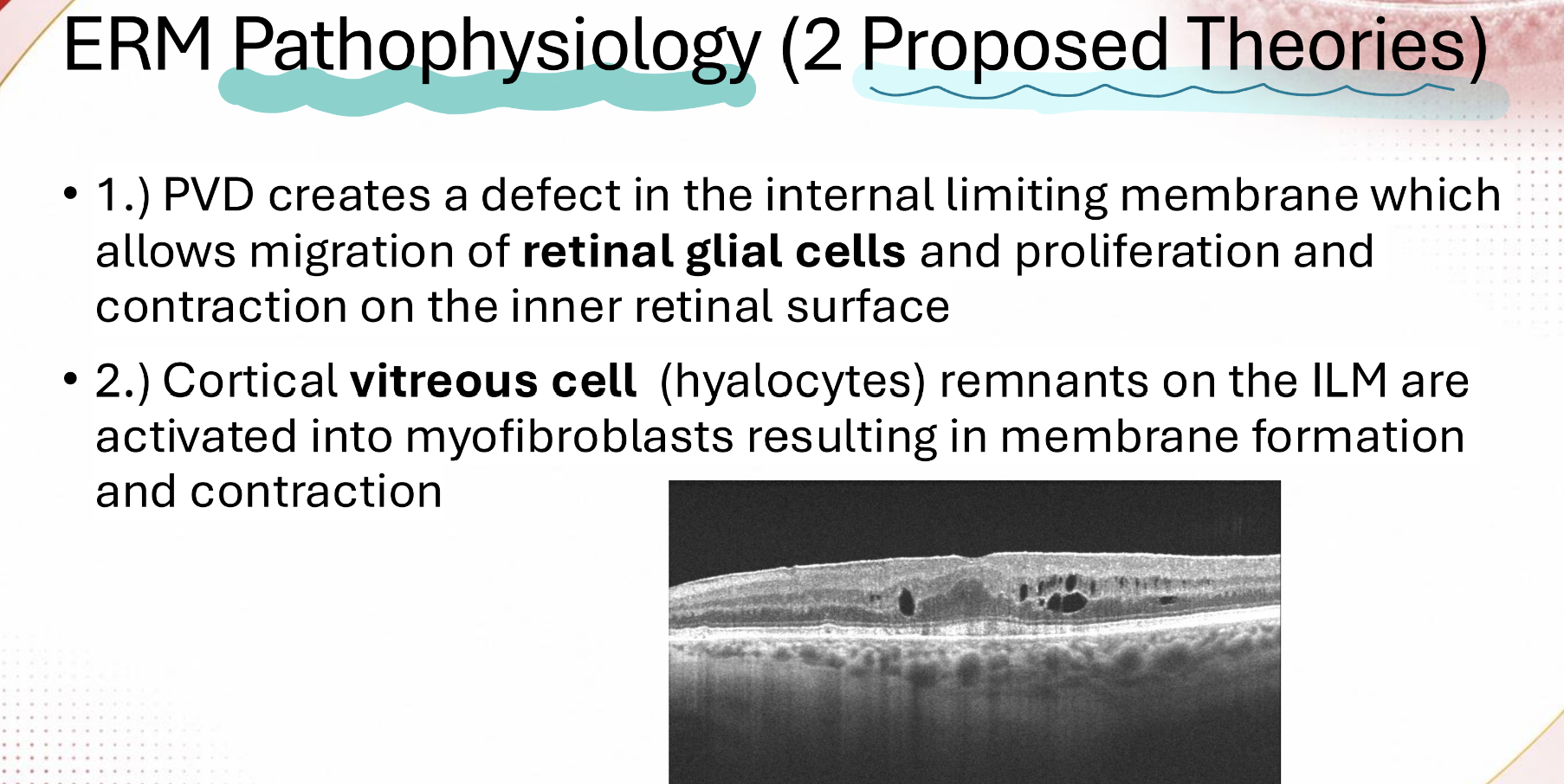
the two ERM pathophys theories involve what cell types?
retinal glial, vitreous cells (hyalocytes)
How will a pseudo-hole appear on Watzke-Allen Beam?
thinning

When might a vitrectomy/ membrane peel surgery be indicated for a patient with an ERM?
decrease in VA

retinal layers are split in a (pseudo macular/ lamellar) hole
lamellar

all retinal layers remain in a (pseudo macular/ lamellar) hole
pseudo-macular

CME think (pathophysiology):
inflammation

ocular dx associated with CME/ causes of CME: (8)
vascular,
post-op,
inflammation,
medication,
retinal dystrophy,
tractional,
anatomical,
neoplastic

medication causes of CME think:
prostaglandin analogs (-prost),
tamoxifen
retinal dystrophy causes of CME think:
retinitis pigmentosa
vascular CME treatment think:
anti-vegf

post-surgical CME treatment think:
Ketorolac (topical NSAID)
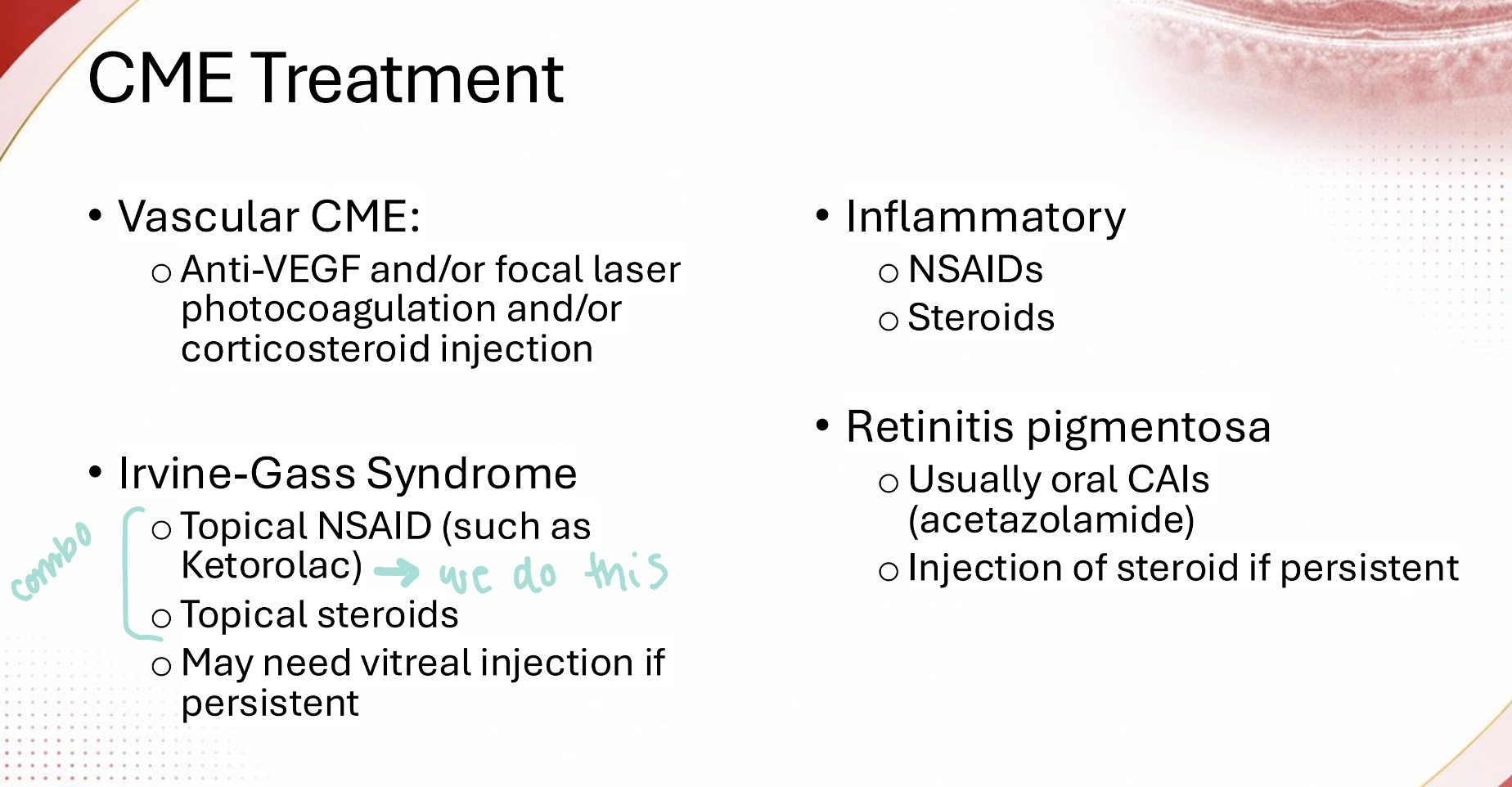
Retinitis Pigmentosa CME treatment think:
oral CAIs (acetazolamide)
