Ch 4 & 5 RAD1060 MIDTERM
 Call Kai
 Learn
Learn Practice Test
 Spaced Repetition
 Match
Match Flashcards
 Knowt Play
1/86

There's no tags or description
Looks like no tags are added yet.
Name | Mastery | Learn | Test | Matching | Spaced | Call with Kai | Chat |
|---|
No analytics yet
Send a link to your students to track their progress
87 Terms
AP Humerus
-greater tubercle is seen in profile laterally
-humeral head partially seen in profile medially
-minimal superimposition of glenoid cavity
-both lateral and medial epicondyles are in profile

AP internal humerus
-epicondyles are directly superimposed
-lesser tubercle shown in profile medially, partially superimposed by lower portion of glenoid cavity


trauma horizontal beam lateral: distal humerus
*fracture and dislocations of midhumerus and distal humerus
-elbow is flexed
-lateral projection of midhumerus and distal humerus, including elbow joint is visible
-long axis of humerus should be aligned with long axis of IR
-elbow is flexed 90*


AP external shoulder: proximal humerus
-greater tuberosity in full profile
-lesser tuberosity superimposed over humeral head


AP internal shoulder: proximal humerus
-lesser tuberosity in full profile on medial aspect of humeral head
-greater tubercle superimposed over humeral head


inferosuperior axial: shoulder (Lawrence Method)
-arm abducted 90* from body
-superior and inferior borders of glenoid cavity should be directly superimposed
-CR medially 25-30* centered horizontally to axilla and humeral head


Inferosuperior axial projection : shoulder (Clements Modification)
-arm abducted 90* from body
-relationship of humeral head and glenoid cavity should be evident
-lateral view of proximal humerus in relationship to scapulohumeral cavity shown


PA axial transaxillary: shoulder (Modified Bernageau Method)
-lateral view of proximal humerus in relationship to scapulohumeral articulated
-coracoid process of scapula seen on end
-arm is seen raised superiorly above the body


AP oblique: shoulder (Grashey Method)
-glenoid cavity seen in profile without superimposition of humeral head
-scapulohumeral joint space should be open
-Anterior and posterior rims of glenoid cavity are superimposed


tangential : Intertubercular (bicipital) sulcus: shoulder (Fisk modification)
-anterior margin of humeral head seen in profile
-humeral tubercles and the intertubercular sulcus are seen in profile
-intertubercular sulcus and tubercles in profile without superimposition of the acromion process

AP neutral: shoulder
-both the greater and lesser tubercles are superimposed by humeral head


Transthoracic lateral: Proximal humerus Trauma (Lawrence method)
-outline of the shaft of proximal humerus should be clearly visualized anterior to thoracic vertebrae
-relationship to humeral head and glenoid cavity


PA oblique: Scapular Y lateral: shoulder (Trauma)
-body of scapula should be seen without rib superimposition
-acromion and coracoid process should appear as symmetric upper limbs of the Y
-humeral head should not appear superimposed over the base of the Y if humerus is not dislocated
-true lateral view of the scapula


Tangential: Supraspinatus: Shoulder (Trauma) Neer Method
-10*-15* CR caudal angle
*demonstrates the coracoaromial arch for supraspinatus outlet region for possible shoulder impingement
-acromion and coracoid process should appear upper limbs of the Y
-humeral head appear superimposed and centered to the glenoid fossa just below supraspinatus outlet


AP Apical oblique axial: shoulder (Garth)
-coracoid process is projected over part of the humeral head, appears elongated
-acromion and AC joint are projected superior to humeral head


AP axial: clavicle
-CR 15-30* cephalad
-projects most of the clavicle above the scapula and second and third ribs
Thin (asthenic) pts require 25-30* CR angle
Thick (hypersthenic) pt require 15-20* CR angle

72in
what is the SID for bilateral AC joints

AP projection :Scapula
-lateral portion of the scapula free of superimposition
-medial portion of the scapula seen through thoracic structures
-arm abducted to 90*
-lateral border of the scapula free of superimposition


Lateral :Scapula
-best shows the acromion and coracoid processes
-

parallel
During Scapular Y: scapula what is the relationship of the acromion and coracoid process
perpendicular
during a scapular Y lateral: scapula what is the relationship of the superior angle of the scapula and AC joint articulation
humerus
largest bone of upper limb
true
AP radiograph of the shoulder taken with external rotation places the humerus in true AP
anteriorly
the lesser tuberosity is located laterally or anteriorly
laterally
the greater tuberosity is located laterally or anteriorly
acromion of scapula
the acromial extremity of clavicle articulates with the
sternoclavicular joint
the sternal extremity articulates with the manubrium called the
clavicle
long bone with double curvature
scapulohumeral joint
ball and socket
plane or gliding
sternoclavicular
plane or gliding
acromiclavicular
synovial/ diarthrodial joints
sternoclavicular, acromioclavicular, and scapulohumeral are what type of joints
parellel
for external rotation of shoulder the epicondyles line is ____ to the image receptor
-the greater tubercle, seen laterally in profile
-the lesser tubercle, located anteriorly, medial to greater tubercle
perpendicular
for internal rotation shoulder epicondyles are ______ to IR
humerus is in true lateral position
greater tuberosity is rotated around the anterior and medial aspect of proximal humerus
lesser tubercle seen in profile medially
45* angle to IR
in Neutral position of shoulder epicondyles are _____
-greater tubercle anteriorly, still lateral to lesser tubercle
female
clavicle is shorter and less curved
male
clavicle is thicker and more curved
costal surface
the anterior surface of the scapula is called
true
adult shoulders than measure 10-15cm require a grid
70-85
kvp range for adult humerus and shoulder
osteroarthritis
degenerative joint disease
-noninflammatory joint disease by gradual deterioration of articular cartilage with hypertrophic bone formation
-most common type of arthritis
hills sachs defect
-compression fracture of articular surface of posterolateral aspect of humeral head than often associated with anterior dislocation of humeral head
head of humerus
What is A?
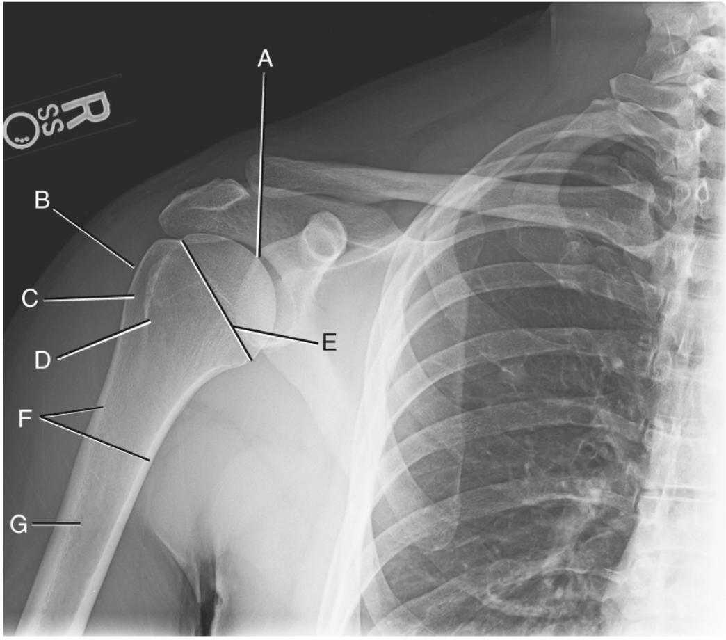
greater tuberosity
what is B?

intertubercular groove
what is C?
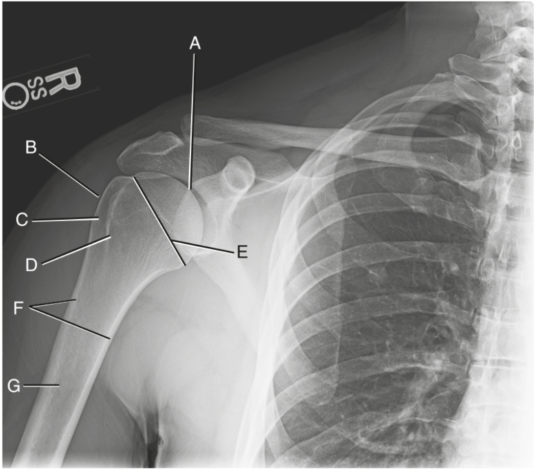
lesser tuberosity
what is D?

anatomic neck
what is E?
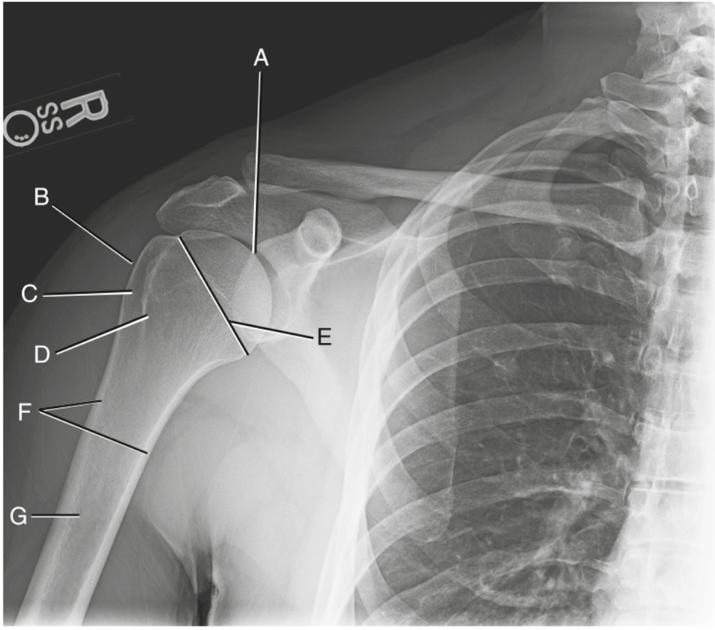
surgical neck
what is F?

body
what is G?

sternoclavicular joint
what is A?

sternal extremity
what is B?

body of clavicle
what is C?

acromical extremity
what is D?

acromioclavicular joint
what is E?

acromion
what is A?

neck of scapula
what is B?

suprascapular notch
what is C?

superior angle
what is D?

medial (vertebral) border
what is E?
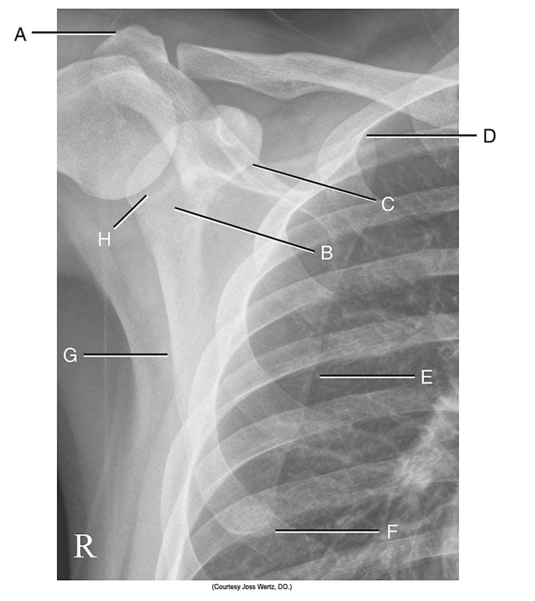
inferior angle
what is F?

lateral (axillary) border
what is G?

glenoid fossa
what is H?"

acromion
A?

coracoid process
B?
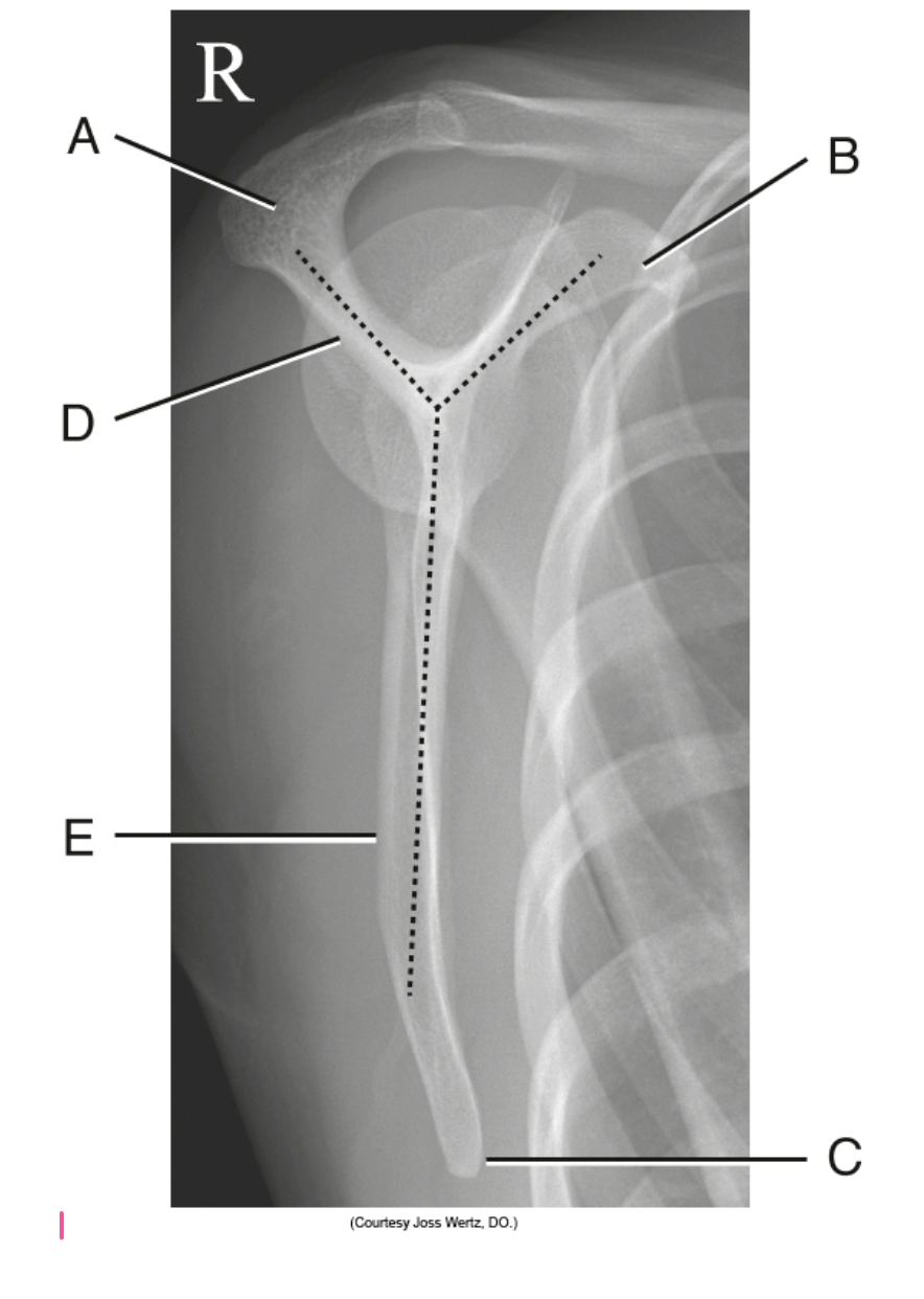
inferior angle
C?
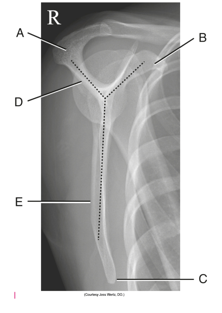
spine of scapula
D?

body of scapula
E?

external
what position or projection

internal
what position?

neutral
what position or projection

lesser tuberosity
A?

body of humerus
B?

capitulum
C?
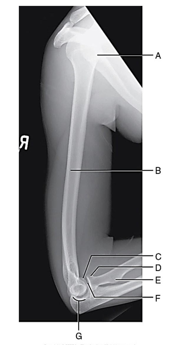
radius head
D?

radial tuberosity
E?

coronoid process
F?

trochlear notch
G?

midscapula, 2in inferior to coracoid process, 2in medial from lateral
what is the CR for the AP scapula
midvertebral border of scapula
what is CR for lateral scapula: scapular Y
30* caudally centered @ level of scapular spine to pass through scapulohumeral joint
what is the CR for PA axial transaxillary: shoulder, Modified Bernageau
scapulohumeral joint, 2in below AC joint
what is the CR AP/PA oblique: scapular Y lateral: shoulder
2in inferior & 2in medial from superolateral border
-midscapulohumeral joint
what is the CR AP oblique: shoulder; Grashey
1in inferior to coracoid process
what is the CR fro AP shoulder External/Internal/Neutral
mid diaphysis
what is the CR transthoracic lateral: Humerus trauma
midpoint between AC joints, 1in above Jugular notch
what is the CR for AC joint pearson method
medially 25*-30*, centered horizontally to axilla and humeral head
what is the CR for Inferosuperior axial: shoulder (Lawrence method)